

Tumor Segmentation in Contrast-Enhanced Magnetic Resonance Imaging for Nasopharyngeal Carcinoma: Deep Learning with Convolutional Neural Network
- PMID: 30417017
- PMCID: PMC6207874
- DOI: 10.1155/2018/9128527
Tumor Segmentation in Contrast-Enhanced Magnetic Resonance Imaging for Nasopharyngeal Carcinoma: Deep Learning with Convolutional Neural Network
Abstract
Objectives: To evaluate the application of a deep learning architecture, based on the convolutional neural network (CNN) technique, to perform automatic tumor segmentation of magnetic resonance imaging (MRI) for nasopharyngeal carcinoma (NPC).
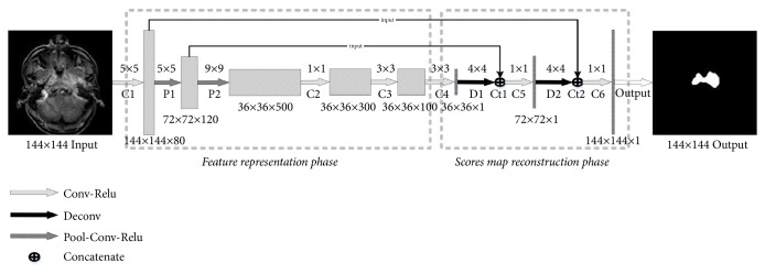
Materials and methods: In this prospective study, 87 MRI containing tumor regions were acquired from newly diagnosed NPC patients. These 87 MRI were augmented to >60,000 images. The proposed CNN network is composed of two phases: feature representation and scores map reconstruction. We designed a stepwise scheme to train our CNN network. To evaluate the performance of our method, we used case-by-case leave-one-out cross-validation (LOOCV). The ground truth of tumor contouring was acquired by the consensus of two experienced radiologists.
Results: The mean values of dice similarity coefficient, percent match, and their corresponding ratio with our method were 0.89±0.05, 0.90±0.04, and 0.84±0.06, respectively, all of which were better than reported values in the similar studies.
Conclusions: We successfully established a segmentation method for NPC based on deep learning in contrast-enhanced magnetic resonance imaging. Further clinical trials with dedicated algorithms are warranted.
Figures


References
-
- Hong Kong Cancer Registry. Hospital Authority of Hong Kong. http://www3.ha.org.hk/cancereg/
-
- International Agency for Research on Cancer. CI5 XI: Cancer incidence in five continents volume XI. http://ci5.iarc.fr/CI5-XI/Default.aspx.
-
- Huang B., Wong C.-S., Whitcher B., et al. Dynamic contrast-enhanced magnetic resonance imaging for characterising nasopharyngeal carcinoma: comparison of semiquantitative and quantitative parameters and correlation with tumour stage. European Radiology. 2013;23(6):1495–1502. doi: 10.1007/s00330-012-2740-7. - DOI - PubMed
MeSH terms
LinkOut - more resources
Full Text Sources
Medical
