. 2018 Oct 24;4(10):eaau3075.
doi: 10.1126/sciadv.aau3075. eCollection 2018 Oct.

Fear extinction requires ASIC1a-dependent regulation of hippocampal-prefrontal correlates

Affiliations

Fear extinction requires ASIC1a-dependent regulation of hippocampal-prefrontal correlates

Qin Wang et al. Sci Adv. .

Abstract

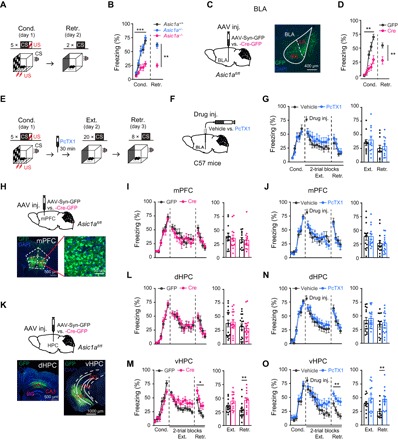
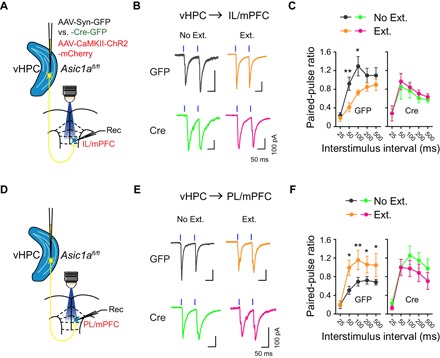
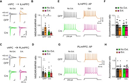
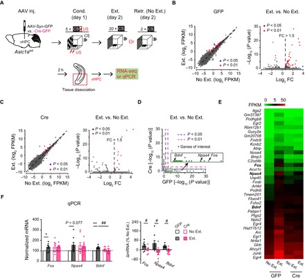
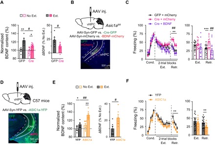
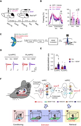
Extinction of conditioned fear necessitates the dynamic involvement of hippocampus, medial prefrontal cortex (mPFC), and basolateral amygdala (BLA), but key molecular players that regulate these circuits to achieve fear extinction remain largely unknown. Here, we report that acid-sensing ion channel 1a (ASIC1a) is a crucial molecular regulator of fear extinction, and that this function requires ASIC1a in ventral hippocampus (vHPC), but not dorsal hippocampus, mPFC, or BLA. While genetic disruption or pharmacological inhibition of ASIC1a in vHPC attenuated the extinction of conditioned fear, overexpression of the channel in this area promoted fear extinction. Channelrhodopsin-2-assisted circuit mapping revealed that fear extinction involved an ASIC1a-dependent modification of the long-range hippocampal-prefrontal correlates in a projection-specific manner. Gene expression profiling analysis and validating experiments identified several neuronal activity-regulated and memory-related genes, including Fos, Npas4, and Bdnf, as the potential mediators of ASIC1a regulation of fear extinction. Mechanistically, genetic overexpression of brain-derived neurotrophic factor (BDNF) in vHPC or supplement of BDNF protein in mPFC both rescued the deficiency in fear extinction and the deficits on extinction-driven adaptations of hippocampal-prefrontal correlates caused by the Asic1a gene inactivation in vHPC. Together, these results establish ASIC1a as a critical constituent in fear extinction circuits and thus a promising target for managing adaptive behaviors.


Figures

Fig. 1
Fig. 1. Brain region–specific roles of ASIC1a in fear acquisition or extinction.
(A to D) Effects of global or BLA-specific KO of Asic1a on cued fear acquisition. (A) Experimental scheme. (C) Left: Schematics of AAV injection. Right: Representative image of GFP expression (green) in a mouse that received AAV-Syn-Cre-GFP injection in BLA. 4′,6-Diamidino-2-phenylindole (DAPI) (blue) was used to label nuclei. (B and D) Time course of freezing response to CS during fear conditioning (Cond.) and retrieval (Retr.). (B) Left: Fear conditioning, two-way repeated-measures analysis of variance (ANOVA), main effect of genotype: F2,29 = 18.043, P < 0.001. Right: Fear retrieval, one-way factorial ANOVA for average freezing during CS of two trials, main effect of genotype, F2,29 = 25.856, P < 0.001. Asic1a+/+, n = 9; Asic1a+/−, n = 12; Asic1a−/−, n = 11. (D) Left: Fear conditioning, two-way repeated-measures ANOVA, main effect of AAV, F1,18 = 14.260, P = 0.001. Right: Fear retrieval, the average freezing during CS of two trials. n = 10 per group. **P < 0.01, unpaired Student’s t test. (E to G, J, N, and O) Effects of pharmacological inhibition of ASIC1a in BLA (G), mPFC (J), dHPC (N), or vHPC (O) on fear extinction. (E) Experimental scheme. (F) Schematics of cannula implantation and drug injection. (G, J, N, and O) Left: Time course of freezing response to CS during fear conditioning, extinction, and retrieval. Two-way repeated-measures ANOVA, main effect of drug, (G) BLA: fear extinction, F1,21 = 0.813, P = 0.377; fear retrieval, F1,21 = 0.236, P = 0.632; (J) mPFC: fear extinction, F1,26 = 0.002, P = 0.964; fear retrieval, F1,26 = 0.162, P = 0.690; (N) dHPC: fear extinction, F1,31 = 0.529, P = 0.472; fear retrieval, F1,31 = 0.143, P = 0.708; and (O) vHPC: fear extinction, F1,24 = 1.574, P = 0.222; fear retrieval, F1,24 = 12.659, P = 0.002. Right: Average freezing response to CS for all trials during fear extinction and retrieval. **P < 0.01, unpaired Student’s t test. n = 11 and 12, 14 and 14, 16 and 17, and 12 and 14 for vehicle and PcTX1 administered in BLA (G), mPFC (J), dHPC (N), and vHPC (O), respectively. (H, I, and K to M) Effects of genetic inactivation of Asic1a in mPFC (I), dHPC (L), or vHPC (M) on fear extinction. (H and K) Schematics of Asic1a gene inactivation in mPFC (H, upper), dHPC, or vHPC (K, upper). Representative images are shown for GFP expression (green) in mPFC (H, bottom), dHPC (K, bottom left), and vHPC (K, bottom right) of AAV-Syn-Cre-GFP–injected mice. DAPI (blue) was used to label nuclei. (I, L, and M) Left: Time course of freezing response to CS during fear conditioning, extinction, and retrieval. Two-way repeated-measures ANOVA, main effect of drug, (I) mPFC: fear conditioning, F1,24 = 0.188, P = 0.668; fear extinction, F1,24 = 0.172, P = 0.682; fear retrieval, F1,24 = 0.020, P = 0.889; (L) dHPC: fear conditioning, F1,32 = 0.029, P = 0.866; fear extinction, F1,32 = 0.079, P = 0.781; fear retrieval, F1,32 = 0.157, P = 0.695; (M) vHPC: fear conditioning, F1,26 = 2.293, P = 0.142; fear extinction, F1,26 = 4.533, P = 0.043; fear retrieval, F1,26 = 6.426, P = 0.018. Right: Average freezing response to CS for all trials during fear extinction and retrieval. **P < 0.01, unpaired Student’s t test. n = 13 and 13, 17 and 17, and 15 and 13 for GFP and Cre in mPFC (J), dHPC (N), and vHPC (O), respectively.
Fig. 2
Fig. 2. ASIC1a in vHPC is important for extinction learning–driven presynaptic changes at the vHPC→mPFC projections.
Effects of fear extinction learning on the PPRs for oEPSCs at vHPC→IL/mPFC (A to C) and vHPC→PL/mPFC (D to F) projections and the dependence of these effects on ASIC1a in vHPC. (A and D) Experimental schemes. (B and E) Representative traces of oEPSCs at vHPC→IL/mPFC (B) or vHPC→PL/mPFC (E) synapses in No Ext. and Ext. groups of GFP (top) and Cre (bottom) mice induced by paired photostimulations (blue vertical bars) with a 100-ms interval. (C and F) Summary of PPRs of oEPSCs plotted against interstimulus intervals at the vHPC→IL/mPFC (C) or vHPC→PL/mPFC (F) synapses. Two-way repeated-measures ANOVA, main effect of behavior, (C) vHPC→IL/mPFC: GFP, F1,118 = 15.33, P < 0.001 (left); Cre, F1,148 = 0.541, P = 0.463 (right); (F) vHPC→PL/mPFC: GFP, F1,178 = 23.17, P < 0.001 (left); Cre, F1,133 = 4.478, P = 0.036 (right). *P < 0.05, **P < 0.01, Ext. versus No Ext., unpaired Student’s t test. (C) vHPC→IL/mPFC: GFP, No Ext., n = 13 neurons of 10 mice; GFP, Ext., n = 11 neurons of 7 mice; Cre, No Ext., n = 18 neurons of 8 mice; Cre Ext., n = 14 neurons of 8 mice. (F) vHPC→PL/mPFC: GFP, No Ext., n = 19 neurons of 11 mice; GFP, Ext., n = 17 neurons of 11 mice; Cre, No Ext., n = 16 neurons of 7 mice; Cre, Ext., n = 12 neurons of 8 mice.
Fig. 3
Fig. 3. ASIC1a in vHPC is required for extinction learning–driven postsynaptic changes at the vHPC→mPFC projections and mPFC neuronal activity pattern.
(A to D) Effects of fear extinction learning on the NMDAR/AMPAR ratios of vHPC→IL/mPFC (A and B) and vHPC→PL/mPFC (C and D) projections and the dependence of these effects on ASIC1a in vHPC. (A and C) Representative traces of AMPAR- and NMDAR-mediated currents in response to photostimulation of vHPC fibers at vHPC→IL/mPFC (A) or vHPC→PL/mPFC (C) synapses in No Ext. and Ext. groups of GFP (left) and Cre (right) mice. (B and D) Ratios of peak NMDAR- to AMPAR-mediated currents at vHPC→IL/mPFC (B) or vHPC→PL/mPFC (D) synapses for individual neurons and the summary. *P < 0.05, Ext. versus No Ext.; #P < 0.05, GFP versus Cre, unpaired Student’s t test. (B) vHPC→IL/mPFC: GFP, No Ext., n = 9 neurons of 7 mice; GFP, Ext., n = 11 neurons of 8 mice; Cre, No Ext., n = 16 neurons of 8 mice; Cre, Ext., n = 6 neurons of 4 mice. (D) vHPC→PL/mPFC: GFP, No Ext., n = 16 neurons of 9 mice; GFP, Ext., n = 13 neurons of 9 mice; Cre, No Ext., n = 12 neurons of 7 mice; Cre, Ext., n = 6 neurons of 6 mice. (E to H) Action potential firing of pyramidal neurons in layer II/III of IL/mPFC (E and F) or PL/mPFC (G and H) to a depolarizing current injection (+250 pA) recorded ex vivo. (E and G) Representative traces of action potential firing recorded from the IL/mPFC (E) or PL/mPFC (G) neurons. (F and H) Accommodation ratios of the action potential number in IL/mPFC (F) or PL/mPFC (H) neurons during the last 100-ms current injection to that during the first 100-ms current injection for individual neurons and the summary. *P < 0.05, Ext. versus No Ext, unpaired Student’s t test. (F) IL/mPFC: GFP, No Ext., n = 16 neurons of 7 mice; GFP, Ext., n = 14 neurons of 5 mice; Cre, No Ext., n = 16 neurons of 6 mice; Cre, Ext., n = 25 neurons of 9 mice. (H) PL/mPFC: GFP, No Ext., n = 19 neurons of 6 mice; GFP, Ext., n = 22 neurons of 6 mice; Cre, No Ext., n = 20 neurons of 6 mice; Cre, Ext., n = 19 neurons of 7 mice.
Fig. 4
Fig. 4. ASIC1a regulates extinction-associated gene expression profiles in vHPC.
(A) Experimental scheme. (B) Left: Differential enrichment calling of vHPC mRNA of GFP mice, showing 38 genes (blue and red dots) enriched in the Ext. group with P < 0.05, after adjusting for multiple comparisons across the detected gene repertoire (Fisher’s exact test for multiple group comparison with Benjamini-Hochberg FDR correction; n = 2 samples from 8 mice per group). Broken lines represent unit slope. Right: Volcano plot showing vHPC mRNA with absolute fold change (FC) values > 1.5 in the Ext. group. (C) Similar to (B) but for vHPC mRNA of Cre mice, showing 14 genes (blue and red dots) enriched in the Ext. group with P < 0.05 (n = 2 samples from 8 mice per group). (D) Scatter plot comparing P values of enrichment in Ext. samples for Cre versus GFP mice. The red dashed lines indicate P = 0.01, and the blue dashed lines represent P = 0.05. Note the genes in the bottom right corner, shown in the inset, which are enriched in the Ext. samples only for GFP mice. These include activity-induced immediate-early gene, Fos, and plasticity- or memory-related genes, Npas4 and Bdnf. (E) Heatmap of 36 genes enriched by fear extinction in GFP, but not Cre, mice. (F) mRNA levels determined by qPCR. Fos, n = 8; Npas4, n = 8; Bdnf, n = 6 to 8 per group. Left: Gene expression levels relative to the control (GFP, No Ext.). Right: Relative difference between Ext. and No. Ext. of mice injected with the same AAV construct. *P < 0.05, **P < 0.01, Ext. versus No Ext; ##P < 0.01, GFP versus Cre, unpaired Student’s t test. FPKM, fragments per kilobase of transcript per million; RNA-seq, RNA sequencing.
Fig. 5
Fig. 5. Extinction learning–dependent BDNF expression mediates the effect of vHPC ASIC1a on cued fear extinction.
(A) BDNF protein levels determined by ELISA in vHPC of Asic1aflox/flox mice infected with AAV-Syn-GFP (GFP) or AAV-Syn-Cre-GFP (Cre) and subjected to fear extinction (Ext.) or retrieval without extinction (No Ext.) (GFP, n = 6; Cre, n = 7 per group). Individual data points and the summary. Left: BDNF protein levels relative to the control (GFP, No Ext.). Right: Relative difference between Ext. and No. Ext. of mice injected with the same AAV construct. *P < 0.05, **P < 0.01, Ext. versus No Ext; #P < 0.05, GFP versus Cre, unpaired Student’s t test. (B and C) In situ BDNF overexpression rescued the deficiency of cued fear extinction in vHPC Asic1a gene–inactivated mice. (B) Top: Schematics of AAV-mediated genetic manipulation on Asic1aflox/flox mice. Bottom: Representative images are shown for the expression of GFP (green) and mCherry (red) in vHPC. (C) Time course of freezing response to CS during fear conditioning, extinction, and retrieval. Left: Two-way repeated-measures ANOVA, main effect of AAV, fear conditioning, F2,44 = 0.173, P = 0.841; fear extinction, F2,44 = 1.949, P = 0.154; fear retrieval, F2,44 = 8.955, P = 0.001, followed by post hoc Bonferroni, P = 0.005 for Cre + mCherry versus GFP + mCherry, P = 0.001 for Cre + BDNF versus Cre + mCherry, P = 1.000 for Cre + BDNF versus GFP + mCherry. Right: One-way factorial ANOVA for average freezing during CS of all trials, main effect of AAV, fear extinction, F2,44 = 1.946, P = 0.155; fear retrieval, F2,44 = 8.272, P = 0.001, followed by post hoc Bonferroni, P = 0.002 for Cre + mCherry versus GFP + mCherry, P = 0.001 for Cre + BDNF versus Cre + mCherry, P = 0.506 for Cre + BDNF versus GFP + mCherry. GFP + mCherry, n = 9; Cre + mCherry, n = 12; Cre + BDNF, n = 11. (D to F) Asic1a gene overexpression in vHPC increased extinction-associated BDNF expression in vHPC and enhanced fear extinction. (D) Top: Schematics of AAV-mediated genetic manipulation on C57BL/6J mice. Bottom: Representative images are shown for the expression of YFP (green) in vHPC. (E) Effects of Asic1a gene overexpression in vHPC on BDNF protein levels. Left: BDNF protein levels relative to the control (YFP, No Ext.). Right: Relative difference between Ext. and No. Ext. of mice injected with the same AAV construct. *P < 0.05, **P < 0.01, Ext. versus No Ext; #P < 0.05, GFP versus Cre, unpaired Student’s t test. n = 6 per group. (F) Time course of freezing response to CS during fear conditioning, extinction, and retrieval. Left: Two-way repeated-measures ANOVA, main effect of AAV, fear conditioning, F1,30 = 4.213, P = 0.050; fear extinction, F1,30 = 1.348, P = 0.255; fear retrieval, F1,30 = 11.183, P = 0.002. Right: Average freezing responses. YFP, n = 19; ASIC1a, n = 13. **P < 0.01, YFP versus ASIC1a, unpaired Student’s t test.
Fig. 6
Fig. 6. ASIC1a in vHPC regulates adaptive changes of vHPC-mPFC synapses via BDNF signaling, which underlies cued fear extinction.
(A) Experimental scheme. (B) Effects of BDNF infusion into mPFC on the cued fear extinction in vHPC Asic1a gene–inactivated mice. Time course of freezing response to CS during fear conditioning, extinction, and retrieval. Left: Two-way repeated-measures ANOVA, main effect of AAV plus drug, fear conditioning, F2,43 = 2.152, P = 0.129; fear extinction, F2,43 = 2.875, P = 0.067; fear retrieval, F2,43 = 9.263, P < 0.001, followed by post hoc Bonferroni, P = 0.005 for Cre + vehicle versus GFP + vehicle, P = 0.001 for Cre + BDNF versus Cre + vehicle, P = 1.000 for Cre + BDNF versus GFP + vehicle. Right: One-way factorial ANOVA for average freezing during CS for all trials, main effect of AAV, fear extinction, F2,43 = 2.883, P = 0.067; fear retrieval, F2,43 = 9.263, P < 0.001, followed by post hoc Bonferroni, P = 0.005 for Cre + vehicle versus GFP + vehicle, P = 0.001 for Cre + BDNF versus Cre + vehicle, P = 1.000 for Cre + BDNF versus GFP + vehicle. GFP + vehicle, n = 15; Cre + vehicle, n = 15; Cre + BDNF, n = 16. (C) Experimental schemes. Animals were treated and brain slides were prepared as in Fig. 2. Brain slices containing mPFC were then incubated with BDNF (200 ng/ml in aCSF) or vehicle (aCSF) for 2 hours before electrophysiological recordings. (D) Representative traces of AMPAR- and NMDAR-mediated oEPSCs at vHPC→IL/mPFC (left) and vHPC→PL/mPFC (right) synapses with or without BDNF pretreatment of mPFC slice. (E) Ratios of peak NMDAR- to AMPAR-mediated oEPSCs at vHPC→IL/mPFC and vHPC→PL/mPFC synapses for individual neurons and the summary. vHPC→IL/mPFC: vehicle, n = 6 neurons of 5 mice; BDNF, n = 28 neurons of 9 mice; vHPC→PL/mPFC: vehicle, n = 5 neurons of 4 mice; BDNF, n = 7 neurons of 5 mice. **P < 0.01, vehicle versus BDNF, unpaired Student’s t test. (F) Working model of vHPC ASIC1a-dependent regulation of fear extinction. ASIC1a activity drives the expression of BDNF in vHPC in response to fear extinction training, which is followed by an antegrade BDNF signaling to modify the synaptic efficacy of vHPC→mPFC projections (see the main text for more details).

References

    1. Milad M. R., Quirk G. J., Fear extinction as a model for translational neuroscience: Ten years of progress. Annu. Rev. Psychol. 63, 129–151 (2012). - PMC - PubMed
    1. Dunsmoor J. E., Niv Y., Daw N., Phelps E. A., Rethinking extinction. Neuron 88, 47–63 (2015). - PMC - PubMed
    1. Milad M. R., Quirk G. J., Neurons in medial prefrontal cortex signal memory for fear extinction. Nature 420, 70–74 (2002). - PubMed
    1. Morgan M. A., Romanski L. M., LeDoux J. E., Extinction of emotional learning: Contribution of medial prefrontal cortex. Neurosci. Lett. 163, 109–113 (1993). - PubMed
    1. Burgos-Robles A., Vidal-Gonzalez I., Santini E., Quirk G. J., Consolidation of fear extinction requires NMDA receptor-dependent bursting in the ventromedial prefrontal cortex. Neuron 53, 871–880 (2007). - PubMed

Publication types

MeSH terms

Substances

LinkOut - more resources