. 2018 Nov 6;3(6):e00083-18.
doi: 10.1128/mSystems.00083-18. eCollection 2018 Nov-Dec.

Multiple Sclerosis-Associated Changes in the Composition and Immune Functions of Spore-Forming Bacteria

Affiliations

Multiple Sclerosis-Associated Changes in the Composition and Immune Functions of Spore-Forming Bacteria

Egle Cekanaviciute et al. mSystems. .

Abstract

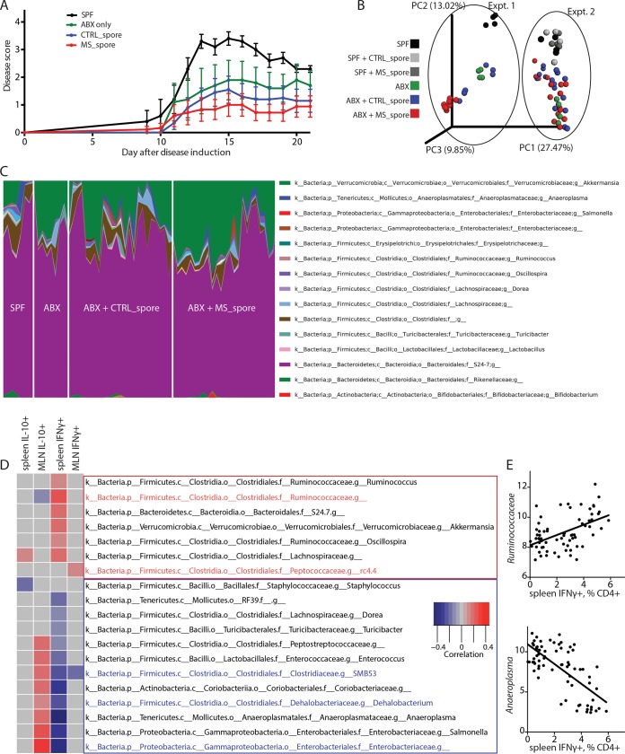
Multiple sclerosis (MS) is an autoimmune disease of the central nervous system characterized by adaptive and innate immune system dysregulation. Recent work has revealed moderate alteration of gut microbial communities in subjects with MS and in experimental, induced models. However, a mechanistic understanding linking the observed changes in the microbiota and the presence of the disease is still missing. Chloroform-resistant, spore-forming bacteria, which primarily belong to the classes Bacilli and Clostridia in the phylum Firmicutes, have been shown to exhibit immunomodulatory properties in vitro and in vivo, but they have not yet been characterized in the context of human disease. This study addresses the community composition and immune function of this bacterial fraction in MS. We identify MS-associated spore-forming taxa (primarily in the class Clostridia) and show that their presence correlates with impaired differentiation of IL-10-secreting, regulatory T lymphocytes in vitro. Colonization of antibiotic-treated mice with spore-forming bacteria allowed us to identify some bacterial taxa favoring IL-10+ lymphocyte differentiation and others inducing differentiation of proinflammatory, IFN-γ+ T lymphocytes. However, when fed into antibiotic-treated mice, both MS and control-derived spore-forming bacteria were able to induce similar IL-10-expressing Treg immunoregulatory responses, thus ameliorating symptoms of experimental allergic encephalomyelitis (EAE). Our analysis also identified Akkermansia muciniphila as a key organism that may interact either directly or indirectly with spore-forming bacteria to exacerbate the inflammatory effects of MS-associated gut microbiota. Thus, changes in the spore-forming fraction may influence T lymphocyte-mediated inflammation in MS. This experimental approach of isolating a subset of microbiota based on its functional characteristics may be useful to investigate other microbial fractions at greater depth. IMPORTANCE To address the impact of microbiome on disease development, it is essential to go beyond a descriptive study and evaluate the physiological importance of microbiome changes. Our study integrates computational analysis with in vitro and in vivo exploration of inflammatory properties of spore-forming microbial communities, revealing novel functional correlations. We specifically show that while small differences exist between the microbiomes of MS patients and healthy subjects, these differences are exacerbated in the chloroform-resistant fraction. We further demonstrate that, when purified from MS patients, this fraction is correlated with impaired immunomodulatory responses in vitro.

Keywords: immune mechanisms; multiple sclerosis; spore-forming bacteria.


Figures

FIG 1
FIG 1
Differences in community composition of spore-forming bacterial fraction in MS patients and healthy controls. (A to C) Comparison of microbial community composition of spore-forming bacterial subset and total stool bacteria in untreated MS patients (n = 25) and controls (n = 24). (A) Chao1 metric of alpha diversity. (B) Median and range of distances (unweighted UniFrac distance matrix) within and between sample groups. (C) Mean relative abundance of microbial genera. (D to F) Comparison of relative abundances of individual microbial taxa in untreated MS patients (n = 25) and controls (n = 24). (D) Volcano plot of relative abundance distribution of microbial OTUs. x axis, log2 fold of relative abundance ratio between MS patients and controls after variance-stabilizing transformation. y axis, negative log10 of P value, negative binomial Wald test, Benjamini-Hochberg correction for multiple comparisons. (E and F) Relative abundances of bacterial classes Bacilli (E) and Clostridia (F) within phylum Firmicutes out of spore-forming bacteria from controls and MS patients. Error bars, mean ± SEM. CTRL, total stool bacteria from controls. CTRL_spore, spore-forming bacteria from controls. MS, total stool bacteria from MS patients. MS_spore, spore-forming bacteria from MS patients.
FIG 2
FIG 2
Spore-forming bacteria from MS patients inhibit IL-10+ Treg differentiation in vitro. (A and B) Representative flow cytometry plots (A) and quantification (B) of CD4+ FoxP3+ Tregs within CD3+ lymphocytes differentiated in response to spore-forming bacteria isolated from controls or untreated MS patients. n = 7 PBMC donors; each dot represents an average response from PBMC donor to isolates from 6 control or MS bacterial donors. **, P < 0.01, two-tailed repeated measures t test. (C and D) Representative flow cytometry plots (C) and quantification (D) of IL-10+ lymphocyte population within CD3+ CD4+ FoxP3+ Tregs differentiated in response to spore-forming bacteria isolated from controls or untreated MS patients. n = 6 bacterial donors per group. *, P < 0.05, two-tailed t test. Error bars, mean ± SEM. The experiment was repeated with nonoverlapping PBMC and bacterial donors and gave the same results. (E) Quantification of T effector cell proliferation in response to Tregs differentiated in the presence of spore-forming bacteria from MS patients or controls. n = 3 bacterial donors per group, each representing an average of 3 technical replicates. (F) Linear correlation between IL-10+ population within CD3+ CD4+ FoxP3+ Tregs and Clostridia-Bacilli relative abundances. R2 = 0.214, P = 0.0459. Black dots, MS patients. Light gray dots, controls.
FIG 3
FIG 3
Spore-forming bacterial composition is correlated with T lymphocyte phenotypes in vivo. (A) Clinical EAE scores of mice that after antibiotic treatment had been colonized with spore-forming bacteria from controls (CTRL_spore) or MS patients (MS_spore) for 2 weeks or kept on antibiotics (ABX) or under SPF conditions as controls, prior to induction of EAE at 9 to 10 weeks of age. n = 5 to 10 mice per group. (B and C) Principal coordinate plot of beta diversity (PCoA; unweighted UniFrac) (B) and genus-level taxonomical distribution (C) of mouse fecal microbiota at 2 weeks of colonization with spore-forming bacteria, 2 separate experiments. (D) Bacterial genera whose abundance is correlated with changes in immune cell differentiation in antibiotic-treated and recolonized mice are shown. The linear correlation between relative abundances of bacterial genera and the percentage of IL-10+ regulatory and IFN-γ+ Th1 out of CD4+ Th lymphocytes from both spleens and mesenteric lymph nodes (MLN) of mice colonized with spore-forming bacteria is depicted as a heat map. Same samples as in panels B and C. Only the genera that show significant linear correlation with immune parameters (P > 0.05 after Benjamini-Hochberg adjustment for multiple comparisons) are included in the heat map. Red rectangle, putative proinflammatory subset. Blue rectangle, putative anti-inflammatory subset. Red font, taxa significantly increased in mice colonized with spore-forming bacteria from MS patients compared to controls. Blue font, taxa significantly reduced in mice colonized with spore-forming bacteria from MS patients compared to controls. (E) Examples of positive and negative correlation between bacteria and Th lymphocyte differentiation from panel D.
FIG 4
FIG 4
Increased Akkermansia is linked with MS-associated changes in spore-forming bacteria and proinflammatory T lymphocytes. (A) Principal coordinate plot of beta diversity (PCoA; unweighted UniFrac) of mouse fecal microbiota excluding Akkermansia at 2 weeks of colonization with spore-forming bacteria, 2 separate experiments, colored by Akkermansia presence (red to green: low to high). P < 0.001, significant contribution of Akkermansia presence to determining distance variation (Adonis method for continuous variables). (B) Relative abundance of Akkermansia in controls and MS patients used for isolation of spore-forming bacteria. P = 1.5E−09, negative binomial Wald test, Benjamini-Hochberg correction for multiple comparisons (across all 144 species detected in the data set). (C) Linear correlation of relative abundance of Akkermansia with IFN-γ+ Th1 lymphocyte differentiation in spleens of mice colonized with spore-forming bacteria. R2 = 0.18, P = 0.0003. (D) Bacterial genera significantly correlated with Akkermansia in vivo.

References

    1. Itoh K, Mitsuoka T. 1985. Comparison of media for isolation of mouse anaerobic faecal bacteria. Lab Anim 19:353–358. doi:10.1258/002367785780887464. - DOI - PubMed
    1. Itoh K, Mitsuoka T. 1985. Characterization of clostridia isolated from faeces of limited flora mice and their effect on caecal size when associated with germ-free mice. Lab Anim 19:111–118. doi:10.1258/002367785780942589. - DOI - PubMed
    1. Kearney SM, Gibbons SM, Poyet M, Gurry T, Bullock K, Allegretti JR, Clish CB, Alm EJ. 2018. Endospores and other lysis-resistant bacteria comprise a widely shared core community within the human microbiota. ISME J 12:2403–2416. doi:10.1038/s41396-018-0192-z. - DOI - PMC - PubMed
    1. Gaboriau-Routhiau V, Rakotobe S, Lecuyer E, Mulder I, Lan A, Bridonneau C, Rochet V, Pisi A, De Paepe M, Brandi G, Eberl G, Snel J, Kelly D, Cerf-Bensussan N. 2009. The key role of segmented filamentous bacteria in the coordinated maturation of gut helper T cell responses. Immunity 31:677–689. doi:10.1016/j.immuni.2009.08.020. - DOI - PubMed
    1. Atarashi K, Tanoue T, Oshima K, Suda W, Nagano Y, Nishikawa H, Fukuda S, Saito T, Narushima S, Hase K, Kim S, Fritz JV, Wilmes P, Ueha S, Matsushima K, Ohno H, Olle B, Sakaguchi S, Taniguchi T, Morita H, Hattori M, Honda K. 2013. Treg induction by a rationally selected mixture of Clostridia strains from the human microbiota. Nature 500:232–236. doi:10.1038/nature12331. - DOI - PubMed