. 2016 Mar 1;6(2):114-122.
doi: 10.3174/ng.2160150.

Central Nervous System and Head and Neck Histiocytoses: A Comprehensive Review on the Spectrum of Imaging Findings

Affiliations

Central Nervous System and Head and Neck Histiocytoses: A Comprehensive Review on the Spectrum of Imaging Findings

S S Hashmi et al. Neurographics (2011). .

Abstract

The histiocytoses are a rare group of varied but related disorders characterized by abnormal tissue proliferation of macrophages and dendritic cells within tissues. The purpose of this article was to review the imaging findings in patients presenting with CNS and with head and neck manifestations of these disorders. Histiocytoses include but are not limited to Rosai-Dorfman disease, Erdheim Chester disease, Langerhans cell histiocytosis, histiocytic sarcoma, and juvenile xanthogranuloma. A review of the literature was performed to determine the sites of disease involvement. This article includes the demographics, histopathologic criteria for diagnosis, and imaging features of these histiocytoses, and describes the manifestations in locations known to harbor disease: intraaxial and extra-axial intracranial regions, the calvaria, skull base, hypothalamopituitary axis, orbits, paranasal sinuses, spine, and the head and neck region. Histiocytoses have variable imaging appearances in the CNS and in the head and neck region, and radiologists should be aware of the spectrum of findings to avoid mistaking them for other disease processes.

Learning objective: To understand the general pathophysiology, clinical presentation, and typical imaging characteristics of the most common histiocytoses; comprehend the morphologic and immunohistochemical characteristics of these histiocytoses and the hallmark findings on pathology; and be able to differentiate between these disorders based on their most common presentations.


Figures

Fig. 1
Fig. 1
ECD, H&E stained tissue section (magnification 100×). Monotonous foamy histiocytes are scattered in a fibrotic background in ECD.
Fig. 2
Fig. 2
Rosai-Dorfman disease, H&E stained tissue section (magnification 100×). Rosai-Dorfman disease, showing sheets of plump histiocytes with prominent engulfing of inflammatory cells (emperipolesis), which remain intact and are identified within the histiocytic cytoplasm (arrows).
Fig. 3
Fig. 3
LCH, H&E stained tissue section (magnification 100×). LCH is composed of slightly elongated histiocytes with nuclear groves in a sea of eosinophils. Immunohistochemical evaluation for CD1a highlights the abnormal histiocytes in LCH.
Fig. 4
Fig. 4
Histiocytic sarcoma, H&E stained tissue section (magnification 100×). The tumor cells are in sheets with numerous mitotic figures. By immunohistochemical evaluation, the neoplastic cells are positive for CD68 and S100, and negative for CD1a (not shown).
Fig. 5
Fig. 5
Juvenile xanthogranuloma, H&E stained tissue section (magnification 100×). A nasal lesion from a 3-year-old with juvenile xanthogranuloma shows squamous epithelium with underlying cellular infiltrate composed of mononuclear cells (histiocytes) and Touton giant cells (the nuclei form ringlike pattern) (arrow).
Fig. 6
Fig. 6
Supratentorial, extra-axial location. (A) A 50-year-old man with Rosai-Dorfman disease. Axial T1 postcontrast MR imaging shows multiple homogenously enhancing extra-axial masses along the cerebral convexities and the skull base (large arrows). The right optic nerve sheath is also involved (small arrows) (B) A 48-year-old woman with ECD. Axial T1 postcontrast MR imaging with multiple enhancing dural-based masses (large arrows), with hypointense spicules radiating outward from the center of each mass (small arrows).
Fig. 7
Fig. 7
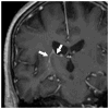
Supratentorial, intra-axial location. A 60-year-old man with ECD. Coronal T1 postcontrast MR imaging shows ependymal involvement, with extension to the right basal ganglia (arrows). This enhancement resolved after treatment with interferon.
Fig. 8
Fig. 8
Infratentorial, intra-axial location. (A) A 14-year-old boy with Rosai-Dorfman disease. Axial T1 postcontrast MR imaging demonstrates an enhancing intra-axial lesion within the right cerebellar hemisphere without mass effect. (B, C) A 60-year-old man with ECD. Axial T1 postcontrast and axial FLAIR MR imaging demonstrates enhancement and FLAIR hyperintensity oriented in a transverse pattern across the pons.
Fig. 9
Fig. 9
Hypothalamopituitary axis. (A) An 8-year-old girl with LCH. Coronal T1 postcontrast MR imaging demonstrates involvement of the pituitary stalk and hypothalamus (arrow). (B) A 53-year-old man with Rosai-Dorfman disease. Sagittal T1 postcontrast MR imaging illustrates disease in the sella and suprasellar region (arrow). (C) The same patient as in (B), coronal T1 postcontrast MR imaging with extension to involve the hypothalamus (large arrows) and laterally into the cavernous sinuses (small arrows).
Fig. 10
Fig. 10
Orbits. A 47-year-old man with ECD. Axial T1 postcontrast MR imaging with disease that involves the retrobulbar orbit (large arrows), with extension through the superior orbital fissure and posteriorly into the cavernous sinuses (small arrows).
Fig. 11
Fig. 11
Eyelids and lacrimal glands. A 53-year-old woman with juvenile xanthogranuloma. Coronal T1 postcontrast MR imaging shows disease that involves the left eyelid, lacrimal gland, and periorbital soft tissues (large arrows). Disease is also present in the sinonasal cavity and the right lacrimal gland (small arrows).
Fig. 12
Fig. 12
Calvaria. A 10-year-old boy with LCH. (A) Plain radiographs show a lytic frontal calvarial lesion (arrow). (B) Coronal T1 postcontrast MR imaging demonstrates a calvarial mass with intracranial extension and involvement of the adjacent dura (arrow).
Fig. 13
Fig. 13
Skull base. A 8-year-old girl with LCH. Axial T1 postcontrast MR imaging demonstrates involvement of the sphenoid bone and left temporal calvaria (large arrows), with extension into the orbits (small arrows).
Fig. 14
Fig. 14
Paranasal sinuses. A 63-year-old woman with Rosai-Dorfman disease. Axial T1 postcontrast MR imaging shows disease filling the maxillary sinuses (arrows).
Fig. 15
Fig. 15
Spine. A 56-year-old woman with Rosai-Dorfman disease. (A) Axial T1 postcontrast MR imaging shows involvement of the T9 vertebral body and paraspinal soft tissues (large arrows), with associated epidural disease (arrow). (B) A 53-year-old man with Rosai-Dorfman disease. Coronal T1 postcontrast MR imaging with involvement of the spinal canal (arrows), extending off the sella and clivus. (C) The same patient as in (B); axial T1 postcontrast MR imaging demonstrates an extra-axial mass with a similar appearance to a meningioma.
Fig. 16
Fig. 16
Soft-tissue neck. A 30-year-old woman with histiocytic sarcoma. (A) Sagittal T1 postcontrast MR imaging, with a lesion that involves the base of the tongue (arrows). (B) Axial contrast-enhanced CT demonstrates recurrent disease (large arrows) after laryngectomy and flap reconstruction (small arrows).

References

    1. Alexiev BA, Sailey CJ, McClure SA, et al. Primary histiocytic sarcoma arising in the head and neck with predominant spindle cell component. Diagn Pathol. 2007;2:7. doi: 10.1186/1746-1596-2-7. - DOI - PMC - PubMed
    1. Cypel TK, Zuker RM. Juvenile xanthogranuloma: case report and review of the literature. Can J Plast Surg. 2008;16:175–77. - PMC - PubMed
    1. Hornick JL, Jaffe ES, Fletcher CD. Extranodal histiocytic sarcoma: clinicopathologic analysis of 14 cases of a rare epithelioid malignancy. Am J Surg Pathol. 2004;28:1133–44. doi: 10.1097/01.pas.0000131541.95394.23. - DOI - PubMed
    1. La Barge DV, III, Salzman KL, Harnsberger HR, et al. Sinus histiocytosis with massive lymphadenopathy (Rosai-Dorfman disease): imaging manifestations in the head and neck. AJR Am J Roentgenol. 2008;191:W299–W306. doi: 10.2214/AJR.08.1114. - DOI - PubMed
    1. Mazor RD, Manevich-Mazor M, Shoenfeld Y. Erdheim-Chester Disease: a comprehensive review of the literature. Orphanet J Rare Dis. 2013;8:137. doi: 10.1186/1750-1172-8-137. - DOI - PMC - PubMed

LinkOut - more resources