. 2019 Jan;29(1):75-84.
doi: 10.1111/bpa.12673.

Clinicopathologic features of anaplastic myxopapillary ependymomas

Affiliations

Clinicopathologic features of anaplastic myxopapillary ependymomas

Julieann C Lee et al. Brain Pathol. 2019 Jan.

Abstract

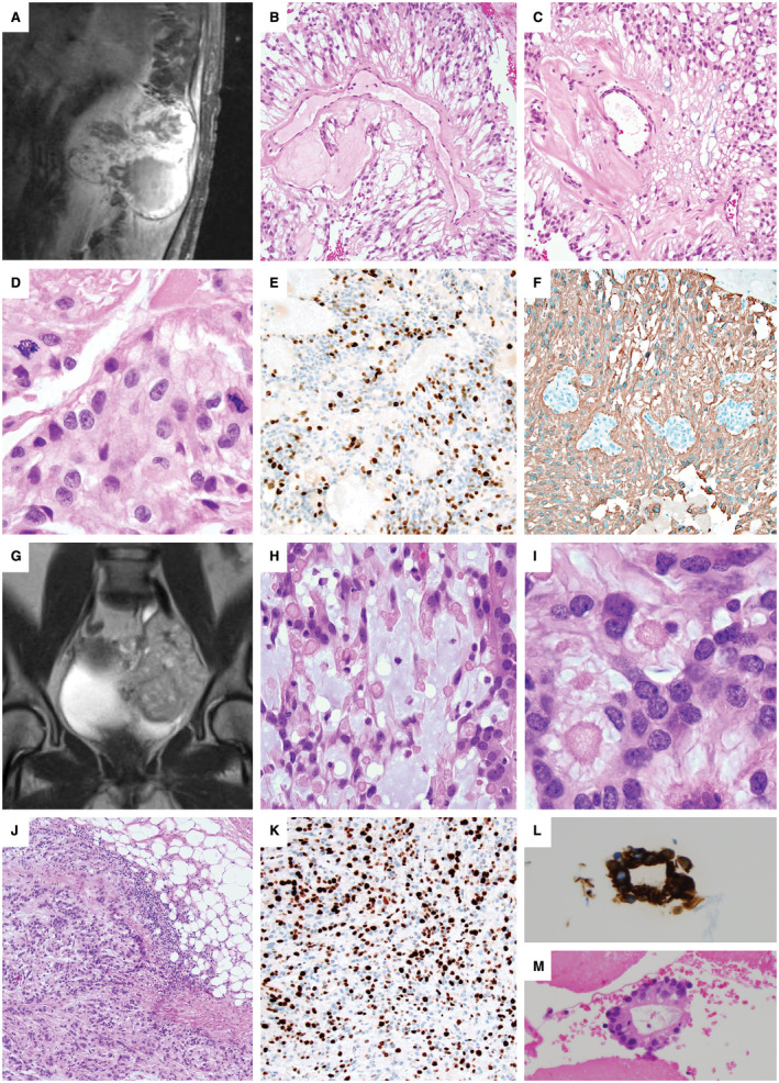
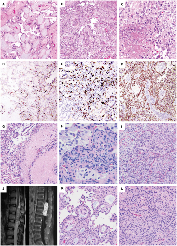
Myxopapillary ependymomas (MPE) are considered benign (World Health Organization (WHO) grade I) neoplasms with favorable prognosis. However, malignant behavior occurs in a small subset. To our knowledge, only five anaplastic MPEs have been reported without consensus on diagnostic criteria. We retrieved 14 anaplastic MPEs from the pathology archives of six institutions. Each tumor included at least two of the following features: ≥5 mitoses per 10 high power fields, Ki-67 labeling index (LI) ≥10%, microvascular proliferation (MVP) and spontaneous necrosis. These features were typically encountered in the foci of hypercellularity and reduced mucin. There were eight male and six female patients (age range 6-57 years, median = 16.5). Ten tumors displayed anaplasia at initial resection, and 4 were anaplastic at a second surgery for recurrence (ranging from 9 months to 14 years following initial resection). The Ki-67 LI ranged between 8% and 40% in the anaplastic foci and <3% in the foci of classic MPE. There was documented cerebrospinal fluid (CSF) dissemination in seven cases, recurrence following an anaplastic diagnosis in three cases and bone or soft tissue invasion in two cases. One patient suffered lung metastases. Two cases evaluated by targeted next-generation sequencing and one evaluated by fluorescence in situ hybridization (FISH) showed nonspecific chromosomal gains. We conclude that although rare, anaplastic MPE occurs in both pediatric and adult patients, similar to other ependymomas. At a minimum, closer follow-up is recommended, given the concern for aggressive biologic potential. Further study is needed to determine WHO grading criteria and genetic indicators of tumor progression.

Keywords: CSF dissemination; anaplastic transformation; malignant neoplasm; metastasis; microvascular proliferation; myxopapillary ependymoma; necrosis; recurrence.


Figures

Figure 1
Figure 1
Anaplastic myxopapillary ependymomas with aggressive clinical features. A–F. Case #12: A 45‐year‐old man underwent subtotal resection and radiation of an 11.5 cm T11‐L2 MPE. (A). Recurrence detected by MRI 12 years later showed destruction of the L1 vertebrae with invasion into the paraspinal soft tissues. Areas of classic MPE histology were present (B,C), but were accompanied by anaplastic features, including increased mitotic activity of 17 mitoses per 10 HPF (D), Ki‐67 LI up to 26% and prominent microvascular proliferation (MVP). There were foci of non‐palisading necrosis in this patient that had previously received radiation therapy. A representative area of increased Ki‐67 staining is shown (E), and foci of MVP are seen among GFAP‐positive neoplastic cells (F). G–M. Case #10: A 10‐cm pelvic recurrence on MR imaging (G) was found in a 45‐year‐old woman 14 years after resection and radiation of a sciatic notch MPE. Careful review of the medical record could not establish if the original mass was intradural or extradural in origin, or if it arose as an extraspinal primary soft tissue MPE. The recurrence contained areas of classic MPE with increased mucin and collagen balloons H–I. The mass was invasive into the adjacent soft tissues (J), with a Ki‐67 LI up to 40% (K), a mitotic index of 7/10 HPF, MVP and palisading necrosis. As the patient had received radiation therapy, the palisading necrosis was not considered an anaplastic feature. The patient was concurrently found to have multiple pulmonary nodules, a large left‐sided pleural effusion and an enhancing pleural‐based mass. Evaluation of the pleural fluid, shown here with GFAP immunostaining (L), and a pleural biopsy (M) demonstrated metastatic MPE. At the time of the pelvic recurrence, a sacral mass was also present, which was resected 4 months later in a separate surgery.
Figure 2
Figure 2
Adult and pediatric intradural anaplastic myxopapillary ependymomas. A–F. Case #14: A 55‐year‐old woman with a 2.7‐cm L1‐L2 mass and CSF dissemination with masses involving the cauda equina at the time of diagnosis underwent gross total resection and radiation therapy. The initial resection included areas of classic MPE (A) intermixed with anaplastic areas demonstrating hypercellularity and reduced mucin (B). The mitotic index reached 7 mitoses per 10 HPF, and there was both non‐palisading necrosis (C) and microvascular proliferation (MVP). A sharp transition between the classic and anaplastic components was present, reflected both by the histology and the variation in Ki‐67 (D), which was 20% in the anaplastic component (E) and <1% in conventional areas (D); left side). There was diffuse nuclear p53 positivity within the areas of anaplastic transformation (F). Recurrence 18 months later as a 2.3‐cm L2 mass also showed anaplastic features. G–I. Case #9: A 31‐year‐old man with an intradural sacral mass and smaller thoracic tumor deposits underwent subtotal resection, chemotherapy and a partial course of radiation. Re‐resection of the sacral MPE 9 months later showed both palisading and non‐palisading necrosis (G), which was not considered an anaplastic feature because of prior chemoradiation therapy. Anaplastic features included a Ki‐67 LI of 10%, a mitotic index of 7/10 HPF and MVP (H). Many areas of the tumor showed hypercellularity with decreased mucin (I). The patient was lost to clinical follow‐up and died at 36 years of age; no autopsy was performed. J–L. Case #4: A 10‐year‐old boy presented with an intradural extramedullary L1‐L2 enhancing mass centered at the conus medullaris by spinal MRI (J). Portions of the tumor showed classic MPE histology with papillary architecture and a myxoid stroma (K), while anaplastic regions were hypercellular with reduced mucin (L). Anaplastic features included a Ki‐67 LI of up to 15%, 6 mitoses per 10 high power fields and small foci of MVP.

References

    1. Akpolat N, Bozlak N, Kazez A, Köseoğullari AA (2003) Sacrococcygeal extraspinal ependymoma: a case report. Turk J Pediatr 45:276–279. - PubMed
    1. Awaya H, Kaneko M, Amatya VJ, Takeshima Y, Oka S, Inai K (2003) Myxopapillary ependymoma with anaplastic features. Pathol Int 53:700–703. - PubMed
    1. Barton VN, Donson AM, Kleinschmidt‐DeMasters BK, Birks DK, Handler MH, Foreman NK (2010) Unique molecular characteristics of pediatric myxopapillary ependymoma. Brain Pathol 20:560–570. - PMC - PubMed
    1. Beschorner R, Wehrmann M, Ernemann U, Bonin M, Horber V, Oehl‐Jaschkowitz B et al (2007) Extradural ependymal tumor with myxopapillary and ependymoblastic differentiation in a case of Schinzel‐Giedion syndrome. Acta Neuropathol 113:339–346. - PubMed
    1. Chakraborti S, Kini H, Pai KG, Upadhyaya V (2012) Sacrococcygeal myxopapillary ependymoma with anaplastic ependymoma component in an infant. J Pediatr Neurosci 7:218–220. - PMC - PubMed