. 2019 Apr 1;104(4):1099-1108.
doi: 10.1210/jc.2018-01922.

Regional Body Fat Changes and Metabolic Complications in Children With Dunnigan Lipodystrophy-Causing LMNA Variants

Affiliations

Regional Body Fat Changes and Metabolic Complications in Children With Dunnigan Lipodystrophy-Causing LMNA Variants

Nivedita Patni et al. J Clin Endocrinol Metab. .

Abstract

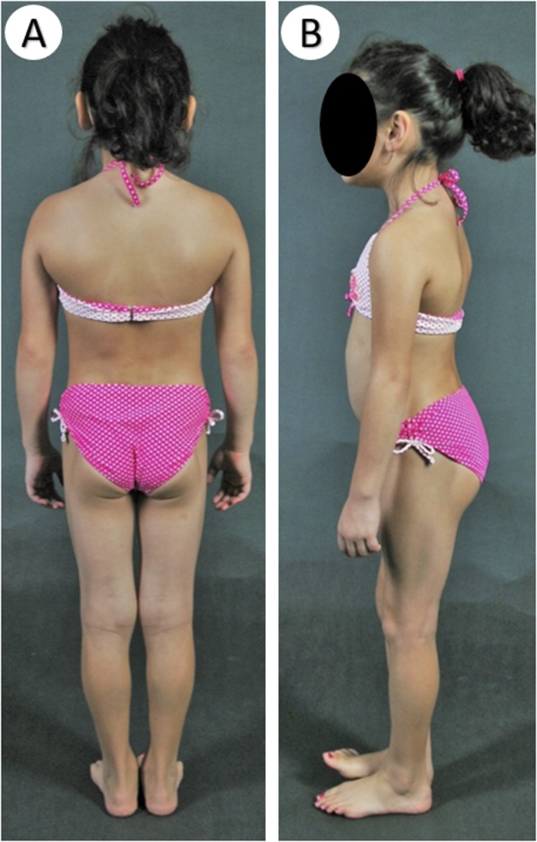
Context: Familial partial lipodystrophy, Dunnigan variety (FPLD2) is a rare autosomal-dominant disorder due to heterozygous missense lamin A/C (LMNA) mutations. Subjects with FPLD2 gradually lose fat from the upper and lower extremities but gain fat in the face and neck around puberty. However, the precise onset of body fat changes and metabolic complications during childhood remains unknown.

Objective: To compare metabolic parameters and regional body fat in children with FPLD2 with the sex- and age-matched controls from the National Health and Nutrition Examination Survey (NHANES) 2005 to 2010.

Methods: We measured fasting serum triglycerides, glucose, and skinfold thicknesses in all children (aged 1 to 18 years) harboring FPLD2-causing LMNA mutations and determined regional body fat by dual-energy X-ray absorptiometry in those aged ≥8 years.

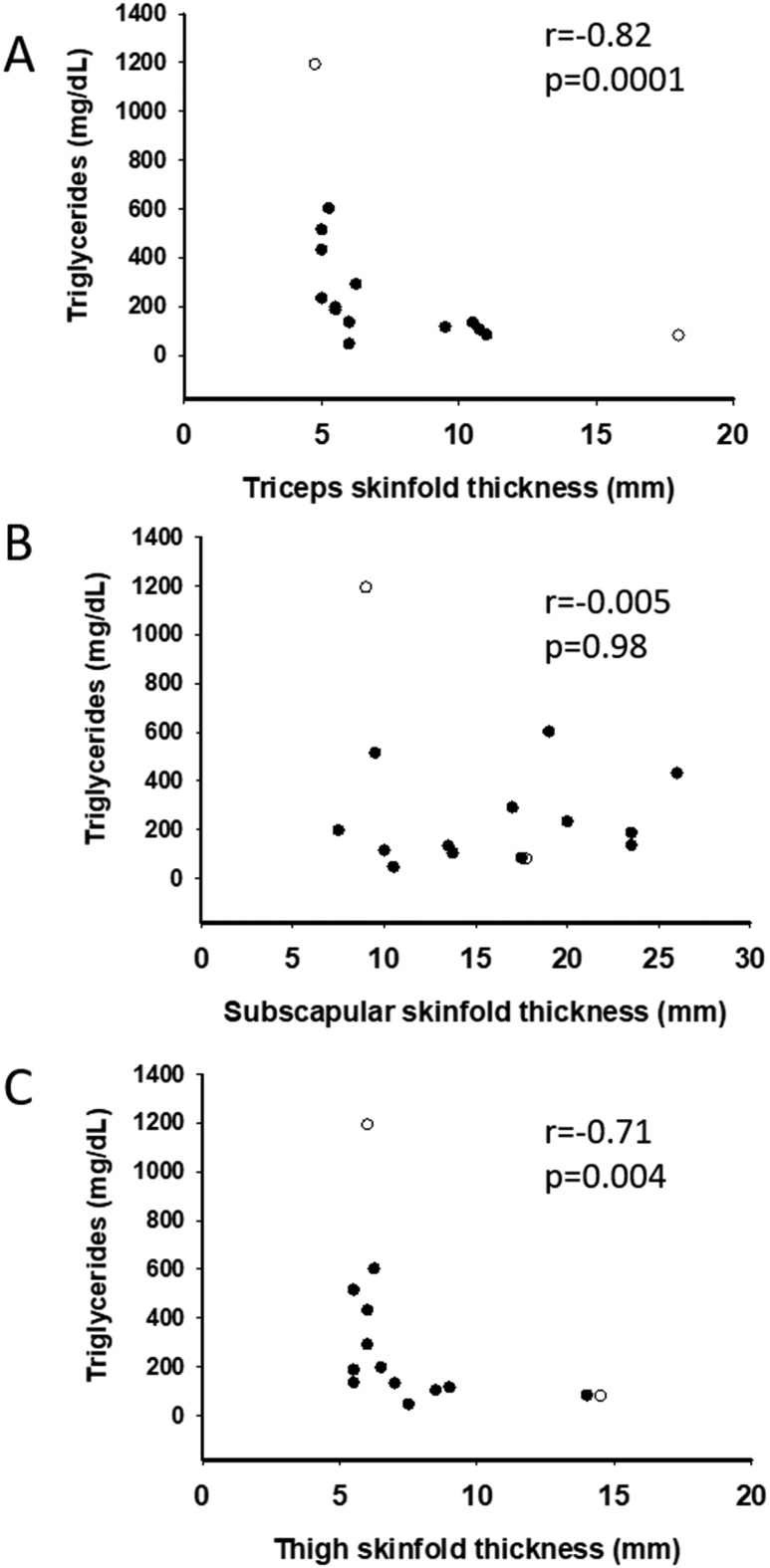
Results: Thirty-two affected females and 14 males participated. The lower limb fat in all affected females, except one, was below or equal to the first percentile and in two affected males was below the fifth percentile for NHANES. One female subject with FPLD2 followed from age 6 to 16 years revealed marked loss of extremity fat much before thelarche. Serum triglycerides were higher in females with FPLD2 aged 7 to 18 years compared with controls (median 208 vs 70 mg/dL; P < 0.0001) and showed inverse correlation with extremity skinfolds. Serum triglycerides in males with FPLD2 were not significantly different than controls.

Conclusions: The onset of fat loss from the extremities, especially in girls with FPLD2, occurs well before the onset of puberty. High serum triglycerides are seen in young females with FPLD2 with severe loss of fat from the extremities.


Figures

Figure 1.
Figure 1.
Comparison of skinfold thickness of FPLD2 children with the NHANES controls. Filled symbols signify subjects with typical LMNA mutations, and unfilled symbols indicate atypical LMNA mutation in females (circles) and males (squares). Skinfolds of two patients with longitudinal data are shown connected with a solid line, with green and red triangles indicating age of onset of thelarche and menarche, respectively. The data from the NHANES controls have been drawn as a percentile chart for comparison. (A) Female children with FPLD2 have subscapular skinfold thickness values in the 5th to 95th percentile, with an upwards trend after 13 years. (B) Male children with FPLD2 have subscapular skin folds in the 5th to 95th percentile with no specific trends until 18 years of age. (C) The triceps skinfold measurements are <25th percentile in the majority of the female subjects with FPLD2 by 8 years of age and <1st percentile in all subjects after 16 years of age. The female with typical LMNA mutation and longitudinal data over a period of 10 years has triceps skinfold thickness values at about the first percentile at the time of thelarche, but the female with atypical LMNA mutation and longitudinal data over a period of 3 years has not had considerable fat loss even after menarche. (D) The majority of males with FPLD2 had triceps skin fold values in 1st to 75th percentile with no specific trends until 18 years of age.
Figure 2.
Figure 2.
Body fat distribution in a 6-year-old Native Hawaiian female with typical heterozygous missense LMNA mutation (p.R482W). (A) Posterior view showing normal SC fat distribution in the trunk and upper extremity but loss of SC fat in the gluteal region. (B) Lateral view showing loss of SC fat and muscular prominence in the lower extremity.
Figure 3.
Figure 3.
Longitudinal skinfold thickness values of the patient with typical FPLD2- causing LMNA mutation. The green and red triangles indicate the onset of thelarche and menarche, respectively. (A) Excessive fat deposition in the chin began ∼11 years of age. (B) The mean truncal (average of subscapular, chest, mid-axillary, suprascapular and abdominal skinfolds) fat loss started ∼1.5 years before thelarche. (C) Peripheral (average of biceps, triceps, thigh, and calf skinfolds) fat loss started ∼1.5 years before thelarche.
Figure 4.
Figure 4.
Comparison of regional body fat (by DEXA scan) of children with FPLD2 to the NHANES controls. Filled symbols signify subjects with typical LMNA mutations, and unfilled symbols indicate those with atypical LMNA mutations in females (circles) and males (squares). Longitudinal data of two patients are shown connected with a solid line, with the green and red triangles indicating the onset of thelarche and menarche, respectively. The data from the NHANES controls have been drawn as age-specific percentile values for comparison. (A) The upper limb fat is <25th percentile in all affected females, with a downward trend after 12 years. The female with a typical LMNA mutation and longitudinal data over a period of 7 years (filled circles connected with a line) has downtrending upper limb fat even before thelarche, but the female with atypical LMNA mutation and longitudinal data over a period of 3 years (unfilled circles connected with a line) has not had considerable fat loss even after menarche. (B) The upper limb fat ranges between the 1st and 25th percentile in affected males. (C) All affected females, except one with atypical FPLD2, had lower limb fat at or below the first percentile after 8 years of age. The female with typical LMNA mutation and longitudinal data had lower limb fat below the first percentile ∼1.5 years before thelarche, but the female with atypical LMNA mutation and longitudinal data has not had marked fat loss even after menarche. (D) Two males with typical FPLD2 had lower limb fat <5th percentile of NHANES, and the two with atypical FPLD2 were <25th percentile. (E) Truncal fat ranged between the 1st and 75th percentile in most females with no specific trends. Longitudinal data on two females showed downtrending truncal fat. (F) The truncal fat ranged between the 1st and 50th percentile in the affected males.
Figure 5.
Figure 5.
Comparison of fasting serum triglycerides, glucose, and HDL cholesterol of children with FPLD2 to NHANES controls. Filled symbols signify subjects with typical LMNA mutations, and unfilled symbols indicate atypical LMNA mutation in females (circles) and males (squares). The data from the NHANES controls have been drawn as percentile age-specific values for comparison. (A) Affected females with both typical and atypical LMNA mutations developed hypertriglyceridemia from age 13 to 18 years. (B) Serum triglyceride levels in the affected males were <99th percentile until 18 years of age. (C) Serum glucose levels in affected females were not significantly different than the NHANES data. Only one girl with atypical FPLD2, harboring a heterozygous p.K486N LMNA variant, developed diabetes mellitus around the age of 14 years. (D) Serum glucose levels in affected males were not significantly different than the NHANES data. (E) Serum HDL-cholesterol levels in females ranged 5th to 95th percentile from 8 to 14 years and decreased to <50th percentile after 14 years of age. (F) Serum HDL-cholesterol levels in affected males were not significantly different than the NHANES data.
Figure 6.
Figure 6.
Relationship of fasting serum triglyceride levels with measures of regional body fat in females with FPLD2-causing LMNA mutations. Filled symbols signify typical LMNA mutations, and unfilled symbols indicate atypical LMNA mutation. Spearman correlation coefficients and P values are shown. (A) Strong inverse relationship of serum triglyceride levels is observed with triceps skinfold thickness (r = −0.82; P = 0.0001), with hypertriglyceridemia in all affected females when triceps skinfold thickness is ≤5 mm. (B) No correlation of serum triglycerides with subscapular skinfold thickness (r = −0.005; P = 0.98). (C) Strong inverse relationship of serum triglyceride levels is observed with thigh skinfold thickness (r = −0.71; P = 0.004), with hypertriglyceridemia in all affected females when thigh skinfold thickness is ≤7 mm.

References

    1. Garg A. Acquired and inherited lipodystrophies. N Engl J Med. 2004;350(12):1220–1234. - PubMed
    1. Peters JM, Barnes R, Bennett L, Gitomer WM, Bowcock AM, Garg A. Localization of the gene for familial partial lipodystrophy (Dunnigan variety) to chromosome 1q21-22. Nat Genet. 1998;18(3):292–295. - PubMed
    1. Cao H, Hegele RA. Nuclear lamin A/C R482Q mutation in canadian kindreds with Dunnigan-type familial partial lipodystrophy. Hum Mol Genet. 2000;9(1):109–112. - PubMed
    1. Shackleton S, Lloyd DJ, Jackson SN, Evans R, Niermeijer MF, Singh BM, Schmidt H, Brabant G, Kumar S, Durrington PN, Gregory S, O’Rahilly S, Trembath RC. LMNA, encoding lamin A/C, is mutated in partial lipodystrophy. Nat Genet. 2000;24(2):153–156. - PubMed
    1. Speckman RA, Garg A, Du F, Bennett L, Veile R, Arioglu E, Taylor SI, Lovett M, Bowcock AM. Mutational and haplotype analyses of families with familial partial lipodystrophy (Dunnigan variety) reveal recurrent missense mutations in the globular C-terminal domain of lamin A/C. Am J Hum Genet. 2000;66(4):1192–1198. - PMC - PubMed

Publication types

MeSH terms