

Identification of Elg1 interaction partners and effects on post-replication chromatin re-formation
- PMID: 30418970
- PMCID: PMC6258251
- DOI: 10.1371/journal.pgen.1007783
Identification of Elg1 interaction partners and effects on post-replication chromatin re-formation
Abstract
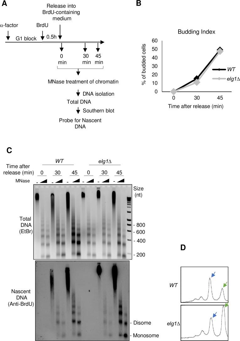
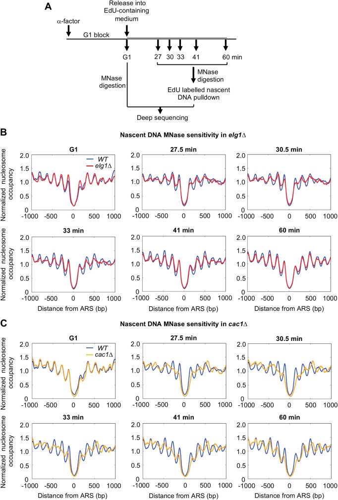
Elg1, the major subunit of a Replication Factor C-like complex, is critical to ensure genomic stability during DNA replication, and is implicated in controlling chromatin structure. We investigated the consequences of Elg1 loss for the dynamics of chromatin re-formation following DNA replication. Measurement of Okazaki fragment length and the micrococcal nuclease sensitivity of newly replicated DNA revealed a defect in nucleosome organization in the absence of Elg1. Using a proteomic approach to identify Elg1 binding partners, we discovered that Elg1 interacts with Rtt106, a histone chaperone implicated in replication-coupled nucleosome assembly that also regulates transcription. A central role for Elg1 is the unloading of PCNA from chromatin following DNA replication, so we examined the relative importance of Rtt106 and PCNA unloading for chromatin reassembly following DNA replication. We find that the major cause of the chromatin organization defects of an ELG1 mutant is PCNA retention on DNA following replication, with Rtt106-Elg1 interaction potentially playing a contributory role.
Conflict of interest statement
The authors have declared that no competing interests exist.
Figures


Similar articles
-
Replication-Coupled PCNA Unloading by the Elg1 Complex Occurs Genome-wide and Requires Okazaki Fragment Ligation.Cell Rep. 2015 Aug 4;12(5):774-87. doi: 10.1016/j.celrep.2015.06.066. Epub 2015 Jul 23. Cell Rep. 2015. PMID: 26212319 Free PMC article.
-
The Elg1 replication factor C-like complex functions in PCNA unloading during DNA replication.Mol Cell. 2013 Apr 25;50(2):273-80. doi: 10.1016/j.molcel.2013.02.012. Epub 2013 Mar 14. Mol Cell. 2013. PMID: 23499004
-
A structure-function analysis of the yeast Elg1 protein reveals the importance of PCNA unloading in genome stability maintenance.Nucleic Acids Res. 2017 Apr 7;45(6):3189-3203. doi: 10.1093/nar/gkw1348. Nucleic Acids Res. 2017. PMID: 28108661 Free PMC article.
-
Is PCNA unloading the central function of the Elg1/ATAD5 replication factor C-like complex?Cell Cycle. 2013 Aug 15;12(16):2570-9. doi: 10.4161/cc.25626. Epub 2013 Jul 10. Cell Cycle. 2013. PMID: 23907118 Free PMC article. Review.
-
New insights into replication clamp unloading.J Mol Biol. 2013 Nov 29;425(23):4727-32. doi: 10.1016/j.jmb.2013.05.003. Epub 2013 May 17. J Mol Biol. 2013. PMID: 23688817 Review.
Cited by
-
Timely termination of repair DNA synthesis by ATAD5 is important in oxidative DNA damage-induced single-strand break repair.Nucleic Acids Res. 2021 Nov 18;49(20):11746-11764. doi: 10.1093/nar/gkab999. Nucleic Acids Res. 2021. PMID: 34718749 Free PMC article.
-
The replisome guides nucleosome assembly during DNA replication.Cell Biosci. 2020 Mar 12;10:37. doi: 10.1186/s13578-020-00398-z. eCollection 2020. Cell Biosci. 2020. PMID: 32190287 Free PMC article. Review.
-
Profilin-1 regulates DNA replication forks in a context-dependent fashion by interacting with SNF2H and BOD1L.Nat Commun. 2022 Nov 1;13(1):6531. doi: 10.1038/s41467-022-34310-9. Nat Commun. 2022. PMID: 36319634 Free PMC article.
-
ATAD5 deficiency alters DNA damage metabolism and sensitizes cells to PARP inhibition.Nucleic Acids Res. 2020 May 21;48(9):4928-4939. doi: 10.1093/nar/gkaa255. Nucleic Acids Res. 2020. PMID: 32297953 Free PMC article.
-
PCNA Loaders and Unloaders-One Ring That Rules Them All.Genes (Basel). 2021 Nov 18;12(11):1812. doi: 10.3390/genes12111812. Genes (Basel). 2021. PMID: 34828416 Free PMC article. Review.
References
-
- Groth A, Rocha W, Verreault A, Almouzni G. Chromatin Challenges during DNA Replication and Repair. Cell. 2007. pp. 721–733. 10.1016/j.cell.2007.01.030 - DOI - PubMed
-
- Mailand N, Gibbs-Seymour I, Bekker-Jensen S. Regulation of PCNA–protein interactions for genome stability. Nat Rev Mol Cell Biol. 2013;14: 269–282. 10.1038/nrm3562 - DOI - PubMed
-
- Bowman GD, O’Donnell M, Kuriyan J. Structural analysis of a eukaryotic sliding DNA clamp–clamp loader complex. Nature. 2004;429: 724–730. 10.1038/nature02585 - DOI - PubMed
-
- Gomes X V., Burgers PMJ. ATP utilization by yeast replication factor C: I. ATP-mediated interaction with DNA and with proliferating cell nuclear antigen. J Biol Chem. 2001;276: 34768–34775. 10.1074/jbc.M011631200 - DOI - PubMed
-
- Kubota T, Nishimura K, Kanemaki MT, Donaldson AD. The Elg1 Replication Factor C-like Complex Functions in PCNA Unloading during DNA Replication. Mol Cell. 2013;50: 273–280. 10.1016/j.molcel.2013.02.012 - DOI - PubMed
Publication types
MeSH terms
Substances
Grants and funding
LinkOut - more resources
Full Text Sources
Molecular Biology Databases
Miscellaneous
