Review
. 2019 Mar:195:172-185.
doi: 10.1016/j.pharmthera.2018.11.001. Epub 2018 Nov 9.

The role of DNA methylation in epigenetics of aging

Affiliations
Review

The role of DNA methylation in epigenetics of aging

Archana Unnikrishnan et al. Pharmacol Ther. 2019 Mar.

Abstract

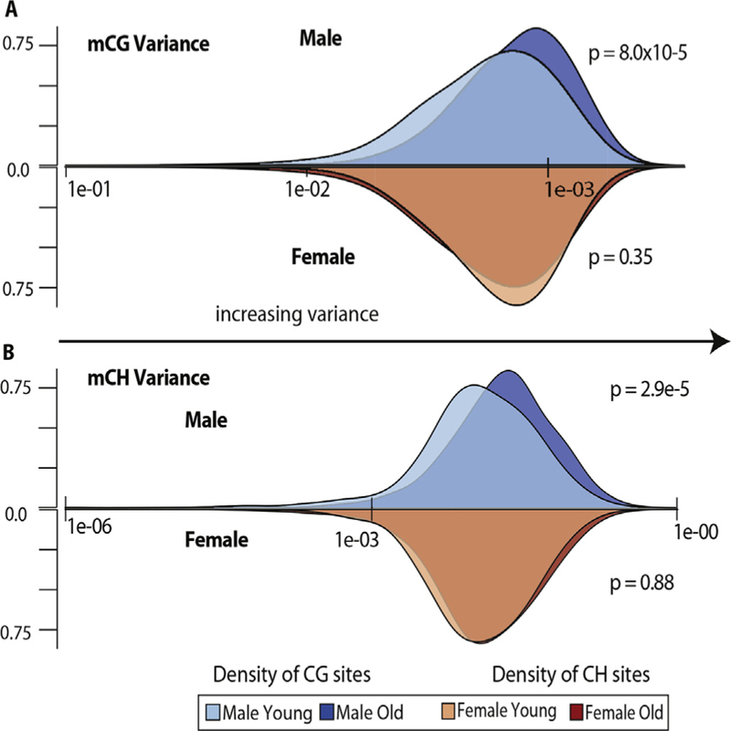
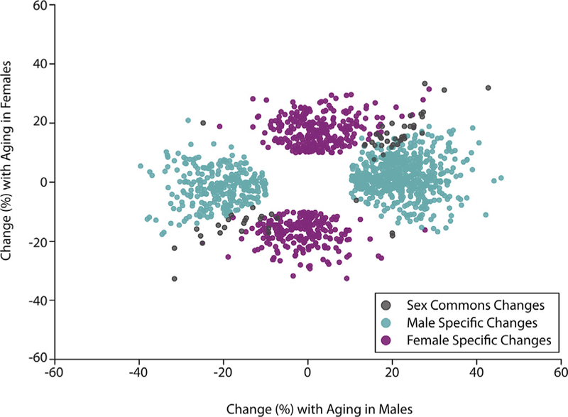
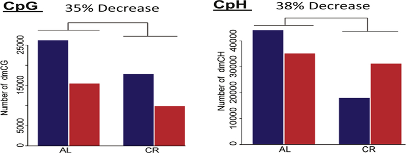
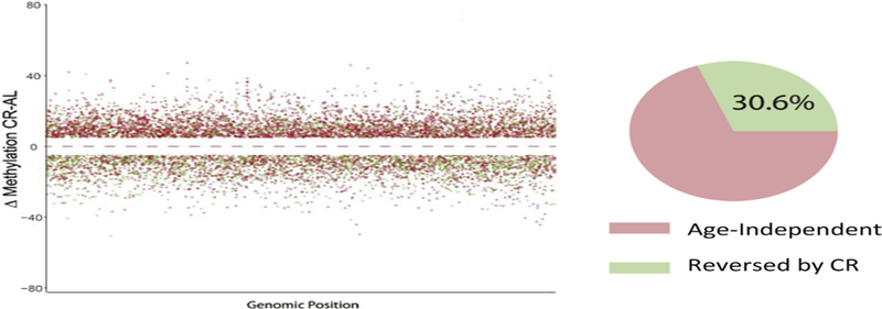
Recent research suggests that epigenetics, especially DNA methylation, plays a mechanistic role in aging. Epigenetic clocks, which measure changes in a few hundred specific CpG sites, can accurately predict chronological age in a variety of species, including humans. These clocks are currently the best biomarkers for predicting mortality in humans. Additionally, several studies have characterized the effects of aging across the methylome in a wide variety of tissues from humans and mice. A small fraction (~2%) of the CpG sites show age-related changes, either hypermethylation or hypomethylation with aging. Evaluation of non-CpG site methylation has only been examined in a few studies, with about ~0.5% of these sites showing a change with age. Therefore, while only a small fraction of cytosines in the genome show changes in DNA methylation with age, this represents 2 to 3 million cytosines in the genome. Importantly, the only study to compare the effect of aging on DNA methylation in male and female mice and humans found that >95% of the age-related changes in DNA methylation in the hippocampus were sexually divergent, i.e., the methylation did not differ between males and females at young age but age-related changes occurred in one sex but not the other. The age-related changes in DNA methylation tend to be enriched and under-represented in specific genomic contexts, with some commonalities between tissues and species that require further investigation. The strongest evidence that the age-related changes in DNA methylation play a role in aging comes from studies of anti-aging interventions (e.g., caloric restriction, dwarfism, and rapamycin treatment) in mice. These anti-aging interventions deaccelerate the epigenetic clocks and reverse/prevent 20 to 40% of the age-related changes in DNA methylation. It will be important in the future to demonstrate that at least some of the age-related changes in DNA methylation directly lead to alterations in the transcriptome of cells/tissues that could potentially contribute to aging.

Keywords: 5-hydroxymethyl cytosine; 5-methyl cytosine; Aging; Caloric restriction; DNA methylation; Epigenetic clocks; Epigenetics; Gene Expression.


Conflict of interest statement

Conflict of interest

The authors declare no conflict of interest.

Figures

Fig. 1.
Fig. 1.
The pathway for DNA methylation. A) The steps involved in the formation and elimination of 5-methyl cytosine (mC) in DNA is shown. Abbreviations: SAM, S-adenosyl methionine; SAH, S-adenosyl homocysteine; hmC, 5-hyroxymethyl cytosine; fC, 5-formyl cytosine; caC, 5-carboxyl cytosine; BER, base excision repair; TDG, thymidine DNA glycosylase. B) Expression of genes involved in methylation varies between cell types and tissues. For example, different cell types in brain express various levels of the mRNA transcripts for DNMTs and TETs. Data taken from Zhang et al. (2014).
Fig. 2.
Fig. 2.
DNA methylation variance with age. A) Mean inter-animal mCG variance of cytosines present at ≥20× sequencing depth in every sample (354,522 CG sites) among young and old males (blue distributions) and females (red distributions) relative to density of sites at a given mean variance. mCG variance distribution in old males (mean variance = 0.157774) compared to young males (mean variance = 0.103581) was significantly higher (p = 8.0 × 10⁻⁵), while comparison of mCG variance in old females (mean variance = 0.112484) to young females (mean variance = 0.107045) was not significant (p = .35). B) Mean inter-animal mCH variance of cytosines present at ≥20× sequencing depth in every sample (5,577,396 CH sites) among young and old males (blue distributions) and females (red distributions) relative to density of sites at a given mean variance. mCH variance distribution in old males (mean variance = 0.050142) compared to young males (mean variance = 0.034209) was significantly higher (p = 2.9 × 10⁻⁵), while comparison of mCH variance in old females (mean variance = 0.038597) to young females (mean variance = 0.033432) was not significant (p = .88).
Fig. 3.
Fig. 3.
– Sex divergence with aging. Hippocampal methylation changes (mCpG) with aging (young = 3 months, old = 24 months, q b 0.01, |Δ%| N 10) are primarily sex-specific. When viewed in this scatterplot there are common changes with age in both males and females (black circle) but the majority of changes occur in only males (blue) or females (pink). Data taken from Masser te al. (2017).
Fig. 4.
Fig. 4.
Comparison of methylation and hydroxymethylation of cytosines in hippocampus. Graph A. The 5mC and 5hmC levels in DNA from the hippocampus is shown when measured by the traditional bisulfite sequencing assay versus the oxidative bisulfite assay. Approximately 25% of the cytosines that would be classified as 5mC by the bisulfite assay are 5hmC residues. Graph B. The distribution of 5mC and 5hmC over a small region (~120 bp of Fkbp6) of the hippocampus genome. Data taken from Messer et al. (2018).
Fig. 5.
Fig. 5.
Effect of age and CR on DNA methylation. DNA was obtained from the hippocampus of 3- and 24-month-old mice fed ad libitum and 24-month-old mice fed a CR diet. The number of CpG and CpH sites that changed with age are shown: blue bars are sites that become hypermethylated and red bars are sites that become hypomethylated. The methylation of ~120 K Cs changed with age (~41 K CpGs and 80 K CpHs). CR reduced the number of sites that changed with age from 35 to 38%. For CpG sites, CR reduced hyper- and hypomethylated sites equally (32 to 36%). However, for CpH sites, CR had had a much greater effect on hypermethylated sites (59%) compared to hypomethylated sites (11%). Data taken from Hadad et al. (2018).
Fig. 6.
Fig. 6.
Effect of CR on DNA methylation. The CpG sites differentially methylated between old mice fed ad libitum and CR is shown. The CpG sites that change with age in the ad libitim fed mice that were prevented by CR are shown in green. The changes in methylation induced by CR that did not change with age in ad libitum fed mice are shown in red. Approximately 70% of the changes in DNA methylation induced by CR are age independent, i.e., do not occur with age in mice fed ad libitum. Data taken from Hadad et al. (2018).

References

    1. Asdell SA, Doornenbal H, Joshi SR, & Sperling GA (1967). The effects of sex steroid hormones upon longevity in rats. Journal of Reproduction and Fertility 14(1), 113–120. - PubMed
    1. Bailey LJ, Cluett TJ, Reyes A, Prolla TA, Poulton J, Leeuwenburgh C, & Holt IJ (2009). Mice expressing an error-prone DNA polymerase in mitochondria display elevated replication pausing and chromosomal breakage at fragile sites of mitochondrial DNA. Nucleic Acids Research 37(7), 2327–2335. 10.1093/nar/gkp091. - DOI - PMC - PubMed
    1. Bird A (2002). DNA methylation patterns and epigenetic memory. Genes & Development 16(1), 6–21. 10.1101/gad.947102. - DOI - PubMed
    1. Bitto A, Ito TK, Pineda VV, Letexier NJ, Huang HZ, Sutlief E, & Kaeberlein M (2016). Transient rapamycin treatment can increase lifespan and healthspan in middle-aged mice. eLife, 5 10.7554/eLife.16351. - DOI - PMC - PubMed
    1. Bize P, Criscuolo F, Metcalfe NB, Nasir L, & Monaghan P (2009). Telomere dynamics rather than age predict life expectancy in the wild. Proceedings of the Biological Sciences 276(1662), 1679–1683. 10.1098/rspb.2008.1817. - DOI - PMC - PubMed

Publication types