. 2018 Nov 12;18(1):174.
doi: 10.1186/s12876-018-0899-3.

Clinical characteristics of hepatic Arterioportal shunts associated with hepatocellular carcinoma

Affiliations

Clinical characteristics of hepatic Arterioportal shunts associated with hepatocellular carcinoma

Huiyong Wu et al. BMC Gastroenterol. .

Abstract

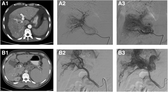
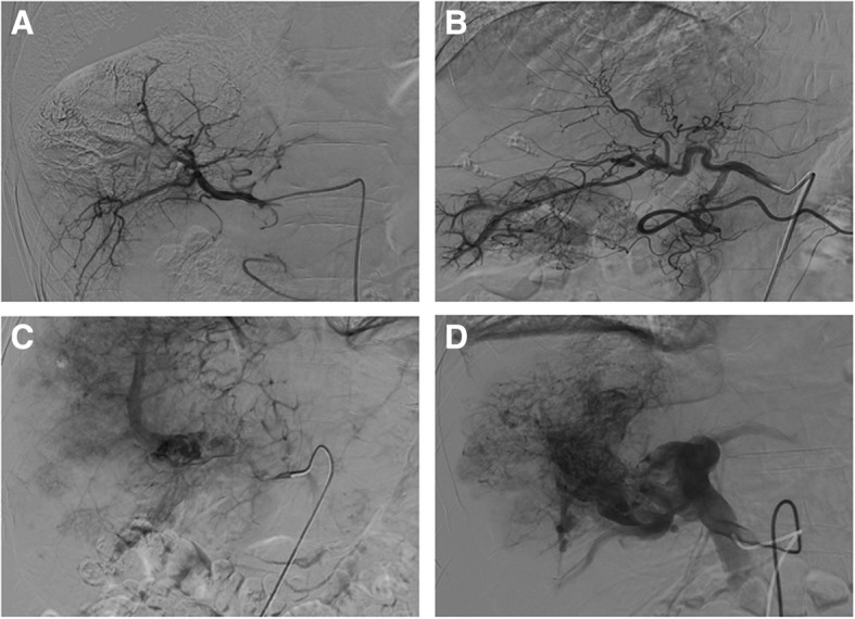
Background: Hepatic arterioportal shunt (A-P shunt) is defined as the direct blood flow established between hepatic artery and portal venous system; it is frequently observed in patients with hepatocellular carcinoma (HCC). Clinically, it is important to diagnose HCC associated A-P shunts, as it may impact the treatment strategy of the patients. In the present study, we described the imaging findings of the HCC associated A-P shunts and discussed the treatments strategy of such patients. From the findings, we also discussed the potential cause of A-P shunts.

Methods: Clinical data of HCC patients (n = 560), admitted to the hospital between April 2012 to April 2014, were reviewed. Hepatic angiography was used to examine the presence of A-P shunts. Of the 137 patients with A-P shunts, grading of the A-P shunts was performed, and statistical analysis of the different grades of A-P shunts and clinical characteristics was performed.

Results: The hepatic angiography confirmed that 99 patients had typical A-P shunts (Grade 1-3), and 38 patients had atypical A-P shunts. Embolization was the main strategy used to treat A-P shunts, in which liquid embolic agents appeared to provide a better treatment outcome. The correlation analysis showed that the grading of portal vein tumor thrombus was significantly associated with the grading of A-P shunt (p = < 0.001, Spearman correlation coefficient was 0.816 ± 0.043).

Conclusions: We characterized A-P shunts and proposed treatment strategy for treating HCC patients with various levels of A-P shunts. The findings supported the hypothesis that the formation of HCC associated A-P shunts was caused by tumor thrombus.

Keywords: Hepatic arterioportal shunts; Hepatocellular carcinoma; Portal vein tumor embolus; Transarterial chemoembolization.


Conflict of interest statement

Ethics approval and consent to participate

This study was carried out in accordance with the approved guidelines by the Ethics Committee of Shandong Tumor Hospital Affiliated to Shandong University (No. SDTHEC20171105) and patients have signed an informed consent.

Consent for publication

Not applicable.

Competing interests

All the authors have no conflict of interest for this study.

Publisher’s Note

Springer Nature remains neutral with regard to jurisdictional claims in published maps and institutional affiliations.

Figures

Fig. 1
Fig. 1
A. A patient with tumor thrombus and A-P shunt. (A1) Liver CT image showed the presence of right portal vein tumor thrombus. Arteries around the tumor thrombus were filled with contrast agent, suggesting the presence of A-P shunt. The intra-luminal filling of embolus indicated the internal blood flow. (A2) Early phase hepatic angiography showed ‘thread and streaks’ signs, corresponding to the blood spaces and vessels, which run longitudinally in and around the tumor thrombus. (A3) Late phase hepatic angiography showed that the blood flow of the tumor thrombus reached the portal vein, leading to the enhancement of portal vein. B. CT images of another patient with tumor thrombus and A-P shunt. (B1) CT image showed tumor thrombus in the right portal vein branch. Staining of the arteries around thrombus suggested the presence of A-P shunt. (B2) Early phase hepatic angiography showed the hepatic arteries run longitudinally in the hepatic portal vein. (B3) Late phase hepatic angiography: blood vessels in the embolus showed ‘tread and streaks’ signs. Enhancement of the left branch of the portal vein and segment of portal vein proximal to the embolus
Fig. 2
Fig. 2
Representative images of hepatic arteriogram showing different grades of A-P shunts. a Grade 0: A-P shunt was not observed. b Grade 1: enhancement of the VI segment of the portal vein in the right lobe of the liver. c Grade 2: enhancement of the right portal vein and portal vein tumor thrombus. d Grade 3: enhancement of right portal vein main branches and trunk, with ill-defined intra-luminal filling defect. A large number of disorganized arteries providing blood supply for the portal vein thrombus was observed

References

    1. Montalto G, Cervello M, Giannitrapani L, Dantona F, Terranova A, Castagnetta LA. Epidemiology, risk factors, and natural history of hepatocellular carcinoma. Ann N Y Acad Sci. 2002;963:13–20. doi: 10.1111/j.1749-6632.2002.tb04090.x. - DOI - PubMed
    1. Chen X, Liu L, Pan X. Portal vein tumor thrombus in advanced hepatocellular carcinoma: a case report. Oncol Lett. 2015;9(6):2495–2498. doi: 10.3892/ol.2015.3115. - DOI - PMC - PubMed
    1. Llovet JM, Bruix J. Systematic review of randomized trials for unresectable hepatocellular carcinoma: chemoembolization improves survival. Hepatology. 2003;37(2):429–442. doi: 10.1053/jhep.2003.50047. - DOI - PubMed
    1. Bruix J, Sherman M. American Association for the Study of liver D, Management of hepatocellular carcinoma: an update. Hepatology. 2011;53(3):1020–1022. doi: 10.1002/hep.24199. - DOI - PMC - PubMed
    1. Lazaridis KN, Kamath PS. Images in hepatology. Arterio-portal fistula causing recurrent variceal bleeding. J Hepatol. 1998;29(1):142. doi: 10.1016/S0168-8278(98)80189-X. - DOI - PubMed

MeSH terms