. 2019 Jan;60(1):19-29.
doi: 10.1194/jlr.M081240. Epub 2018 Nov 12.

Cholesterol intake and statin use regulate neuronal G protein-gated inwardly rectifying potassium channels

Affiliations

Cholesterol intake and statin use regulate neuronal G protein-gated inwardly rectifying potassium channels

Anna N Bukiya et al. J Lipid Res. 2019 Jan.

Abstract

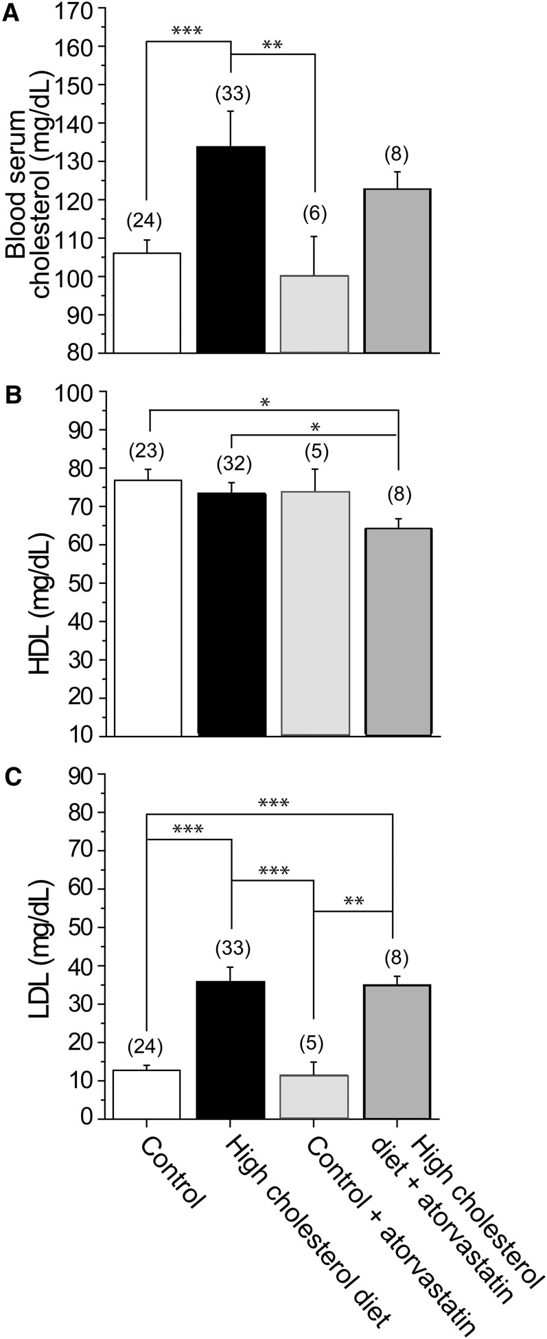
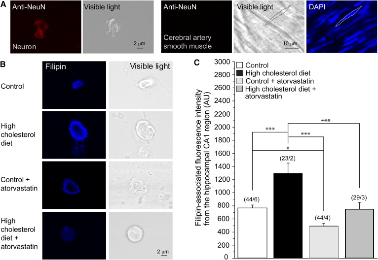
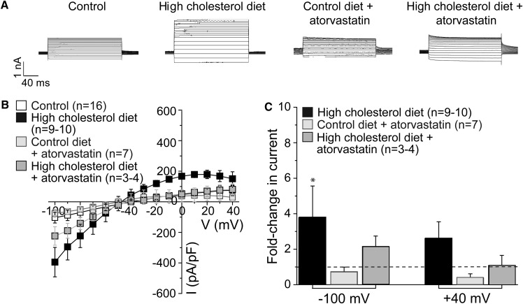
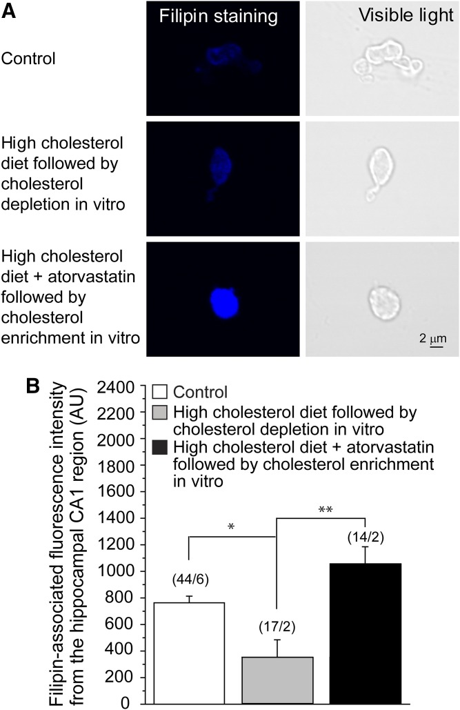
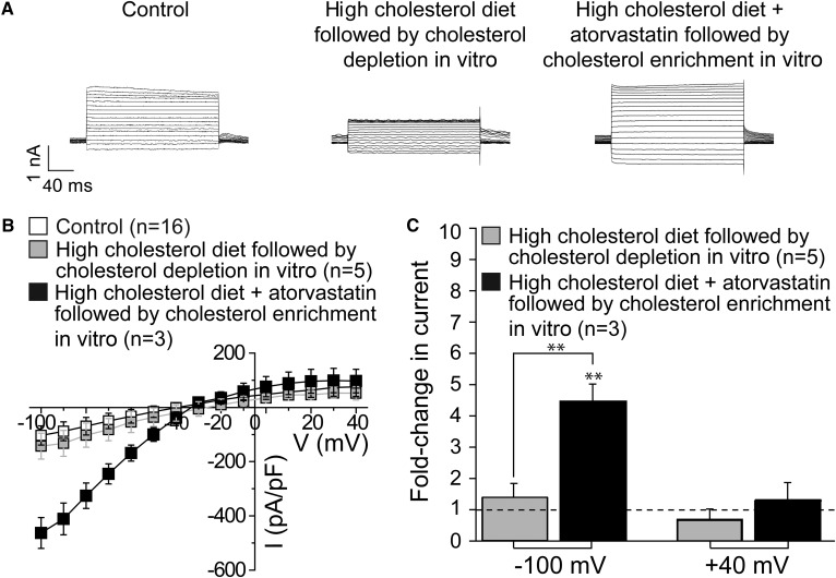
Cholesterol, a critical component of the cellular plasma membrane, is essential for normal neuronal function. Cholesterol content is highest in the brain, where most cholesterol is synthesized de novo; HMG-CoA reductase controls the synthesis rate. Despite strict control, elevated blood cholesterol levels are common and are associated with various neurological disorders. G protein-gated inwardly rectifying potassium (GIRK) channels mediate the actions of inhibitory brain neurotransmitters. Loss of GIRK function enhances neuron excitability; gain of function reduces neuronal activity. However, the effect of dietary cholesterol or HMG-CoA reductase inhibition (i.e., statin therapy) on GIRK function remains unknown. Using a rat model, we compared the effects of a high-cholesterol versus normal diet both with and without atorvastatin, a widely prescribed HMG-CoA reductase inhibitor, on neuronal GIRK currents. The high-cholesterol diet increased hippocampal CA1 region cholesterol levels and correspondingly increased neuronal GIRK currents. Both phenomena were reversed by cholesterol depletion in vitro. Atorvastatin countered the high-cholesterol diet effects on neuronal cholesterol content and GIRK currents; these effects were reversed by cholesterol enrichment in vitro. Our findings suggest that high-cholesterol diet and atorvastatin therapy affect ion channel function in the brain by modulating neuronal cholesterol levels.

Keywords: 3-hydroxy-3-methylglutaryl-CoA reductase; CA1 hippocampal neuron; brain lipids; dietary cholesterol; inwardly rectifying potassium channel; lipid mediators; lipid signaling.


Figures

Fig. 1.
Fig. 1.
Modulation of blood lipid levels by a high-cholesterol diet and atorvastatin therapy. Averaged data show blood serum total cholesterol (A), HDL (B), and LDL (C). LDL levels were obtained by subtracting HDL readings from serum total cholesterol. (n) is the number of blood serum samples. Each sample was collected from a separate animal. Statistically significant difference is indicated. * P < 0.05; ** P < 0.01; *** P < 0.001.
Fig. 2.
Fig. 2.
High-cholesterol diet and atorvastatin therapy modulate neuronal cholesterol content in the CA1 hippocampal region. A: Validation of the neuronal origin of the cellular content in the CA1 hippocampal region using neuron-specific immunostaining followed by confocal microscopy imaging. Immunofluorescence staining of neuronal tissue with an anti-NeuN Ab resulted in a fluorescence signal (leftmost snapshot). This staining failed to yield a signal from rat cerebral artery vasculature (three snapshots on the right). For cerebral artery vasculature, the silhouette of an individual myocyte is highlighted in the visible light spectrum and in the DAPI-stained specimen (rightmost snapshot). The sharp fluorescence image of the myocyte nucleus confirms that the vasculature was in the focal plane during imaging. B: Original representative snapshots showing filipin staining of isolated neuronal cells from the CA1 hippocampal brain region of rats on control diet, high-cholesterol diet, control diet supplemented by atorvastatin, and high-cholesterol diet supplemented by atorvastatin. C: Averaged data of filipin-associated fluorescence intensity from the hippocampal CA1 region of rats on control diet, high-cholesterol diet, and high-cholesterol diet supplemented by atorvastatin. AU, arbitrary units. (n/N) is the number of cells/number of animal donors. Statistically significant difference is indicated. * P < 0.05; *** P < 0.001.
Fig. 3.
Fig. 3.
High-cholesterol diet and atorvastatin therapy modulate neuronal GIRK currents in the CA1 hippocampal region. A: Representative traces of tertiapin-sensitive GIRK whole-cell currents from neurons of rats subjected to control rodent food, high-cholesterol diet, control diet supplemented by atorvastatin, and high-cholesterol diet supplemented by atorvastatin. Vholding = −80 mV. B: Summary data of neuronal GIRK current-voltage curves. C: Averaged data showing fold change in GIRK current amplitude at −100 and +40 mV on high-cholesterol diet in the absence versus presence of atorvastatin supplementation. n is the number of neurons. GIRK currents were recorded from no more than two neurons isolated from the same animal donor. The horizontal dashed line highlights the lack of change in current amplitude. Statistically significant difference with respect to control diet is indicated. * P < 0.05.
Fig. 4.
Fig. 4.
Manipulations with cholesterol level in vitro reverse high-cholesterol diet- and atorvastatin-driven changes in neuronal cholesterol level. A: Original representative snapshots showing filipin staining of isolated neuronal cells from the CA1 hippocampal brain region of rats on control rodent food, on high-cholesterol diet following cholesterol depletion in vitro, and on high-cholesterol diet supplemented by atorvastatin followed by cholesterol enrichment in vitro. B: Averaged data of filipin-associated fluorescence intensity from the hippocampal CA1 region of rats on control diet and on high-cholesterol diet in the absence versus presence of atorvastatin followed by cholesterol manipulations in vitro. AU, arbitrary units. (n/N) is the number of cells/number of animal donors. Statistically significant difference is indicated. * P < 0.05; ** P < 0.01.
Fig. 5.
Fig. 5.
Manipulations with cholesterol level in vitro reverse high-cholesterol diet- and atorvastatin-driven changes in neuronal GIRK current amplitude. A: Representative traces of tertiapin-sensitive GIRK whole-cell currents from neurons isolated from rats subjected to control rodent food and high-cholesterol diet in the absence versus presence of atorvastatin followed by cholesterol modifications in vitro. Vholding = –80 mV. B: Summary data of neuronal GIRK current-voltage curves. C: Averaged data showing fold change in GIRK current amplitude at –100 and +40 mV on high-cholesterol diet in the absence versus presence of atorvastatin supplementation followed by cholesterol manipulations in vitro. n is the number of neurons; GIRK currents were recorded from no more than two neurons isolated from the same animal donor. The horizontal dashed line highlights lack of change in current amplitude. Statistically significant difference is indicated. ** P < 0.01.

References

    1. Yeagle P. L. 1985. Cholesterol and the cell membrane. Biochim. Biophys. Acta. 822: 267–287. - PubMed
    1. Yeagle P. L. 1991. Modulation of membrane function by cholesterol. Biochimie. 73: 1303–1310. - PubMed
    1. Rosenhouse-Dantsker A., Mehta D., and Levitan I.. 2012. Regulation of ion channels by membrane lipids. Compr. Physiol. 2: 31–68. - PubMed
    1. Ramprasad O. G., Srinivas G., Rao K. S., Joshi P., Thiery J. P., Dufour S., and Pande G.. 2007. Changes in cholesterol levels in the plasma membrane modulate cell signaling and regulate cell adhesion and migration on fibronectin. Cell Motil. Cytoskeleton. 64: 199–216. - PubMed
    1. Goluszko P., and Nowicki B.. 2005. Membrane cholesterol: a crucial molecule affecting interactions of microbial pathogens with mammalian cells. Infect. Immun. 73: 7791–7796. - PMC - PubMed

MeSH terms

Substances