. 2019 Jan 4;294(1):71-88.
doi: 10.1074/jbc.RA118.005914. Epub 2018 Nov 12.

Tri-arginine exosite patch of caspase-6 recruits substrates for hydrolysis

Affiliations

Tri-arginine exosite patch of caspase-6 recruits substrates for hydrolysis

Derek J MacPherson et al. J Biol Chem. .

Abstract

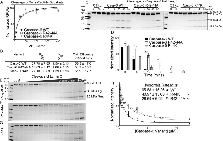
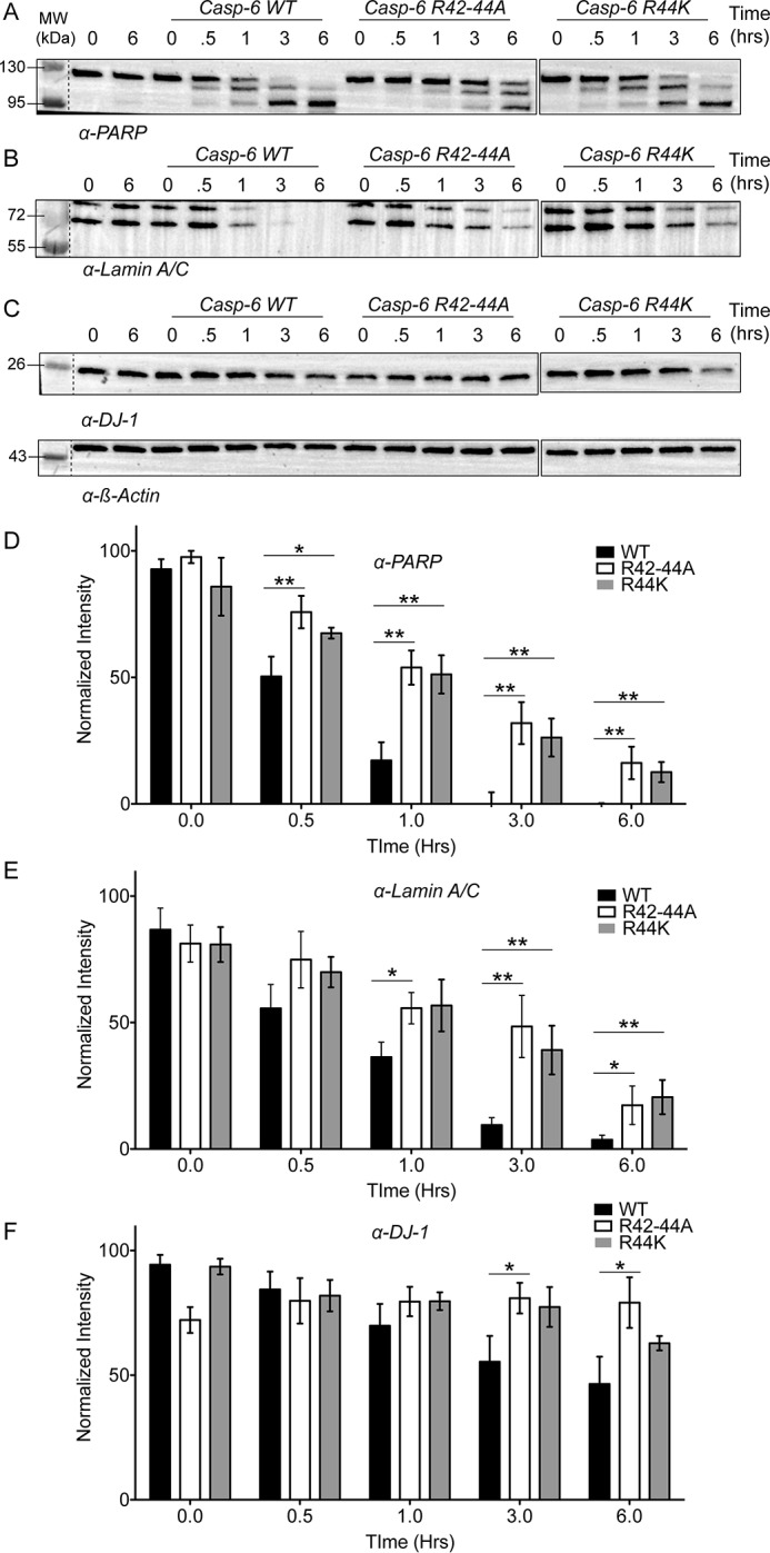
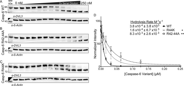
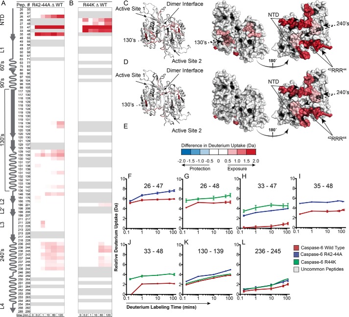
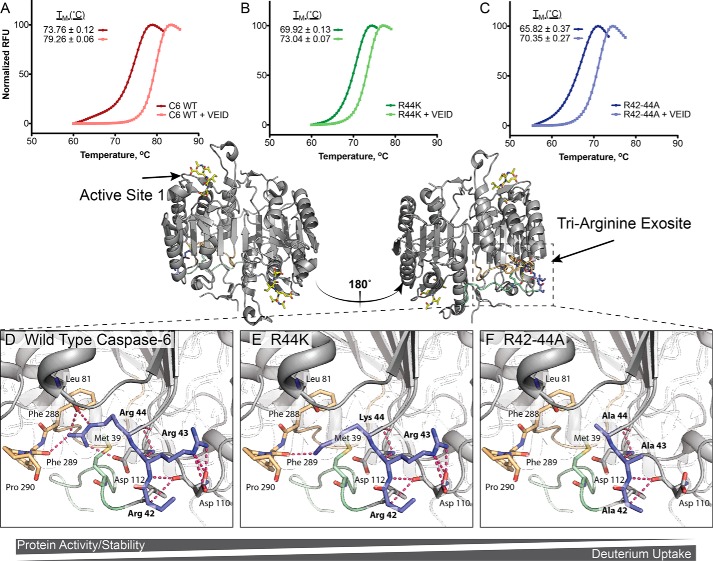
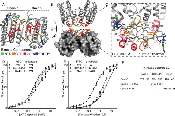
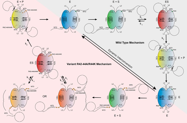
Caspases are cysteine-aspartic proteases involved in the regulation of programmed cell death (apoptosis) and a number of other biological processes. Despite overall similarities in structure and active-site composition, caspases show striking selectivity for particular protein substrates. Exosites are emerging as one of the mechanisms by which caspases can recruit, engage, and orient these substrates for proper hydrolysis. Following computational analyses and database searches for candidate exosites, we utilized site-directed mutagenesis to identify a new exosite in caspase-6 at the hinge between the disordered N-terminal domain (NTD), residues 23-45, and core of the caspase-6 structure. We observed that substitutions of the tri-arginine patch Arg-42-Arg-44 or the R44K cancer-associated mutation in caspase-6 markedly alter its rates of protein substrate hydrolysis. Notably, turnover of protein substrates but not of short peptide substrates was affected by these exosite alterations, underscoring the importance of this region for protein substrate recruitment. Hydrogen-deuterium exchange MS-mediated interrogation of the intrinsic dynamics of these enzymes suggested the presence of a substrate-binding platform encompassed by the NTD and the 240's region (containing residues 236-246), which serves as a general exosite for caspase-6-specific substrate recruitment. In summary, we have identified an exosite on caspase-6 that is critical for protein substrate recognition and turnover and therefore highly relevant for diseases such as cancer in which caspase-6-mediated apoptosis is often disrupted, and in neurodegeneration in which caspase-6 plays a central role.

Keywords: allosteric regulation; apoptosis; cancer; caspase-6; cysteine protease; hydrogen–deuterium exchange; mass spectrometry; neurodegeneration; protein dynamic; substrate recognition; substrate specificity; tri-arginine exosite.


Conflict of interest statement

The authors declare that they have no conflicts of interest with the contents of this article

Figures

Figure 1.
Figure 1.
Caspases show conserved folds but different surface charges. The overall caspase fold is largely conserved across the family with minimal divergent structural differences concentrated largely to the mobile loops. A, surface topology of caspase-6 (blue, PDB code 3OD5 (80)) and caspase-7 (green, PDB code 1F1J (87)). B, calculated electrostatics highlight key differences in positive (blue) and negative (red) regions distal from the active site. Unique surface characteristics may tune caspase substrate recognition.
Figure 2.
Figure 2.
Conservation analysis of arginine patch in caspase-6 orthologues. A, human caspases 1–14 in the N-terminal domain, the region post prodomain, curated from CaspBase (40), aligned by Clustal Omega, and colored to depict the degree of conservation calculated by ConSurf (42–46) across all caspase homologues and species. Conservation ranges from least conserved (blue) to most conserved (magenta). Arg-44 is conserved within the 205 caspase-6 sequences from a wide collection of phyla, classes, and taxa. B, sequence conservation across the 12 human caspases analyzed by ConSurf mapped onto the human caspase-6 sequence. The tri-arginine patch is not conserved across human caspases. C, conservation results of 205 caspase-6 sequences aligned and analyzed as in A and mapped onto the human caspase-6 structure. D, conservation of residues across the 12 human caspases analyzed as in B and mapped onto the human caspase-6 structure. Caspase-6 PDB code is 3OD5. ‡ denotes the caspase-7 exosite; * denotes the 42RRR44 patch in caspase-6.
Figure 3.
Figure 3.
POOL results show distal sites are important for biochemical function. POOL analysis performed on an ensemble of caspase-6 crystal structures in various activation states and conformations indicated by PDB codes in the figure. The top 12% of residues identified and predicted to be important for enzymatic function for each structure are highlighted in gray. Residues further clustered based on their conformational state: helical or strand, as well as their proximity to the caspase-6 active-site, 130's region, or distal positions.
Figure 4.
Figure 4.
Disruptions in the 42RRR44 patch retain activity for peptide substrates but alter kinetics for protein substrates. A, representative Michaelis-Menten kinetics of caspase-6 exosite variants compared with the WT enzyme. B, catalytic parameters of caspase-6 and exosite knockout variants R42–44A and R44K demonstrate that the active-site affinity is not altered by exosite modifications. C, cleavage of purified substrate caspase-6 C163S FL by the R42–44A or R44K variants over a time course of 0, 5, 15, 30, 45, and 60 min shows the rate of protein hydrolysis is severely delayed compared with the WT. D, quantification of caspase-6 C163S FL cleavage by WT or caspase-6 variants. E–G, hydrolysis of a constant amount of purified substrate lamin C from FL 66-kDa protein into 38/28-kDa fragments by caspase-6 (E), R42–44A (F), or R44K (G) in a 2-fold serial dilution from 25 to 0 μm for 4 h. H, quantification of the cleaved caspase-6 C163S FL band relative to the 0 μm point fit to a single-phase decay to calculate the CF50 used to calculate the hydrolysis rate of caspase-6 or variants R42–44A and R44K. Experiments performed in triplicate. *, ** denotes 90 or 95% confidence interval by unpaired two-tailed t test of the variants individually compared with the WT. In all cases, the caspase-6 variants are in the ΔN D179CT form. Only caspase-6 C163S as a substrate was in the full-length form.
Figure 5.
Figure 5.
42RRR44 patch is required for general protein hydrolysis in cell lysates. A–C, lysates generated from SK-N-AS neuroblastoma cells were incubated with 100 nm of either WT caspase-6, R42–44A, or R44K to assess hydrolysis over time by Western blotting of substrate proteins. A, PARP; B, lamin A/C; or C, DJ-1 normalized to loading control β-actin. D–F, quantification of the data from WT (black), R42–44A (white), or R44K (gray) normalized to the loading control for protein substrates PARP (D), lamin A/C (E), or DJ-1 (F) demonstrates that the hydrolysis of several biologically relevant substrates of caspase-6 are impacted by the removal of the 42RRR44 exosite. Changes in substrate processing of multiple proteins suggest they are engaging caspase-6 through a similar mechanism. Experiments were performed in triplicate. *, ** denotes 90 or 95% confidence interval by unpaired two-tailed t test of the variants individually compared with the WT.
Figure 6.
Figure 6.
Cleavage of key caspase-6 substrates occurs using the 42RRR44 exosite. A–C, hydrolysis of DVL3, which is one of the substrates most quickly cleaved after caspase-6 activation, was assessed by added caspase-6 variants in the ΔN D179CT form. A 2-fold serial dilution of caspase-6 (A), R42–44A (B), or R44K (C) from 250 to 0 nm incubated with a constant amount of SK-N-AS cell lysate over a 1-h time course at 37 °C imaged by Western blotting and normalized to the loading control β-actin. D, quantification of bands fit to a single-phase decay to find the CF50 used to calculate the hydrolysis rate of DVL3 by either caspase-6 (black), R42–44A (white), R44K (gray). Experiments were performed in triplicate. *, ** denotes 90% or 95% confidence interval by unpaired two-tailed t test of the variants individually compared with the WT.
Figure 7.
Figure 7.
Disruption of the 42RRR44 exosite increases dynamics of caspase-6 variants in NTD, 240's regions. A and B, caspase-6, R42–44A, or R44K incubated in D2O and monitored for exchange over time. Difference plots generated by subtracting the deuterium uptake profiles of common peptides of WT caspase-6 from peptides from either R42–44A (A) or R44K (B) variants. Regions depicted in blue (negative) are less exchanging in the variant than in the WT, whereas regions in red (positive) were more exchanging in the variant than in WT based on the error of the data sets ±0.42 or ±0.73 for the R42–44A or R44K based on the 98 and 95% confidence interval, respectively. C and D, exchange differences mapped to the structure of caspase-6 (predicted with MODELLER to incorporate all amino acids not visible in known structures (55)) of the R42–44A (C) or R44K (D) variants. E, relative deuterium uptake difference scale. F–L, important peptides identified as significantly different upon exchange compared with the WT peptides. Experiments were repeated on 2 different days. Δ indicates the difference, wherein the data from WT is always subtracted from the data from the indicated variant. In all cases, the caspase-6 variants are the ΔN D179CT form.
Figure 8.
Figure 8.
42RRR44 exosite functions as a hinge between NTD and core, stabilizing caspase-6. A–C, differential scanning fluorimetry was used to observe how substitutions in the 42RRR44 hinge impact the stability of caspase-6 (A), R44K (B), or R42–44A (C) in the unliganded state or liganded to the caspase-6 cognate inhibitor Ac-VEID-cho state. D–F, changes in the observed thermal stability may be attributed to sequential loss of interactions as side chains are removed. D, WT; E, 42RRK44 (R44K), and F, 42AAA44 (R42–44A). Models (E and F) were produced using the PyMOL mutagenesis wizard to demonstrate potential loss in contacts. In all cases the caspase-6 variants are the ΔN D179CT form.
Figure 9.
Figure 9.
Exosite is critical for stabilization of putative protein-binding interface. A, regions identified by H/Dx-MS are displayed on the caspase-6 structure 3K7E. The NTD (green) containing the exosite patch (blue) and 240's regions (red) make contacts with the CTD (orange) on the reverse face of caspase-6. B, rotating caspase-6 90° reveals a potential binding interface that occurs in the tetrameric helical structures of caspase-6, incorporating all regions identified by H/Dx. C, putative binding interface analyzed by PISA (58), containing large portions of buried surface area primarily consisting of buried hydrophobics. Residue label subscripts correspond to the chain of the crystal structure. D, fluorescence polarization assay of FITC-labeled caspase-6 C163S FL constructs of either the WT (black), R42–44A (white), or R44K (gray) variants incubated with the WT unlabeled protein as a substrate to assess KD apparent. E as in D except incubated with their respective unlabeled R42–44A C163S FL or R44K C163S FL variants. F, KD values of all labeled proteins incubated with the unlabeled WT substrate are not statistically significantly different, as compared to those incubated with R42–44A or R44K unlabeled proteins, which have a weaker KD value. In all cases, the caspase-6 variants are the ΔN D179CT form.
Figure 10.
Figure 10.
Model of caspase-6 protein substrate recognition. In the mature state, caspase-6 dynamically interconverts between the unique helical conformation and the canonical strand conformations (blue) (30). Herein, we propose, consistent with other models, that the intrinsically disordered NTD of caspase-6 fishes for protein substrates within the context of the cell, utilizing the unique sequences and charges of the domain to tune protein substrate recognition (green). The intrinsically disordered domain of caspase-6 is likely binding to the disordered domains of the substrate proteins (21, 55). The NTD orients the protein substrate, facilitating formation of the enzyme–substrate complex, by docking with the protein-binding interface of caspase-6. This step also coincides with the transition to the obligate strand state, which is required for enzymatic processing (red). Once assembled, chemistry proceeds producing cleaved product (yellow), wherein the remaining substrate is released allowing transition back to the dynamic resting state of mature caspase-6 (blue). Substitutions of the exosite at 42RRR44 are not proposed to affect the dynamic resting state of caspase-6, as it will still undergo helix-to-strand transitions, albeit at a different rate (blue) (Fig. 7A). However, upon substrate recruitment (green) and transition into the ES complex (red), the substitutions at the exosite of caspase-6 impact the dynamics of the NTD (Fig. 7), unhinging it, leading to perturbations of the protein docking face of the enzyme (Fig. 9, B and C) and the decreased rate of full substrate engagement, observed indirectly through the increased KD apparent of substrate binding (orange) (Fig. 9, D–F). Once the ES complex is successfully assembled (red), chemistry will proceed as expected (Fig. 4, A and B) leading to product formation (yellow) and back to the resting state of caspase-6 (blue).

References

    1. Elmore S. (2007) Apoptosis: a review of programmed cell death. Toxicol. Pathol. 35, 495–516 10.1080/01926230701320337 - DOI - PMC - PubMed
    1. Pistritto G., Trisciuoglio D., Ceci C., Garufi A., and D'Orazi G. (2016) Apoptosis as anticancer mechanism: function and dysfunction of its modulators and targeted therapeutic strategies. Aging 8, 603–619 10.18632/aging.100934 - DOI - PMC - PubMed
    1. Greenblatt M. S., Bennett W. P., Hollstein M., and Harris C. C. (1994) Mutations in the p53 tumor suppressor gene: clues to cancer etiology and molecular pathogenesis. Cancer Res. 54, 4855–4878 - PubMed
    1. Vaux D. L., Cory S., and Adams J. M. (1988) Bcl-2 gene promotes haemopoietic cell survival and cooperates with c-myc to immortalize pre-B cells. Nature 335, 440–442 10.1038/335440a0 - DOI - PubMed
    1. Plati J., Bucur O., and Khosravi-Far R. (2008) Dysregulation of apoptotic signaling in cancer: molecular mechanisms and therapeutic opportunities. J. Cell. Biochem. 104, 1124–1149 10.1002/jcb.21707 - DOI - PMC - PubMed

Publication types

MeSH terms