. 2018 Nov 12;8(1):16693.
doi: 10.1038/s41598-018-34257-2.

Coherent Tabletop EUV Ptychography of Nanopatterns

Affiliations

Coherent Tabletop EUV Ptychography of Nanopatterns

Nguyen Xuan Truong et al. Sci Rep. .

Abstract

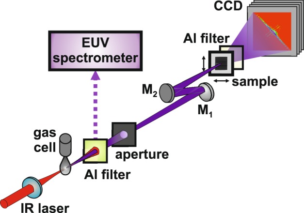
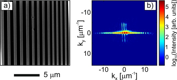
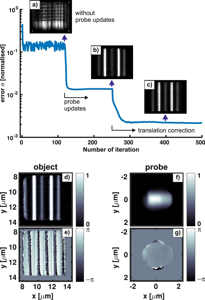
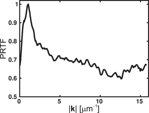
Coherent diffraction imaging (CDI) or lensless X-ray microscopy has become of great interest for high spatial resolution imaging of, e.g., nanostructures and biological specimens. There is no optics required in between an object and a detector, because the object can be fully recovered from its far-field diffraction pattern with an iterative phase retrieval algorithm. Hence, in principle, a sub-wavelength spatial resolution could be achieved in a high-numerical aperture configuration. With the advances of ultrafast laser technology, high photon flux tabletop Extreme Ultraviolet (EUV) sources based on the high-order harmonic generation (HHG) have become available to small-scale laboratories. In this study, we report on a newly established high photon flux and highly monochromatic 30 nm HHG beamline. Furthermore, we applied ptychography, a scanning CDI version, to probe a nearly periodic nanopattern with the tabletop EUV source. A wide-field view of about 15 × 15 μm was probed with a 2.5 μm-diameter illumination beam at 30 nm. From a set of hundreds of far-field diffraction patterns recorded for different adjacent positions of the object, both the object and the illumination beams were successfully reconstructed with the extended ptychographical iterative engine. By investigating the phase retrieval transfer function, a diffraction-limited resolution of reconstruction of about 32 nm is obtained.


Conflict of interest statement

The authors declare no competing interests.

Figures

Figure 1
Figure 1
Schematic view of the tabletop EUV source for high-numerical aperture ptychography. An 1 kHz Ti:sa amplifier delivers 35 fs–FWHM optical pulses with up to 8 mJ pulse energy and at an 800 nm central wavelength. The IR beam was focused with a 500 mm focal length lens into an 8 mm long gas cell fed with argon gas through a piezo-driven valve at a backing pressure of 1.5 bars. The resulting HHG beam passed from the source through an 1 mm–inner diameter differential pumping tube to the characterisation chamber, and then was characterised with a flat-field EUV spectrometer. To separate the IR beam, a 300 nm–thick Al foil was used as a spectral filter. At about 80 cm downstream from the HHG source, an 1 mm–diameter aperture was inserted as a spatial filter, resulting in a desired illumination profile for ptychography. A single harmonic at 30 nm was selected and focused with a pair of multilayer mirrors in a z-configuration to minimise astigmatism, including a flat bending mirror (M1) and an f2 = 110 mm spherical mirror (M2). A sample with lithographed nanopatterns was mounted on an xyz-translation stage and located at the focus of the 30 nm probe beam. An SEM image of the sample is provided in Fig. 2a. Diffraction patterns of the adjacent areas on the sample were recorded with an in-vacuum X-ray CCD camera at a distance z = 16.5 mm. A second 200 nm–thick Al filter was installed in front of the CCD camera to block the residual stray light. The lateral positioning and data recording were synchronised with a home-built LabVIEW program. Image reconstruction was performed with the extended ptychographical iterative engine (ePIE) on an NVIDIA Tesla K40 computing processor.
Figure 2
Figure 2
(a) An SEM image of the nanopatterned sample. (b) A representative diffraction pattern of the sample after binning 2 × 2 pixels into 1 pixel and performing curvature correction. An almost six orders of magnitude high dynamic range image was obtained without the use of a beamstop.
Figure 3
Figure 3
(Top) Typical normalised error as a function of the number of iteration (Eq. (7)). The ePIE algorithm performed 500 iterations with probe updates after 120 iterations and translation refinement after 250 iterations. (Top, insets) The reconstructed images illustrate the visual quality of the object without probe updates (a), with probe updates (b), and with translation correction (c), taken at the iteration marked by vertical arrows. (Bottom) The final reconstructed amplitude and phase of the object (d,e) and probe (f,g), respectively.
Figure 4
Figure 4
Phase retrieval transfer function (PRTF) computed from five hundred independent ePIE reconstructions. The resolution cutoff of the ePIE, determined by the spatial frequency at which the PRTF reaches a value of 1/e, is greater than the experimental cutoff kmax ≈ 16 μm−1.
Figure 5
Figure 5
Amplitude and phase of the object (a,b) and probe (c,d), respectively, obtained after a thousand ePIE iterations from a collection of 900 far-field diffraction patterns of the sample. (c) An intensity line profile (blue-filled) is extracted along the green line in (a) compared to its corresponding part of the SEM image (yellow-filled) shown in Fig. 2a. Note the different imaging methods between the ePIE image (transmission) and the SEM (reflection).

References

    1. Sakdinawat A, Attwood D. Nanoscale X-ray imaging. Nat. Photonics. 2010;4:840–848. doi: 10.1038/nphoton.2010.267. - DOI
    1. Mimura H, et al. Breaking the 10 nm barrier in hard-X-ray focusing. Nat. Phys. 2010;6:122–125. doi: 10.1038/Nphys1457. - DOI
    1. Sayre D. Some Implications of a Theorem Due to Shannon. Acta Crystallogr. 1952;5:843–843. doi: 10.1107/S0365110x52002276. - DOI
    1. Gerchberg RW, Saxton WO. Practical Algorithm for Determination of Phase from Image and Diffraction Plane Pictures. Optik. 1972;35:237–246.
    1. Fienup JR. Phase Retrieval Algorithms - a Comparison. Appl. Opt. 1982;21:2758–2769. doi: 10.1364/AO.21.002758. - DOI - PubMed