. 2018 Nov;119(11):1347-1357.
doi: 10.1038/s41416-018-0289-1. Epub 2018 Nov 13.

Bcl-2-dependent synthetic lethal interaction of the IDF-11774 with the V0 subunit C of vacuolar ATPase (ATP6V0C) in colorectal cancer

Affiliations

Bcl-2-dependent synthetic lethal interaction of the IDF-11774 with the V0 subunit C of vacuolar ATPase (ATP6V0C) in colorectal cancer

Bo-Kyung Kim et al. Br J Cancer. 2018 Nov.

Abstract

Background: The IDF-11774, a novel clinical candidate for cancer therapy, targets HSP70 and inhibits mitochondrial respiration, resulting in the activation of AMPK and reduction in HIF-1α accumulation.

Methods: To identify genes that have synthetic lethality to IDF-11774, RNA interference screening was conducted, using pooled lentiviruses expressing a short hairpin RNA library.

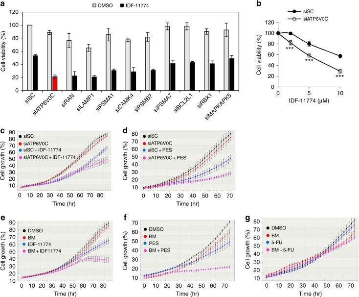
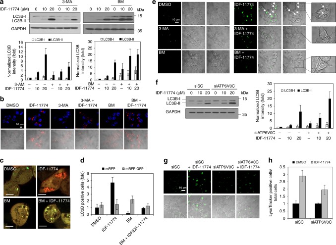
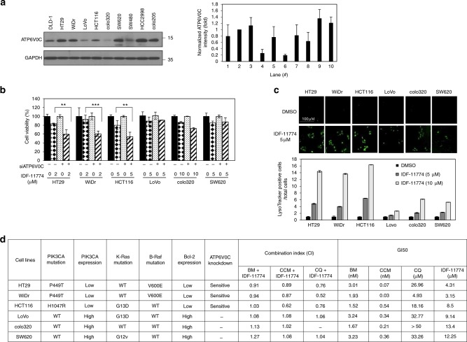
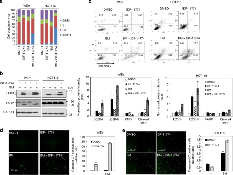
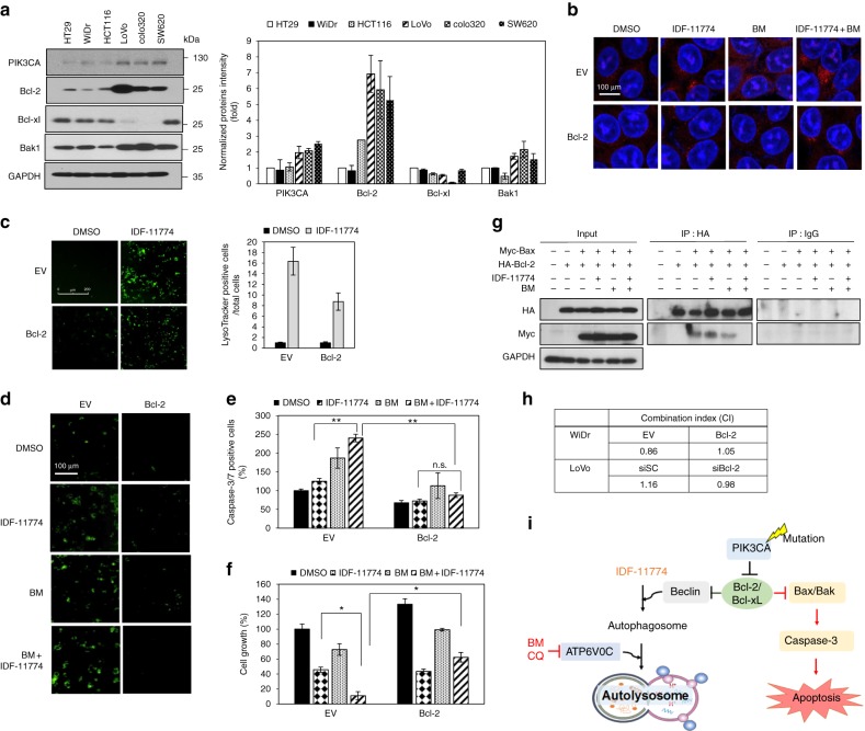
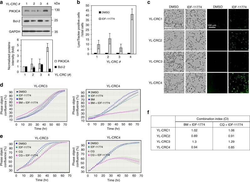
Results: We identified ATP6V0C, encoding the V0 subunit C of lysosomal V-ATPase, knockdown of which induced a synergistic growth-inhibitory effect in HCT116 cells in the presence of IDF-11774. The synthetic lethality of IDF-11774 with ATP6V0C possibly correlates with IDF-11774-mediated autolysosome formation. Notably, the synergistic effect of IDF-11774 and the ATP6V0C inhibitor, bafilomycin A1, depended on the PIK3CA genetic status and Bcl-2 expression, which regulates autolysosome formation and apoptosis. Similarly, in an experiment using conditionally reprogramed cells derived from colorectal cancer patients, synergistic growth inhibition was observed in cells with low Bcl-2 expression.

Conclusions: Bcl-2 is a biomarker for the synthetic lethal interaction of IDF-11774 with ATP6V0C, which is clinically applicable for the treatment of cancer patients with IDF-11774 or autophagy-inducing anti-cancer drugs.


Conflict of interest statement

The authors declare no competing interests.

Figures

Fig. 1
Fig. 1
RNA interference (RNAi) screening to identify candidate genes sensitizing HCT116 cells to IDF-11774. a Knockdown effect of candidate genes on cell growth of HCT116 cells treated with 10 µM IDF-11774. b Effect of ATP6V0C knockdown on the growth of HCT116 cells treated with different concentrations of IDF-11774 for 72 h. Cell viability was analyzed by the sulforhodamine B (SRB) assay. Results are shown as the mean ± S.D. of the three independent experiments. c, d Effect of ATP6V0C knockdown on the growth of HCT116 cells treated with 5 μM IDF-11774 or 5 μM 2-phenylethynesulfonamide (PES). eg Effect of bafilomycin A1 (BM, 1 nM) on the growth of HCT116 cells treated with 5 μM IDF-11774, 5 μM PES or 5 μM fluorouracil (5-FU). Cell growth was evaluated by live-cell imaging (IncuCyte ZOOM system). Data are presented as the single representative result of three experiments as the mean ± S.D. In each experiment, each treatment was repeated in triplet wells, and each well was analyzed in 4 frames
Fig. 2
Fig. 2
Synthetic lethal interaction of IDF-11774 with ATP6V0C in HCT116 cells. a, b Effect of autophagy inhibitor on LC3B accumulation in cells treated with IDF-11774 as analyzed by western blot analysis (a) and immunofluorescence (b). HCT116 cells were pre-incubated with 5 mM 3-methyladenine (3-MA) or 1 nM bafilomycin A1 (BM) for 3 h and then treated with IDF-11774 for 18 h. Scale bar, 10 µm. Western blot band intensities were quantified using ImageJ. The signal intensity of LC3B was normalized to that of GAPDH. c, d Autolysosome formation of cells treated with IDF-11774 and 1 nM bafilomycin A1 (BM). Representative images of confocal fluorescence microscopy (c) and quantification of positive cells for mRFP (red) and mRFP-GFP (yellow) in HCT116 cells transfected with ptfLC3 (d). Positive cells were counted using fluorescence microscopy (n = ~70). Scale bar, 5 µm. e Effect of autophagy inhibitor on autolysosome formation in cells treated with 5 μM IDF-11774 and/or 1 nM bafilomycine A1 for 18 h. Autolysosome formation was assessed by LysoTracker staining. Arrows indicate autolysosomes. Scale bar, 10 µm. f Effect of ATP6V0C knockdown using siATP6V0C on LC3B accumulation in cells treated with 5 μM IDF-11774, as analyzed by western blotting. g, h Effect of ATP6V0C knockdown on autolysosome formation, as analyzed by LysoTracker staining (g) and the ratio of LysoTracker-positive to total HCT116 cells (h). Scale bar, 10 µm
Fig. 3
Fig. 3
Synergistic effect of ATP6V0C and IDF-11774 on the growth of various colorectal cancer cells. a Western blot analysis of ATP6V0C levels in human colorectal cancer cells. The signal intensity of ATP6V0C was normalized to that of GAPDH. b Effect of ATP6V0C knockdown on the sensitivity of human colorectal cancer cells to IDF-11774. Cell viability was analyzed using the sulforhodamine B (SRB) assay. Results are shown as the mean ± S.D. of three independent experiments. c LysoTracker staining for autolysosomes in various colorectal cancer cells. LysoTracker-positive cells as observed by fluorescence microscopy. Ratios of LysoTracker-positive to total cells are shown in the graph. Scale bar, 100 µm. d Correlation of mutation status or expression of genes and combination index of two compounds. BM (bafilomycin A1), CCM (concanamycin A), and CQ (chloroquine) were used
Fig. 4
Fig. 4
Combined treatment of bafilomycin A1 and IDF-11774 induces apoptosis in colorectal cancer cells. a FACScan analysis of cells treated with IDF-11774 and 1 nM bafilomycin A1 for 72 h by propidium iodide (PI) staining. b Western blot analysis of cells treated with IDF-11774 and 1 nM bafilomycin A1 for 72 h. The signal intensity of each protein was normalized to that of GAPDH. c Scatter-grams of fluorescein isothiocyanate (FITC)-Annexin V/propidium iodide (PI) staining of WiDr and HCT116 cells treated with IDF-11774 and 1 nM bafilomycin A1 for 72 h. d, e Measurement of caspase-3/7 activity using the CellPlayer kinetic caspase-3/7 reagent. Caspase-3/7-positive cells appeared green under a fluorescence microscope. WiDr (d) and HCT116 cells (e) treated with IDF-11774 and 1 nM bafilomycin A1 for 72 h using the IncuCyte ZOOM system. Scale bar, 200 µm. The concentration of IDF-11774 was 5 μM in WiDr and 10 μM in HCT116 cells
Fig. 5
Fig. 5
Bcl-2 expression level is crucial for the synthetic lethality of IDF-11774 and bafilomycin A1. a Western blot analysis of Bcl-2 family protein levels in human colorectal cancer cells. The signal intensity of each protein was normalized to that of GAPDH. bf HCT116 cells transfected with pCMV-SPORT6- BCL2 were treated with 10 µM IDF-11774 and 1 nM bafilomycin A1. b Immunofluorescence of LC3B. Scale bar, 10 µm. c Autolysosome formation. Caspase-3/7 activity (d, e) and cell growth (f) were measured by IncuCyte ZOOM system. Graphs are shown as the mean ± S.D. in different regions (n = 4) of the well. g Evaluation of the change in the interaction between Bcl-2 and Bax induced by bafilomycin A1 using immunoprecipitation assay. HCT116 cells transfected with pCMV3-HA-BCL2 and pCMV3-Myc-BAX were treated with 10 µM IDF-11774 and 1 nM bafilomycin A1. h Combination index of IDF-11774 with bafilomycin A1 in WiDr cells transfected with pCMV-SPORT6-BCL2 and in LoVo cells transfected with siBCL2. i Model depicting Bcl-2-dependent synthetic lethal interaction of IDF-11774 and ATP6V0C
Fig. 6
Fig. 6
Regulation of IDF-1174 sensitivity to conditionally reprogramed cells (CRCs) derived from patients with colon cancer. a Western blot analysis of Bcl-2 expression in the CRCs. The signal intensity of each protein was normalized to that of GAPDH. b, c Autolysosome formation in CRCs treated with 10 µM IDF-11774. Scale bar, 200 µm. Bars indicate the ratio of the average of LysoTracker-positive cells in different regions (n = 9) of the well. d, e Growth inhibition of CRCs expressing different levels of Bcl-2 in the presence of 1 nM bafilomycin A1 and 5 µM IDF-11774 or 5 µM chloroquine, as measured using IncuCyte ZOOM system. Data are presented as the single representative result of three experiments and as the mean ± S.D. In each experiment, each treatment was repeated in triplet wells, and each well was analyzed in 4 frames. f Combination index of IDF-11774 with bafilomycin A1 (BM) or chloroquine (CQ) in CRCs

Similar articles

Cited by

References

    1. Leung AW, de Silva T, Bally MB, Lockwood WW. Synthetic lethality in lung cancer and translation to clinical therapies. Mol. Cancer. 2016;15:61. doi: 10.1186/s12943-016-0546-y. - DOI - PMC - PubMed
    1. Srivas R, et al. A network of conserved synthetic lethal interactions for exploration of precision cancer therapy. Mol. Cell. 2016;63:514–525. doi: 10.1016/j.molcel.2016.06.022. - DOI - PMC - PubMed
    1. Diehl P, Tedesco D, Chenchik A. Use of RNAi screens to uncover resistance mechanisms in cancer cells and identify synthetic lethal interactions. Drug Discov. Today Technol. 2014;11:11–18. doi: 10.1016/j.ddtec.2013.12.002. - DOI - PMC - PubMed
    1. Baratta MG, et al. An in-tumor genetic screen reveals that the BET bromodomain protein, BRD4, is a potential therapeutic target in ovarian carcinoma. Proc. . Natl Acad. Sci. USA. 2015;112:232–237. doi: 10.1073/pnas.1422165112. - DOI - PMC - PubMed
    1. Schonherr M, et al. Genome-wide RNAi screen identifies protein kinase Cb and new members of mitogen-activated protein kinase pathway as regulators of melanoma cell growth and metastasis. Pigment. Cell. Melanoma Res. 2014;27:418–430. doi: 10.1111/pcmr.12216. - DOI - PubMed

Publication types

MeSH terms