. 2019 Jan;51(1):151-162.
doi: 10.1038/s41588-018-0270-1. Epub 2018 Nov 12.

Subtype-specific regulatory network rewiring in acute myeloid leukemia

Affiliations

Subtype-specific regulatory network rewiring in acute myeloid leukemia

Salam A Assi et al. Nat Genet. 2019 Jan.

Abstract

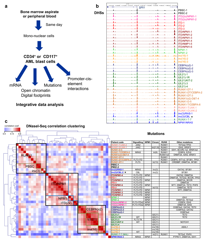
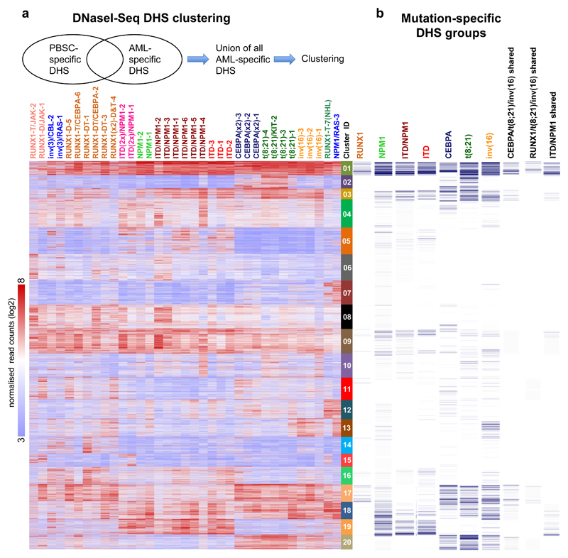
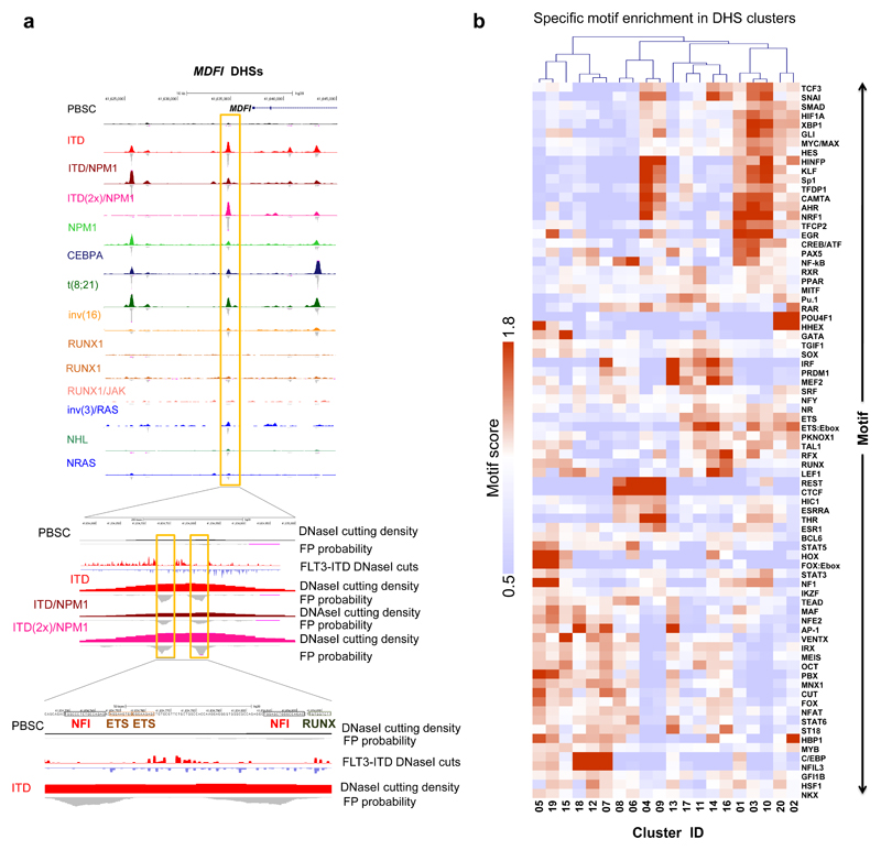
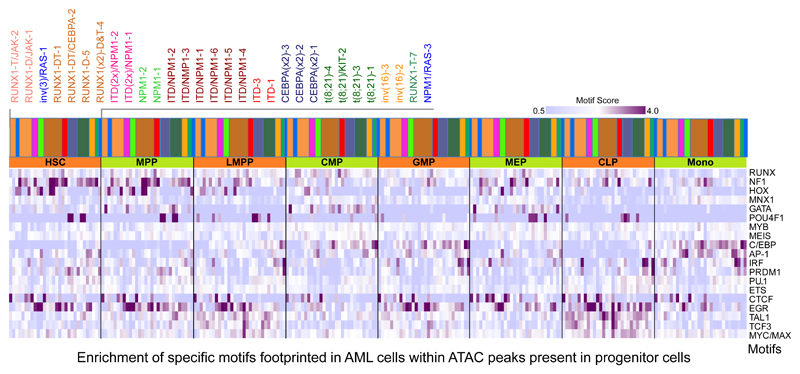
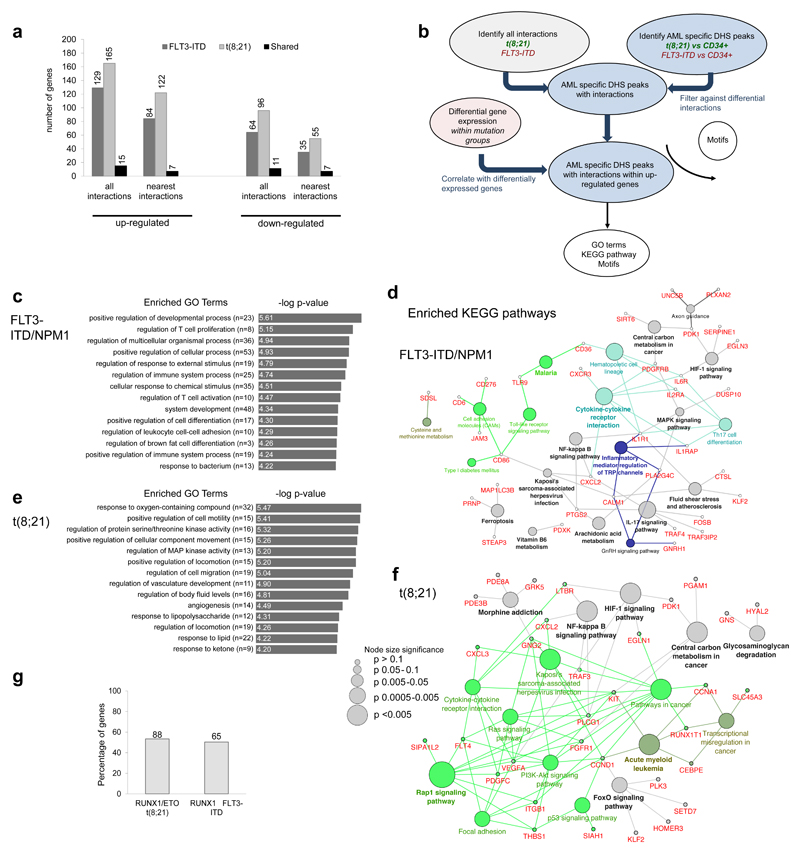
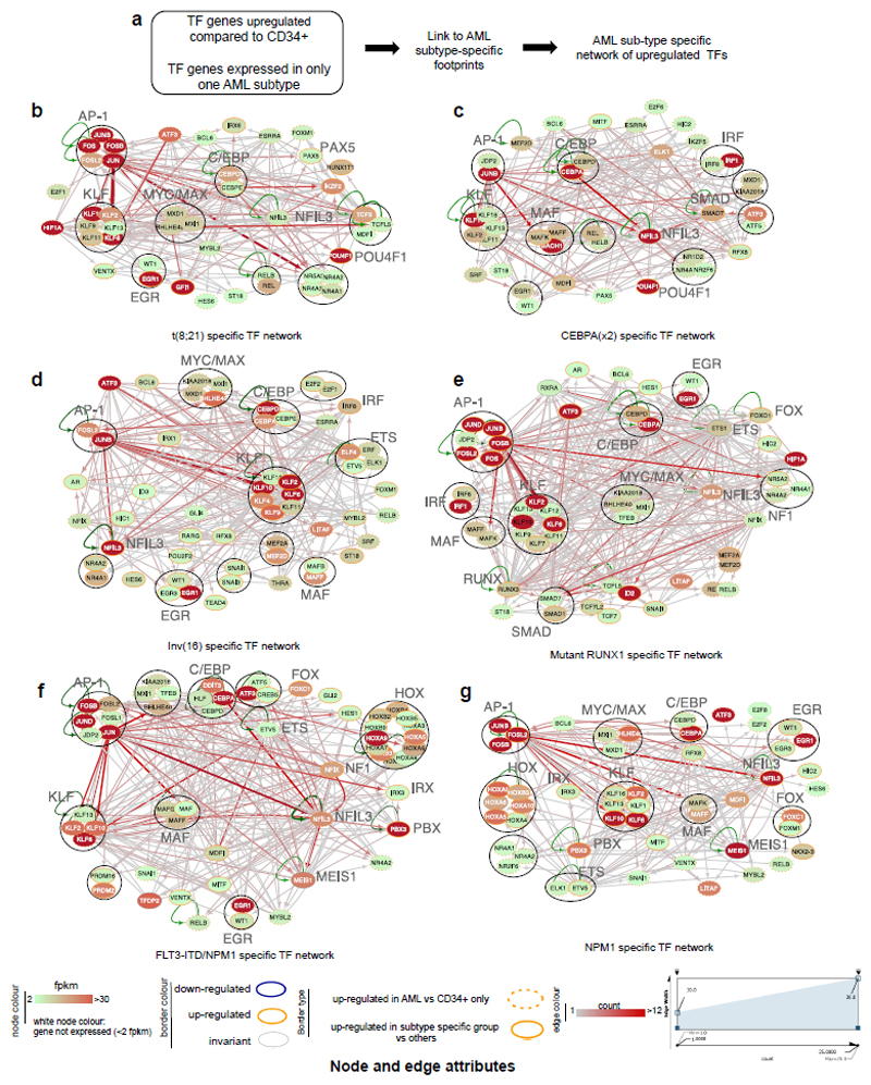
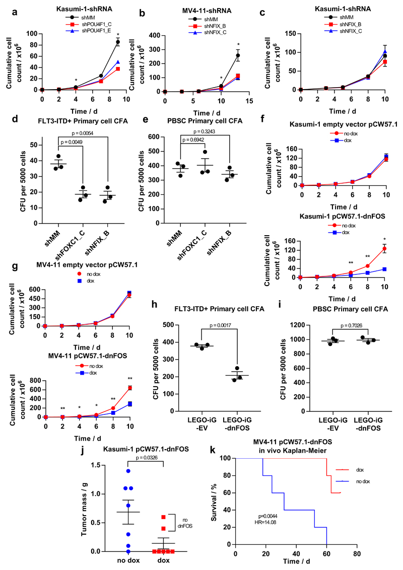
Acute myeloid leukemia (AML) is a heterogeneous disease caused by a variety of alterations in transcription factors, epigenetic regulators and signaling molecules. To determine how different mutant regulators establish AML subtype-specific transcriptional networks, we performed a comprehensive global analysis of cis-regulatory element activity and interaction, transcription factor occupancy and gene expression patterns in purified leukemic blast cells. Here, we focused on specific subgroups of subjects carrying mutations in genes encoding transcription factors (RUNX1, CEBPα), signaling molecules (FTL3-ITD, RAS) and the nuclear protein NPM1). Integrated analysis of these data demonstrates that each mutant regulator establishes a specific transcriptional and signaling network unrelated to that seen in normal cells, sustaining the expression of unique sets of genes required for AML growth and maintenance.


Conflict of interest statement

Competing financial interests

The authors declare no competing financial interests.

Figures

Figure 1
Figure 1. Different types of AML adopt unique transcriptome and chromatin landscapes.
(a) Experimental strategy. (b) UCSC Genome browser tracks of DNaseI-seq mapping in purified AML cells. (c) Hierarchical clustering of Pearson correlation coefficients of DNaseI accessible sequences from all patient samples with normalized read counts of DNaseI-Seq data for the different classes of mutations (left panel), right panel: list of mutations in cells from each patient
Figure 2
Figure 2. AML-specifically active cis-regulatory elements cluster into common and unique chromatin landscapes.
(a) Heatmap depicting unsupervised K-mean clustering of the DNaseI-Seq log2 signals seen in each AML specific distal DHS peak in each AML sample compared to PBSCs. Clustering was done only on rows (DHS peaks) while samples were ranked based on the clustering in Fig, 1c. The diagram on top of the heatmap shows the DHS peak population used for clustering. (b) Binary heatmap showing the overlap between the clusters from A and the DHSs of the 7 mutation classes which are deregulated compared to CD34+ PBSCs as described in Supplementary Fig 4a.
Figure 3
Figure 3. AML-specifically active cis-regulatory elements display AML type-specific transcription factor occupancy patterns.
(a) UCSC browser screen shot of the MDFI locus zooming in on an AML type-specific DHS (box) with occupied NFI, ETS and RUNX sites. (b) Heatmap depicting the degree of motif enrichment after hierarchical clustering of motif occupancy in each of the 20 AML DHS clusters. Enrichment score was calculated by the level of motif enrichment in all the footprints of all high read-depth samples for each cluster, as compared to union of footprints in all experiments.
Figure 4
Figure 4. AML cells show occupied motif patterns unrelated to normal progenitor cells.
Enrichment analysis of footprinted motifs in AML subgroups which overlap with ATAC-Seq peaks present in precursor cells. The bars with the colour code above the heatmap reflect the type of mutation and the order of the different patients is depicted in the enlarged panel above the colour bars. Additional explanations are provided in Supplementary methods.
Figure 5
Figure 5. Capture HiC shows differences in locus-specific cis-regulatory interactions between different types of AML and normal cells.
(a) Percentage of up- and down-regulated genes with differential interactions from the FLT3-ITD and the t(8;21) compared to CD34+ PBSCs. The bar figure shows also the percentage of the common genes for the FLT3-ITD and the t(8;21), the number of differentially expressed genes (DEG) is shown on top of each bar. (b) Flow diagram showing the steps for identification of differential interactions and the downstream analysis. (c) Top enriched GO terms for the up-regulated genes of the FLT3-ITD compared to CD34+ PBSCs as outlined in (a). (d) Network diagram of top KEGG pathways for the up-regulated genes of the FLT3-ITD compared to CD34+ PBSCs as outlined in (a). (e) Top enriched GO terms for the up-regulated genes of the t(8;21) compared to to CD34+ PBSCs shown as outlined in (a) Network diagram of top KEGG pathways for the up-regulated genes of the t(8;21) compared to CD34+ PBSCs as outlined in (b). (h): Percentage of RUNX1-ETO and RUNX1 targets amongst up-regulated genes with differential interactions, the exact number of genes is shown above the bars.
Figure 6
Figure 6. Identification of transcription factor networks driving the expression of AML type-specific up-regulated TF genes
(a) Outline of analysis strategy. (b) t(8;21)-specific TF network, (c) CEBPA(x2)-specific TF, (d) Inv(16) specific TF network, (e) Mutant RUNX1-specific TF network, (f) FLT3-ITD/NPM1 specific TF network, (g) NPM1-specific TF network Factor families binding to the same motif as shown in Supplementary table 2 form a node contained within a circle. Arrows going outwards from the entire node highlight footprinted motifs in individual genes generated by any member of this factor family whereby the footprint was annotated to the gene using the CHiC data where possible, otherwise to the nearest gene. For selected nodes, the name of the underlying motif is highlighted in large letters. The expression level (FKPM) for the individual genes is depicted in white (low)/red (high) colour. An orange smooth ring around the circle indicates that this gene is specifically up-regulated in this type of AML compared to CD34+ PBSCs and/or other AML types, a dotted circle indicates a gene that is up-regulated as compared to CD34+ cells. Genes without outgoing arrows due to a lack of know binding motifs are highlighted by their octagon shapes.
Figure 7
Figure 7. Identification of AML type-specific TFs required for maintaining leukemic growth and colony forming ability.
(a - c) Scatter plot showing the growth curves of (a) Kasumi-1 cells after transduction with shPOU4F1 or (c) shNFIX and (b) of MV4-11 cells after transduction with shNFIX. (d, e) Dot plot showing the number of colonies formed by a FLT3-ITD+ primary AML cell samples (d) or PBSCs (e) after transduction with shRNA targeting FOXC1, NFIX or a mismatch control. (f) Scatter plot showing the growth curve of Kasumi-1 cells transduced with either a doxycycline-inducible dnFOS or an empty vector control with and without 1.5 mcg/ml doxycycline. (g) Dot plots showing the growth curve of MV4-11 cells transduced with either a doxycycline-inducible dnFOS or an empty vector control (right panel) with and without 1.5 μg/ml doxycycline. (h,i) The expression of a dnFOS causes a reduction in the colony forming ability of CD34+ FLT3-ITD+ primary AML cells (H) but not CD34+ PBSCs (i (j) Granulosarcoma formation in RG mice by Kasumi-1 expressing a doxycycline-inducible dnFOS. dnFOS was induced by intraperitoneal injection of doxycycline. (k) Survival curve for RG mice transplanted with MV4-11 cells expressing doxycycline-inducible dnFOS. The induction of dnFOS significantly increased the survival time of mice. Significance in all experiments was tested using a two-tailed Student’s t-test (n=3) with * p<0.05, **p<0.01 in both samples compared to the mismatch control where indicated. Error bars show standard error of the mean. Further detail on statistical analysis can be found in Online Methods.

Comment in

References

    1. Cancer Genome Atlas Research N. Genomic and epigenomic landscapes of adult de novo acute myeloid leukemia. N Engl J Med. 2013;368:2059–74. - PMC - PubMed
    1. Papaemmanuil E, et al. Genomic Classification and Prognosis in Acute Myeloid Leukemia. New England Journal of Medicine. 2016;374:2209–2221. - PMC - PubMed
    1. Bonifer C, Cockerill PN. Chromatin Structure Profiling Identifies Crucial Regulators of Tumor Maintenance. Trends Cancer. 2015;1:157–160. - PubMed
    1. Rosenbauer F, Tenen DG. Transcription factors in myeloid development: balancing differentiation with transformation. Nature reviews. Immunology. 2007;7:105–17. - PubMed
    1. Cockerill PN. Receptor Signaling Directs Global Recruitment of Pre-existing Transcription Factors to Inducible Elements. Yale J Biol Med. 2016;89:591–596. - PMC - PubMed

Publication types