. 2018 Dec;24(12):1859-1866.
doi: 10.1038/s41591-018-0233-1. Epub 2018 Nov 12.

Venetoclax with azacitidine disrupts energy metabolism and targets leukemia stem cells in patients with acute myeloid leukemia

Affiliations

Venetoclax with azacitidine disrupts energy metabolism and targets leukemia stem cells in patients with acute myeloid leukemia

Daniel A Pollyea et al. Nat Med. 2018 Dec.

Abstract

Acute myeloid leukemia (AML) is the most common acute leukemia in adults. Leukemia stem cells (LSCs) drive the initiation and perpetuation of AML, are quantifiably associated with worse clinical outcomes, and often persist after conventional chemotherapy resulting in relapse1-5. In this report, we show that treatment of older patients with AML with the B cell lymphoma 2 (BCL-2) inhibitor venetoclax in combination with azacitidine results in deep and durable remissions and is superior to conventional treatments. We hypothesized that these promising clinical results were due to targeting LSCs. Analysis of LSCs from patients undergoing treatment with venetoclax + azacitidine showed disruption of the tricarboxylic acid (TCA) cycle manifested by decreased α-ketoglutarate and increased succinate levels, suggesting inhibition of electron transport chain complex II. In vitro modeling confirmed inhibition of complex II via reduced glutathionylation of succinate dehydrogenase. These metabolic perturbations suppress oxidative phosphorylation (OXPHOS), which efficiently and selectively targets LSCs. Our findings show for the first time that a therapeutic intervention can eradicate LSCs in patients with AML by disrupting the metabolic machinery driving energy metabolism, resulting in promising clinical activity in a patient population with historically poor outcomes.


Conflict of interest statement

Competing interests Statement

The authors declare no competing interests.

Figures
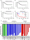
Figure 1:
Figure 1:. Clinical responses for the 33 patients treated with venetoclax + azacitidine at a single institution. Median follow up time was 580 days
a) Duration of response for the 33 responding patients. Median response duration was not reached. b) Progression free survival for the 30 responding patients. Median time to progression or death was not reached. c) Overall survival for all treated patients. Median overall survival was not reached. d) Overall survival comparison of 33 treated patients with venetoclax + azacitidine (blue line, venetoclax + azacitidine) versus any other treatment in 88 older previously untreated AML patients treated consecutively at a single institution (red line, control patients). Control patients had a significantly worse survival in comparison to venetoclax + azacitidine patients. Log-rank (Mantel-Cox) test p=0.0003 e) Measurable residual disease (MRD) monitoring of 22 patients who had a clinical response and an amenable mutation for droplet digital PCR performed with each bone marrow biopsy. Each patient had one to four mutations monitored; each number represents a patient, each gene monitored is shown as a bar beneath the patient number and annotated along the X-axis. The length of the bar represents the maximal decrease in variant allele frequency (VAF); 100% is equivalent to MRD negativity. Genes that achieved MRD negativity are annotated with an asterisk. Green bars represent patients who achieved MRD negativity and have not progressed. Blue bars represent patients who have not achieved MRD negativity and have not progressed. Red bars represent patients who did not achieve MRD negativity and subsequently progressed. No patients who achieved MRD negativity in all genes analyzed have progressed. All patients who have progressed did not achieve MRD negativity in at least one measurable gene.
Figure 2:
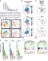
Figure 2:. Mass cytometry and single cell transcriptomics at early time points show significant decreases in blasts and LSCs.
a) Temporal blast counts during the first week of therapy for nine patients who began treatment with >20% peripheral blasts at diagnosis (assessed by complete blood count with manual differential). Table below figure shows the proportion of patients at 0% and the average percent reduction of blasts for each day of analysis. For summary of blast counts on all available patients, please see supplemental Table 5. b) Mass cytometry analysis of peripheral blood at Day 0, 2, and 4 for a representative patient. AML blasts are indicated by the red shaded area, with blast percentages shown (n=1) . c) Mass cytometry analysis of peripheral blood at baseline and 24 hours for patients 7 and 16. Red circles indicate the phenotypically-defined LSC population (CD34+, CD38-, Lin-, CD123+) (n=1) . d) tSNE plots of single cell transcriptomics measured at baseline, day two and four post-treatment with blasts as defined by gene signature in red circle(n=1) . e) Number of cells in clusters of tSNE plot by time point. Blast cluster (indicated by red arrow) shows a decrease at day two and disappearance of cells by day four (n=1) . f) GSEA analysis of blast cluster compared to all other clusters in baseline sample tSNE plots shows the cluster is significantly enriched for two different LSC gene signatures.
Figure 3:
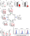
Figure 3:. Venetoclax + Azacitidine reduces oxidative phosphorylation in AML LSCs.
a) Oxygen consumption rate as a measure of OXPHOS was quantified pre and 24 hours post venetoclax + azacitidine treatment in the leukemia cells isolated from an AML patient, revealing a significant reduction in OXPHOS levels. N=3–5, technical replicates. Significance was measure by unpaired two-tailed Student’s t-test. b) Using FCCP to uncouple the electron transport chain and maximize oxidative phosphorylation, the oxygen consumption rate was measured in the leukemia cells of one patient before and 24 hours after treatment, revealing a significant decrease in the ability of the post-treatment leukemia cells to maximize oxygen consumption. N=3–4 technical replicates. Significance was measure by unpaired two-tailed Student’s t-test. c) Oxygen consumption levels were measured in ROS-low LSCs isolated from 4 primary AML specimens A, B, C, and D treated with 500nM venetoclax + 2.5 μm azacitidine in vitro for 4 hours. n=4 (biological)/n=5 (technical) replicates. Significance was measure by paired two-tailed Student’s t-test. d) ATP production was measured in ROS-low LSCs isolated from 3 primary AML specimens A, B, and C treated with 500nM venetoclax + 2.5 μm azacitidine in vitro for 4 hours. n=3 (biological)/ n=5 (technical) replicates. Significance was measure by paired two-tailed Student’s t-test. (e-g) Three patients with circulating blasts sampled from the peripheral blood at baseline and 24 hours after treatment with venetoclax + azacitidine show evidence of decreased electron transport chain complex II activity. e) Levels of TCA cycle intermediates, alpha-ketoglutarate, succinate, citrate, fumarate, malate, oxaloacetate in patients pre and 24 hours post venetoclax + azacitidine treatment. n=3 (biological)/ n=2–5 (technical) replicates. Significance was measure by unpaired two-tailed Student’s t-test. f) Levels of glutathione in patients pre and 24 hours post venetoclax + azacitidine treatment. n=3 (biological)/ n=2–5 (technical) replicates. Significance was measure by unpaired two-tailed Student’s t-test. g) Cellular reactive oxygen species (ROS) in leukemia cells from patients pre (red shaded histogram) and 24 hours post (blue shaded histogram) venetoclax + azacitidine treatment (as determined by labeling with CellROX™). ROS was measured in each individual patient once. Bars represent the mean of the replicates and error bars represent standard deviation for each bar graph. Statistical tests were performed on technical replicates only when assay was measuring metabolic responses of individual patients on therapy. NS= not significant, AU= arbitrary units
Figure 4:
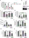
Figure 4:. Venetoclax + azacitidine reduces complex II activity through reduction in sdhA glutathionylation.
a) Glutathione levels were measured in ROS-low LSCs isolated from 3 primary AML specimens A, B, and C treated with 500nM venetoclax + 2.5 μm azacitidine in vitro for 4 hours. n=3 (biological)/n=4 (technical) replicates. Significance was measure by paired two-tailed Student’s t-test. b) Immune precipitation of succinate dehydrogenase (sdhA) followed by western blot for GSH modification of sdhA isolated from leukemia cells treated with 500nM venetoclax + 2.5 μm azacitidine in vitro for 4 hours. Blot shown here is a representative image using specimen A (n=3). c) Relative complex II activity measured in 3 AML specimens A, B, and C pretreated with cell permeable glutathione or vehicle and then treated with 500nM venetoclax + 2.5 μm azacitidine in vitro for 4 hours. N=3 (biological)/3 (technical) replicates. Significance was measure by multiple paired two-tailed Student’s t-test. d) Levels of glutathione measured in LSCs isolated from 3 AML specimens A, B, and C, pretreated with cell permeable glutathione or vehicle and then treated with 500nM venetoclax + 2.5 μm azacitidine in vitro for 4 hours. N=3 (biological)/n=4 (technical) replicates. Significance was measured using multiple two-tailed paired Student’s t-test. e) Levels of succinate measured in LSCs isolated from 3 AML specimens A, B, and C, pretreated with cell permeable glutathione or vehicle and then treated with 500nM venetoclax + 2.5 μm azacitidine in vitro for 4 hours. N=3 (biological)/4 (technical) replicates. Significance was measure by multiple paired two-tailed Student’s t-test. f) Relative oxygen consumption measured in 3 AML specimens A, B, and C pretreated with cell permeable glutathione or vehicle and then treated with 500nM venetoclax + 2.5 μm azacitidine in vitro for 4 hours. N=3 (biological)/5 (technical) replicates. Significance was measure by multiple paired two-tailed Student’s t-test. g) Relative ATP production measured in 3 AML specimens A, B, and C pretreated with cell permeable glutathione or vehicle and then treated with 500nM venetoclax + 2.5 μm azacitidine in vitro for 4 hours. N=3 (biological)/5 (technical) replicates. Significance was measure by multiple paired two-tailed Student’s t-test. h) Relative cell viability measured in 3 AML specimens A, B, and C pretreated with cell permeable glutathione or vehicle and then treated with 500nM venetoclax + 2.5 μm azacitidine in vitro for 4 hours. N=3 (biological)/3 (technical) replicates. Significance was measure by multiple paired two-tailed Student’s t-test. Bars represent the mean of the replicates and error bars represent standard deviation for all bar graphs. Statistical tests were performed on technical replicates only when assay was measuring metabolic responses of individual patients. AU= arbitrary units

Comment in

References

    1. Bonnet D & Dick JE Human acute myeloid leukemia is organized as a hierarchy that originates from a primitive hematopoietic cell. Nat Med 3, 730–737 (1997). - PubMed
    1. van Rhenen A, et al. High stem cell frequency in acute myeloid leukemia at diagnosis predicts high minimal residual disease and poor survival. Clin Cancer Res 11, 6520–6527 (2005). - PubMed
    1. Shlush LI, et al. Identification of pre-leukaemic haematopoietic stem cells in acute leukaemia. Nature 506, 328–333 (2014). - PMC - PubMed
    1. Lapidot T, et al. A cell initiating human acute myeloid leukaemia after transplantation into SCID mice. Nature 367, 645–648 (1994). - PubMed
    1. Jordan CT, Guzman ML & Noble M Cancer stem cells. N Engl J Med 355, 1253–1261 (2006). - PubMed

References (Methods only)

    1. DiNardo CD, et al. Safety and preliminary efficacy of venetoclax with decitabine or azacitidine in elderly patients with previously untreated acute myeloid leukaemia: a non-randomised, open-label, phase 1b study. Lancet Oncol 19, 216–228 (2018). - PubMed
    1. Slovak ML, et al. Karyotypic analysis predicts outcome of preremission and postremission therapy in adult acute myeloid leukemia: a Southwest Oncology Group/Eastern Cooperative Oncology Group Study. Blood 96, 4075–4083 (2000). - PubMed
    1. Cheson BD, et al. Revised recommendations of the International Working Group for Diagnosis, Standardization of Response Criteria, Treatment Outcomes, and Reporting Standards for Therapeutic Trials in Acute Myeloid Leukemia. J Clin Oncol 21, 4642–4649 (2003). - PubMed
    1. Ho T-C, et al. Evolution of acute myelogenous leukemia stem cell properties following treatment and progression. Blood (2016). - PMC - PubMed
    1. Pei S, et al. Rational Design of a Parthenolide-based Drug Regimen That Selectively Eradicates Acute Myelogenous Leukemia Stem Cells. Journal of Biological Chemistry 291, 21984–22000 (2016). - PMC - PubMed

Publication types

MeSH terms