. 2018 Dec;24(12):1804-1808.
doi: 10.1038/s41591-018-0238-9. Epub 2018 Nov 12.

Fecal microbiota transplantation for refractory immune checkpoint inhibitor-associated colitis

Affiliations

Fecal microbiota transplantation for refractory immune checkpoint inhibitor-associated colitis

Yinghong Wang et al. Nat Med. 2018 Dec.

Erratum in

Abstract

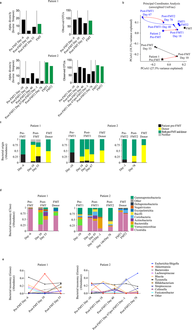
We report the first case series of immune checkpoint inhibitors (ICI)-associated colitis successfully treated with fecal microbiota transplantation, with reconstitution of the gut microbiome and a relative increase in the proportion of regulatory T-cells within the colonic mucosa. These preliminary data provide evidence that modulation of the gut microbiome may abrogate ICI-associated colitis.


Conflict of interest statement

Competing Interests Declaration:

J.A.W and V.G. are inventors on a US patent application (PCT/US17/53,717) submitted by The University of Texas MD Anderson Cancer Center that covers methods to enhance checkpoint blockade therapy by the microbiome. J.A.W. is a clinical and scientific advisor at Microbiome DX and a consultant at Biothera Pharma, Merck Sharp, and Dohme. J.A.W. has honoraria from speakers’ bureau of Dava Oncology, Bristol-Myers Squibb, Gilead, Illumina, Omniprex, Imedex and is an advisory board member for GlaxoSmithKline, Novartis, and Roche/Genentech, Astra-Zeneca. V.G. is a consultant at Microbiome DX, and reports honoraria from ExpertConnect. R.R.J. is on the scientific advisory board for Seres Therapeutics, Inc., has consulted for Ziopharm Oncology and Microbiome Dx, and holds patents licensed to Seres Therapeutics, Inc. M.T.T. serves on the advisory board for Novartis, Seattle Genetics and Myriad Genetics. J.A.W., P.S., and J.P.A. are members of the Parker Institute for Cancer Immunotherapy at MD Anderson Cancer Center. P.S. is a consultant for Bristol-Myers Squibb, Jounce Therapeutics, Helsinn, and GlaxoSmithKline and is also a stockholder from Jounce Therapeutics. J.P.A. is a consultant and stockholder for Jounce Therapeutics, receives royalties from Bristol-Myers Squibb, and has intellectual property with Bristol-Myers Squibb and Merck. The other authors declare no competing interests.

Figures

Figure 1:
Figure 1:
Endoscopic changes and characterization of colonic mucosal infiltrate throughout clinical course for Patient 1 (a-c) and Patient 2 (d-f). (a) Changes in colonic mucosa as assessed by full colonoscopy. Near the time of diagnosis (row 1), multiple large ulcers and diffuse inflammatory exudate are present (in the distal 40cm of the colon only, with the normal appearing proximal colon) and remain despite months of treatment with steroids and biologic immunosuppressive agents (steroid + 2 doses infliximab + 1 dose vedolizumab) (row 2). Approximately one month after FMT (row 3), colonic mucosa exhibits grossly normal vasculature, minimal patchy erythema, and near-complete healing of prior ulcers. Yellow arrows point to ulcerative lesions. This patient had full colonoscopic evaluations that examined every segment of the colon (ascending, transverse, descending, sigmoid and rectum). Endoscopy was performed once at each time point. Given the qualitative nature of endoscopic data collection and inability to provide true statistical analyses, we chose to include multiple other representative photos from the same colonoscopic evaluations. Additional representative photos are presented in Supplemental Figure 4a. (b) Immunohistochemical analysis of mucosal biopsies of the colon/rectum prior to, and following, FMT. A single slide representative of the endoscopic biopsy specimen as a whole was stained for each patient for each time point. Representative slides from additional time points are included in Supplemental Figure 5a. (c) Analysis of changes in the density immune cell subsets (CD8 red squares, CD4 blue circles, FOXP3 black triangles) over time, expressed as a fold change from baseline based on total densities of cells expressing these markers (absolute densities are presented in Supplemental Figure 6a). Timepoints include time of diagnosis, prior to FMT, following steroids and biologic immunosuppression and following FMT. Date of FMT is represented by dotted vertical line and is designated Day 0. These data represent the average cell density from 4 regions of interest (ROIs) per sample (single slide per patient at each time point) with each ROI measuring 500 μm x 500 μm for a total of 0.25 mm2. We report the mean # IHC-positive cells/mm2 divided by the mean # IHC-positive cells/mm2 at baseline. (d) Changes in colonic mucosa as assessed by full colonoscopy. Near the time of diagnosis (row 1), multiple large ulcers and inflammatory exudate is present (throughout the entire colon) and remains after unsuccessful treatment with steroids and biologic immunosuppressive agents (steroid + 2 doses infliximab + 4 doses vedolizumab) (row 2). There is notable improvement following first FMT (row 3) but residual ulcers remain. Following second FMT (row 4) we note near complete resolution of all ulcerative lesions. Again full endoscopic examinations were performed, once for each time point. Additional representative photos from the colonoscopic evaluation are shown in Supplemental Figure 4b. (e) Immunohistochemical analysis of mucosal biopsies of the colon/rectum taken prior to first FMT and following first FMT. A single slide representative of the endoscopic biopsy specimen was stained for each patient for each time point. Representative slides from additional time points are included in Supplemental Figure 5b. (f) Analysis of changes in the density immune cell subsets (CD8 red squares, CD4 blue circles, FOXP3 black triangles) over time, expressed as a fold change from baseline based on total densities of cells expressing these markers (absolute densities are presented in Supplemental Figure 6b). These data represent the average cell density from 4 regions of interest (ROIs) per sample (single slide per patient at each time point) with each ROI measuring 500 μm x 500 μm for a total of approximately 1 mm2. We report the mean # IHC-positive cells/mm2 divided by the mean # IHC-positive cells/mm2 at baseline. Date of first (Day 0) and second FMT (Day 67) are represented by dotted vertical lines.
Figure 2:
Figure 2:
Microbiome analysis of patient and donor intestinal bacteria by 16S deep sequencing. The patients’ stool microbiomes were longitudinally sampled at indicated time points before and after FMT, along with samples from the FMT donor. Between 3380 and 42,776 sequences were obtained for each sample (average 10,003). (a) Alpha diversity, quantified by the inverse Simpson index after rarefying to 3000 sequences, as well as total observed OTU numbers, was evaluated for patient and FMT donor samples. (b) Using principal coordinate analysis of unweighted UniFrac distances, microbiome samples from (a) are depicted in space with more similar samples located closer together. (c) Bacterial 16S sequences from samples in (a) were classified by origin (unique to patient baseline, unique to donor, present in both patient baseline and donor, or absent in both patient baseline and donor). (d) Sequences were classified by taxonomy at the Class level. (e) Changes in the abundances of top varying 10 bacterial genera over time.

Comment in

References

    1. Michot JM, et al. Immune-related adverse events with immune checkpoint blockade: a comprehensive review. Eur J Cancer 54, 139–148 (2016). - PubMed
    1. Cramer P & Bresalier RS Gastrointestinal and Hepatic Complications of Immune Checkpoint Inhibitors. Curr Gastroenterol Rep 19, 3 (2017). - PubMed
    1. Chen JH, Pezhouh MK, Lauwers GY & Masia R Histopathologic Features of Colitis Due to Immunotherapy With Anti-PD-1 Antibodies. Am J Surg Pathol 41, 643–654 (2017). - PubMed
    1. Dadu R, Zobniw C & Diab A Managing Adverse Events With Immune Checkpoint Agents. Cancer J 22, 121–129 (2016). - PubMed
    1. Bertrand A, Kostine M, Barnetche T, Truchetet ME & Schaeverbeke T Immune related adverse events associated with anti-CTLA-4 antibodies: systematic review and meta-analysis. BMC Med 13, 211 (2015). - PMC - PubMed

Publication types

MeSH terms