

Time-lapse imaging reveals delayed development of embryos carrying unbalanced chromosomal translocations
- PMID: 30421343
- PMCID: PMC6420524
- DOI: 10.1007/s10815-018-1361-8
Time-lapse imaging reveals delayed development of embryos carrying unbalanced chromosomal translocations
Abstract
Purpose: The purpose of the study was to compare the morphokinetic parameters of embryos carrying balanced chromosomal translocations with those carrying unbalanced chromosomal translocations using time-lapse microscopy.
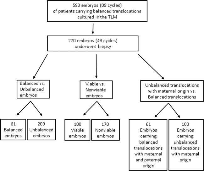
Methods: The study group included 270 embryos that underwent biopsies on day 3 for preimplantation genetic diagnosis (PGD) for chromosomal translocations in our unit between 2013 and 2015. All embryos were incubated under time-lapse microscopy and evaluated for timing of developmental events up to day 5. The timing of these events was compared between balanced and unbalanced embryos, potentially viable and nonviable variants, and maternal versus paternal inheritance of the translocation.
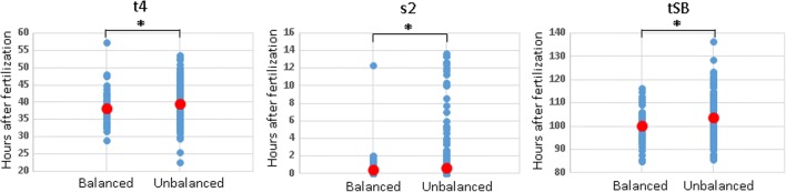
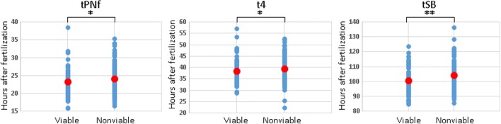
Results: The PGD analysis found that 209 (77%) of the 270 biopsied embryos carried an unbalanced translocation. Embryos carrying unbalanced translocations, which are expected to lead to implantation failure or miscarriage, cleaved less synchronously and were delayed in time of cleavage to the 4-cell stage (t4) and in time of start of blastulation (tSB) compared with balanced embryos (P < 0.05). Furthermore, embryos carrying nonviable translocations demonstrated a significant delay at the time of pronuclei fading (tPNf) compared with those carrying potentially viable translocations (P < 0.05). Embryos whose unbalanced translocations were of maternal origin were significantly delayed in most of the morphokinetic parameters (including tPNf, t2, t3, t4, t6, t7, t8, cc2, s2, and tSB) compared with embryos carrying balanced translocations (P < 0.05).
Conclusions: Embryos carrying unbalanced chromosomal translocations mainly of maternal origin undergo delayed development and asynchronous cleavage that may lead to implantation failure or miscarriage.
Keywords: Chromosomal translocations; Morphokinetic parameters; Preimplantation development; Time-lapse imaging.
Conflict of interest statement
Conflict of interest
The authors declare that they have no conflict of interest.
Informed consent
Informed consent was obtained from all individual participants included in the study.
Figures
References
-
- Loh SF, Wong PC, Jiang B, Yeo GH, Tan AS, Prasath EB, Mathew J, Chan ML, Tan WC, Choolani M, Yap CH, Chong SS. Preimplantation genetic diagnosis of chromosome translocations by analysis of polymorphic short tandem repeats. Singap Med J. 2012;53:648–654. - PubMed
MeSH terms
LinkOut - more resources
Full Text Sources
Miscellaneous
