. 2019 Feb;23(2):954-966.
doi: 10.1111/jcmm.13996. Epub 2018 Nov 12.

CD147 promotes progression of head and neck squamous cell carcinoma via NF-kappa B signaling

Affiliations

CD147 promotes progression of head and neck squamous cell carcinoma via NF-kappa B signaling

Binbin Yu et al. J Cell Mol Med. 2019 Feb.

Abstract

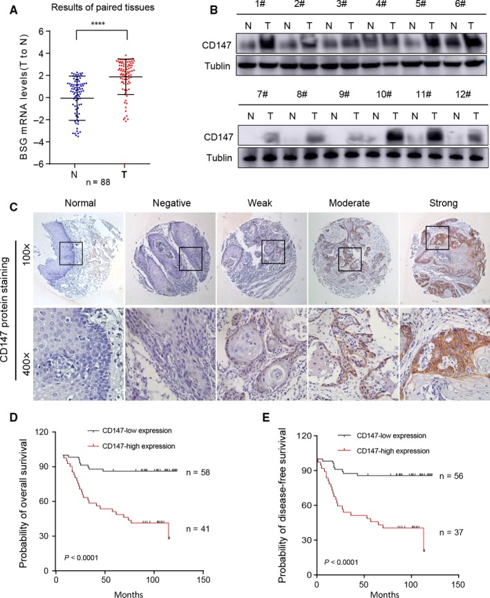
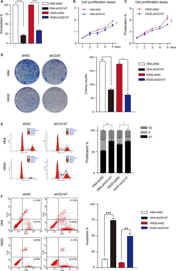
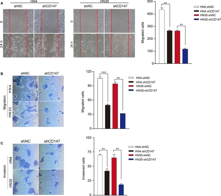
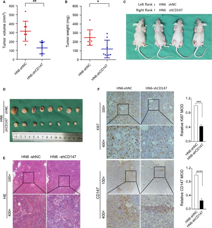
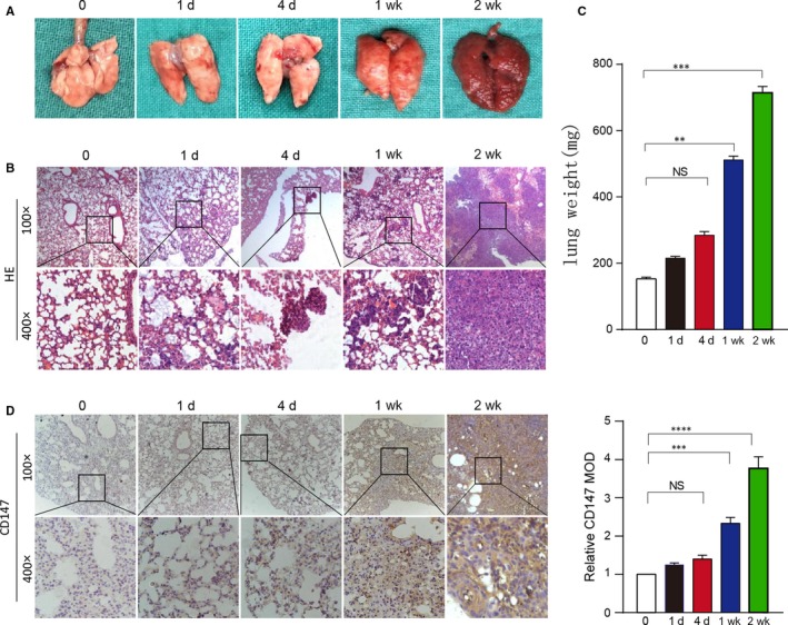
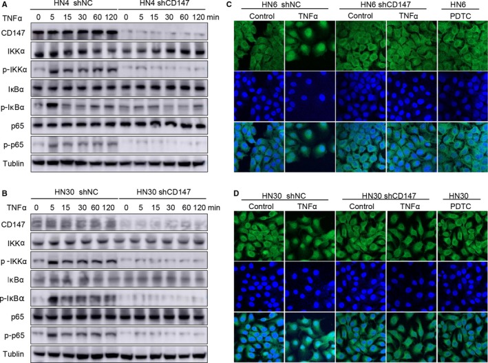
CD147/basigin (BSG) is highly upregulated in many types of cancer, our previous study has found that CD147/BSG is highly expressed in head and neck squamous cell carcinoma (HNSCC) stem cells, but its role in HNSCC and the underlying mechanism is still unknown. In this study, we investigated the role of CD147 in the progression of HNSCC. Real-time PCR, western blot and immunohistochemistry were used to detect the expression of CD147 in total 189 HNSCC tissues in compared with normal tissues. In addition, we used proliferation, colony formation, cell cycle and apoptosis, migration and invasion as well as wound-healing assay to determine the biological roles of CD147 in HNSCC. Then, a xenograft model was performed to evaluate tumor-promoting and metastasis-promoting role of CD147 in HNSCC. The results showed that upregulated CD147 expression was associated with aggressive clinicopathologic features in HNSCC. In addition, CD147 promoted proliferation, migration and reduced the apoptosis phenotype of HNSCC cells in vitro as well as tumor initiation and progression in vivo. Furthermore, we demonstrated that CD147 promoted HNSCC progression through nuclear factor kappa B signaling. Therefore, we concluded that CD147 promoted tumor progression in HNSCC and might be a potential prognostic and treatment biomarker for HNSCC.

Keywords: CD147; NF-kappa B; head and neck squamous cell carcinoma.


Figures

Figure 1
Figure 1
Upregulated CD147 expression was associated with aggressive clinicopathologic features and poor prognosis in HNSCC. (A) The relative CD147 mRNA levels of 88 paired tumor and adjacent non‐tumor tissues in HNSCC is shown. ****P < 0.0001, based on Student's t test. (B) The CD147 protein expression levels in 12 paired tumor and adjacent non‐tumor tissues in HNSCC is shown. Tublin was used as a loading control. (C) Immunohistochemistry (IHC) of CD147 expression level on tissue microarrays containing 101 paired HNSCC and 10 normal tissues is shown. Relative negative, weak, moderate and strong PTK7 stain images compared with normal tissues are shown. (D) The probability of overall survival for CD147‐low expression group and CD147‐high expression group of the 101 patients were significantly different (P < 0.05). (E) The probability of disease free survival for CD147‐low expression group and CD147‐high expression group of the 101 patients were significantly different (P < 0.05). The life status of some patients are missing. Therefore, the total patients are not 101
Figure 2
Figure 2
CD147 promoted the proliferation and reduced the apoptosis phenotype of HNSCC cells. (A) The knockdown rate of CD147 in HN4 and HN30 is shown. (B) Proliferation assays showed that proliferation was reduced in HN4 cells after transfected with shCD147 and negative control lentivirus. **P < 0.01, based on Student's t test. (C) Proliferation assays showed that proliferation was reduced in HN30 cells after transfected with shCD147 and negative control lentivirus. ***P < 0.001, based on Student's t test. (D) The negative control and shCD147 of HN4 and HN30 cells were performed by colony formation assay. **P < 0.01, ***P < 0.001, based on Student's t test. (E) Flow cytometry analysis of cell cycle progression in HN4 and HN30 cells were determined after transfected with shCD147 and negative control lentivirus. *P < 0.05, **P < 0.01, based on Student's t test. (F) Flow cytometry analysis of apoptosis in HN4 and HN30 cells were performed after transfected with shCD147 and negative control lentivirus. **P < 0.01, ***P < 0.001, based on Student's t test
Figure 3
Figure 3
CD147 promoted the migration and invasion phenotype of HNSCC cells. (A) HN4 and HN30 cells were transfected with shCD147 and negative control lentivirus, subsequently analysed with scratch wound healing assay. **P < 0.01, based on Student's t test. (B) HN4 and HN30 cells were transfected with shCD147 and negative control lentivirus, subsequently analysed with migration assays. **P < 0.01, ***P < 0.001, based on Student's t test. (C) HN4 and HN30 cells were transfected with shCD147 and negative control lentivirus, subsequently analysed with invasion assays. **P < 0.01, based on Student's t test
Figure 4
Figure 4
Knockdown of CD147 in HNSCC cells reduced tumor growth in vivo. (A) A total of 1 × 10⁶ HN6 negative control cells and shCD147 cells in 100 μL fresh DMEM medium were subcutaneously injected into the left or right flank of the BALB/C nude mice. Tumor volume of two groups are presented. **P < 0.01, based on Student's t test. (B) Tumor weight of two groups are presented. *P < 0.05, based on Student's t test. (C) Tumor growth 20 days after injection of the BALB/C nude mice are shown. (D) Mice were killed and the tumors are isolated. (E) Representative H&E staining of tumor tissues is shown. (F) Immunohistochemical staining of Ki67 and CD147 in shNC and shCD147 xenografted tumors is shown. ***P < 0.001, ****P < 0.0001, based on Student's t test
Figure 5
Figure 5
CD147 expression was higher lung metastasis tissues in vivo. (A) Whole mount pictures of the lung tissues are shown. (B) H&E staining of lung tissue sections at 0, 1 day, 4 days, 1 week and 2 weeks is shown. (C) The lung weight of BALB/C nude mice killed at 0, 1 day, 4 days, 1 week and 2 weeks are shown. ****P < 0.0001, based on Student's t test. (D) Immunohistochemical staining of CD147 in lung tissue sections at 0, 1 day, 4 days, 1 week and 2 weeks is shown. ****P < 0.0001, based on Student's t test
Figure 6
Figure 6
CD147 activated NF‐kappa B signaling in HNSCC. (A) After being starved with serum‐free DMEM overnight, HN4 shCD147 and negative control cells were treated with 5 ng/mL TNFα for 0, 5, 15, 30, 60, 120 min. Lysates were prepared, and CD147, IKKα, p‐IKKα, IκBα, p‐IκBα, p65, p‐p65 were analysed by western blotting. Tublin was used as a loading control. (B) After being starved with serum‐free DMEM overnight, HN30 shCD147 and negative control cells were treated with 5 ng/mL TNFα for 0, 5, 15, 30, 60, 120 min. Lysates were prepared, and CD147, IKKα, p‐IKKα, IκBα, p‐IκBα, p65, p‐p65 were analysed by western blotting. Tublin was used as a loading control. (C) After being starved with serum‐free DMEM overnight, HN6 shNC and shPTK7 cells were treated with 5 ng/mL TNFα or serum‐free DMEM for 5 min, and HN6 was treated with PDTC for 5 min, then the location of p65 was detected by laser scanning microscopy. (D) After being starved with serum‐free DMEM overnight, HN30 shNC and shPTK7 cells were treated with 5 ng/mL TNFα or serum‐free DMEM for 5 min, and HN30 was treated with PDTC for 5 min, then the location of p65 was detected by laser scanning microscopy

References

    1. Rothenberg SM, Ellisen LW. The molecular pathogenesis of head and neck squamous cell carcinoma. J Clin Invest. 2012;122:1951‐1957. - PMC - PubMed
    1. Ren ZH, Xu JL, Li B, Fan TF, Ji T, Zhang CP. Elective versus therapeutic neck dissection in node‐negative oral cancer: evidence from five randomized controlled trials. Oral Oncol. 2015;51:976‐981. - PubMed
    1. Tan DSW, Chong FT, Leong HS, et al. Long noncoding RNA EGFR‐AS1 mediates epidermal growth factor receptor addiction and modulates treatment response in squamous cell carcinoma. Nat Med. 2017;23:1167‐1175. - PubMed
    1. Worsham MJ, Ali H, Dragovic J, Schweitzer VP. Molecular characterization of head and neck cancer: how close to personalized targeted therapy? Mol Diagn Ther. 2012;16:209‐222. - PMC - PubMed
    1. Yan M, Yang X, Wang L, et al. Plasma membrane proteomics of tumor spheres identify CD166 as a novel marker for cancer stem‐like cells in head and neck squamous cell carcinoma. Mol Cell Proteomics. 2013;12:3271‐3284. - PMC - PubMed

Publication types

MeSH terms