

Neurotheranostics as personalized medicines
- PMID: 30421721
- PMCID: PMC6486471
- DOI: 10.1016/j.addr.2018.10.011
Neurotheranostics as personalized medicines
Abstract
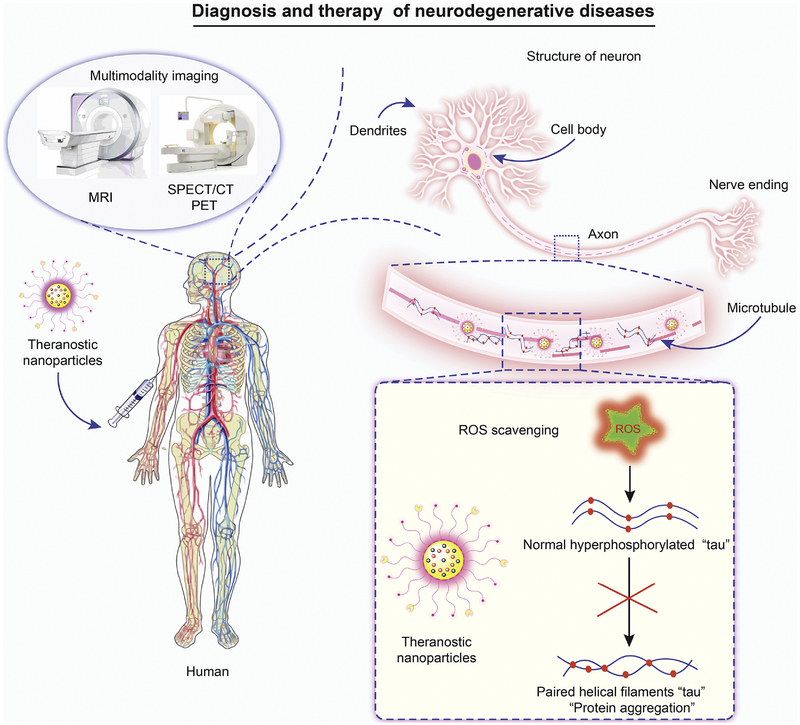
The discipline of neurotheranostics was forged to improve diagnostic and therapeutic clinical outcomes for neurological disorders. Research was facilitated, in largest measure, by the creation of pharmacologically effective multimodal pharmaceutical formulations. Deployment of neurotheranostic agents could revolutionize staging and improve nervous system disease therapeutic outcomes. However, obstacles in formulation design, drug loading and payload delivery still remain. These will certainly be aided by multidisciplinary basic research and clinical teams with pharmacology, nanotechnology, neuroscience and pharmaceutic expertise. When successful the end results will provide "optimal" therapeutic delivery platforms. The current report reviews an extensive body of knowledge of the natural history, epidemiology, pathogenesis and therapeutics of neurologic disease with an eye on how, when and under what circumstances neurotheranostics will soon be used as personalized medicines for a broad range of neurodegenerative, neuroinflammatory and neuroinfectious diseases.
Keywords: Alzheimer's disease (AD); Blood brain barrier (BBB); Brain-targeted nanoparticles; Magnetic resonance imaging (MRI); Nanomedicine; Neurodegenerative disorders; Neuroimaging; Neurotheranostics; Parkinson's disease (PD); Single photon emission computed tomography (SPECT/CT); Theranostics.
Copyright © 2018 The Author(s). Published by Elsevier B.V. All rights reserved.
Figures
Similar articles
-
Dendrimer Architectonics to Treat Cancer and Neurodegenerative Diseases with Implications in Theranostics and Personalized Medicine.ACS Appl Bio Mater. 2021 Feb 15;4(2):1115-1139. doi: 10.1021/acsabm.0c01319. Epub 2021 Jan 13. ACS Appl Bio Mater. 2021. PMID: 35014470 Review.
-
Alzheimer's Disease and Parkinson's Disease: A Review of Current Treatment Adopting a Nanotechnology Approach.Curr Pharm Des. 2018;24(1):22-45. doi: 10.2174/1381612823666170828133059. Curr Pharm Des. 2018. PMID: 28847307 Review.
-
Nanotheranostics: The Fabrication of Theranostics with Nanoparticles and their Application to Treat the Neurological Disorders.Recent Pat Nanotechnol. 2025;19(1):17-34. doi: 10.2174/1872210517666230718115651. Recent Pat Nanotechnol. 2025. PMID: 37464820 Review.
-
Nanomedicine-based neuroprotective strategies in patient specific-iPSC and personalized medicine.Int J Mol Sci. 2014 Mar 4;15(3):3904-25. doi: 10.3390/ijms15033904. Int J Mol Sci. 2014. PMID: 24599081 Free PMC article. Review.
-
Nanotechnology enabled regenerative medicine for neurological disorders.Adv Drug Deliv Rev. 2019 Aug;148:1-2. doi: 10.1016/j.addr.2019.11.006. Adv Drug Deliv Rev. 2019. PMID: 31787167 Free PMC article. No abstract available.
Cited by
-
New Drug Design Avenues Targeting Alzheimer's Disease by Pharmacoinformatics-Aided Tools.Pharmaceutics. 2022 Sep 9;14(9):1914. doi: 10.3390/pharmaceutics14091914. Pharmaceutics. 2022. PMID: 36145662 Free PMC article. Review.
-
Metabolic Priming as a Tool in Redox and Mitochondrial Theragnostics.Antioxidants (Basel). 2023 May 10;12(5):1072. doi: 10.3390/antiox12051072. Antioxidants (Basel). 2023. PMID: 37237939 Free PMC article. Review.
-
Manipulation of Axonal Outgrowth via Exogenous Low Forces.Int J Mol Sci. 2020 Oct 28;21(21):8009. doi: 10.3390/ijms21218009. Int J Mol Sci. 2020. PMID: 33126477 Free PMC article. Review.
-
GLP-1 Analogs, SGLT-2, and DPP-4 Inhibitors: A Triad of Hope for Alzheimer's Disease Therapy.Biomedicines. 2023 Nov 12;11(11):3035. doi: 10.3390/biomedicines11113035. Biomedicines. 2023. PMID: 38002034 Free PMC article. Review.
-
Bioimaging predictors of rilpivirine biodistribution and antiretroviral activities.Biomaterials. 2018 Dec;185:174-193. doi: 10.1016/j.biomaterials.2018.09.018. Epub 2018 Sep 14. Biomaterials. 2018. PMID: 30245386 Free PMC article.
References
-
- Nie S, Xing Y, Kim GJ, Simons JW, Nanotechnology applications in cancer, Annual review of biomedical engineering, 9 (2007) 257–288. - PubMed
-
- Ross JS, Fletcher JA, The HER-2/neu Oncogene in Breast Cancer: Prognostic Factor, Predictive Factor, and Target for Therapy, The Oncologist, 3 (1998) 237–252. - PubMed
-
- Lukashov VV, Goudsmit J, HIV heterogeneity and disease progression in AIDS: a model of continuous virus adaptation, AIDS (London, England), 12 Suppl A (1998) S43–52. - PubMed
-
- Bouzid D, Kammoun A, Amouri A, Mahfoudh N, Haddouk S, Tahri N, Makni H, Masmoudi H, Inflammatory bowel disease: susceptibility and disease heterogeneity revealed by human leukocyte antigen genotyping, Genetic testing and molecular biomarkers, 16 (2012) 482–487. - PubMed
Publication types
MeSH terms
Substances
Grants and funding
- R01 NS036126/NS/NINDS NIH HHS/United States
- P01 DA028555/DA/NIDA NIH HHS/United States
- P30 MH062261/MH/NIMH NIH HHS/United States
- P01 NS031492/NS/NINDS NIH HHS/United States
- T32 NS105594/NS/NINDS NIH HHS/United States
- R56 AI138613/AI/NIAID NIH HHS/United States
- P30 GM127200/GM/NIGMS NIH HHS/United States
- P01 MH064570/MH/NIMH NIH HHS/United States
- R01 MH104147/MH/NIMH NIH HHS/United States
- R13 NS083315/NS/NINDS NIH HHS/United States
- P20 GM103480/GM/NIGMS NIH HHS/United States
- R01 NS034239/NS/NINDS NIH HHS/United States
- R24 OD018546/OD/NIH HHS/United States
- R01 MH115860/MH/NIMH NIH HHS/United States
- P01 NS043985/NS/NINDS NIH HHS/United States
- R37 NS036126/NS/NINDS NIH HHS/United States
- R01 AG043540/AG/NIA NIH HHS/United States
LinkOut - more resources
Full Text Sources
Medical
