. 2019 Feb 1;129(2):647-658.
doi: 10.1172/JCI123878. Epub 2019 Jan 7.

Cardiac CaV1.2 channels require β subunits for β-adrenergic-mediated modulation but not trafficking

Affiliations

Cardiac CaV1.2 channels require β subunits for β-adrenergic-mediated modulation but not trafficking

Lin Yang et al. J Clin Invest. .

Abstract

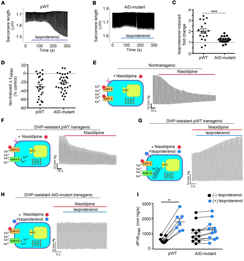
Ca2+ channel β-subunit interactions with pore-forming α-subunits are long-thought to be obligatory for channel trafficking to the cell surface and for tuning of basal biophysical properties in many tissues. Unexpectedly, we demonstrate that transgenic expression of mutant α1C subunits lacking capacity to bind CaVβ can traffic to the sarcolemma in adult cardiomyocytes in vivo and sustain normal excitation-contraction coupling. However, these β-less Ca2+ channels cannot be stimulated by β-adrenergic pathway agonists, and thus adrenergic augmentation of contractility is markedly impaired in isolated cardiomyocytes and in hearts. Similarly, viral-mediated expression of a β-subunit-sequestering peptide sharply curtailed β-adrenergic stimulation of WT Ca2+ channels, identifying an approach to specifically modulate β-adrenergic regulation of cardiac contractility. Our data demonstrate that β subunits are required for β-adrenergic regulation of CaV1.2 channels and positive inotropy in the heart, but are dispensable for CaV1.2 trafficking to the adult cardiomyocyte cell surface, and for basal function and excitation-contraction coupling.

Keywords: Calcium; Calcium channels; Cardiology; Excitation contraction coupling; Muscle Biology.


Conflict of interest statement

Conflict of interest: The authors have declared that no conflict of interest exists.

Figures

Figure 1
Figure 1. AID-mutant α1C channels trafficking and function in cardiomyocytes.
(A) Schematic of rabbit cardiac α1C subunit topology showing β-subunit binding to α-interacting domain (AID) motif in I-II loop. WT and mutant-AID motif in the I-II loop of α1C. (B) Schematic representation of the binary transgene system. The αMHCMOD construct is a modified αMHC promoter containing the tet-operon for regulated expression of FLAG-tagged DHP-resistant (DHP*) α1C. (C) Anti-FLAG (upper) and anti-β immunoblots (lower) of anti–FLAG antibody immunoprecipitation of cardiac homogenates of nontransgenic (NTG), pWT α1C, and AID-mutant α1C mice. Representative of 3 experiments. (D) Immunostaining of pWT and AID-mutant α1C cardiomyocytes. Anti-FLAG and FITC-conjugated secondary antibodies, and nuclear labeling with Hoechst stain. Negative control omitted anti–FLAG antibody. Images obtained with confocal microscopy at ×40. Scale bars: 20 μm. (E) Exemplar whole-cell CaV1.2 currents recorded from freshly dissociated cardiomyocytes of NTG, pWT, and AID-mutant α1C transgenic mice. Pulses from –60 mV to 0 mV before (black traces) and 3 minutes after (red traces) administration of 300 nM nisoldipine. (F) Scatter plot showing current densities before and after administration of 300 nM nisoldipine. Mean ± SEM. *P < 0.05 NTG versus transgenic pWT α1C, ****P < 0.0001 NTG versus transgenic AID-mutant α1C and also NTG pre- versus post-nisoldipine, ***P < 0.001 pWT or AID-mutant α1C pre- versus post-nisoldipine. One-way ANOVA and Dunnett’s multiple comparison test. NTG, n = 8 cardiomyocytes from 5 mice; pWT, n = 21 cardiomyocytes from 7 mice; AID-mutant, n = 45 cardiomyocytes from 9 mice. (GI) Representative time courses of changes in sarcomere length after superfusion of 300 nM nisoldipine-containing solution for cardiomyocytes isolated from NTG mice (G) and pWT (H) and AID-mutant transgenic α1C mice. Cardiomyocytes were field-stimulated at 1 Hz. (J) Scatter plot showing percentage of contraction of sarcomere length in the absence and presence of nisoldipine for cardiomyocytes isolated from NTG mice and pWT and AID-mutant α1C transgenic mice. NTG, n = 12 cells from 3 mice; pWT, n = 16 cells from 3 mice; AID-mutant, n = 18 cells from 3 mice.
Figure 2
Figure 2. AID-mutant CaV1.2 channels lack β-adrenergic regulation.
(A) Normalized CaV1.2 current-voltage relationships for transgenic pWT and AID-mutant α1C cardiomyocytes in the presence of nisoldipine (n = 19 cardiomyocytes from 3 pWT α1C transgenic mice; n = 18 cardiomocytes from 6 AID-mutant α1C transgenic mice). (B and C) Bar graphs of Boltzmann function parameters Vmid and slope (Vc). **P < 0.01, ANOVA and Sidak’s multiple comparison test; n = 19 cardiomyocytes from 3 pWT α1C transgenic mice; n = 18 cardiomocytes from 6 AID-mutant α1C transgenic mice. (D) Summary of time constants of inactivation at the indicated potentials obtained from a single exponential fit (n = 24 pWT α1C cardiomyocytes from 4 mice and n = 24 AID-mutant α1C cardiomyocytes from 4 mice). P > 0.05 pWT versus AID-mutant for all voltages using Sidak’s multiple comparison test. (E and F) Exemplar nisoldipine-resistant current-voltage relationships of transgenic pWT α1C (E) and AID-mutant α1C (F) acquired in the absence (black trace) and presence of 200 nM isoproterenol (red trace). (G) Diary plot of normalized nisoldipine-resistant ICa amplitude at 0 mV (normalized to 1 at 50 seconds prior to isoproterenol) of pWT and AID-mutant α1C cardiomyocytes. Cells exposed to 300 nM nisoldipine followed by 200 nM isoproterenol in the continued presence of nisoldipine. pWT, n = 30 cardiomyocytes from 5 mice; AID-mutant, n = 45 cardiomyocytes from 7 mice. P < 0.0001 by 1-way ANOVA/multiple comparison at all time points 30 seconds after isoproterenol. (H) Diary plot of normalized nisoldipine-resistant ICa amplitude at +10 mV (normalized to 1 at 50 seconds, prior to forskolin) of pWT and AID-mutant α1C cardiomyocytes. Cells exposed to 300 nM nisoldipine followed by 10 μM forskolin in the continued presence of nisoldipine. pWT: n = 15 cardiomyocytes from 2 mice; AID-mutant: n = 20 cardiomyocytes from 6 mice. P < 0.0001 by 1-way ANOVA/multiple comparison at all time points 30 seconds after forskolin. (I) Bar graph of isoproterenol- or forskolin-induced fold increase in nisoldipine-resistant ICa. Mean ± SEM. ***P < 0.001; ****P < 0.0001 by t test. (J) Graph of isoproterenol- and forskolin-induced increase in nisoldipine-resistant current stratified by total basal current density before nisoldipine for pWT α1C and AID-mutant α1C transgenic mice. Lines fitted by linear regression for pWT cells for isoproterenol (black) and forskolin (red). For isoproterenol, pWT α1C, n = 29 cardiomyocytes; AID-mutant α1C, n = 45 cardiomyocytes. For forskolin, pWT α1C, n = 17 cardiomyocytes; AID-mutant α1C, n = 9 cardiomyocytes.
Figure 3
Figure 3. β-less WT endogenous CaV1.2 channels are not stimulated by PKA.
(AC) Adenovirus-induced GFP, AID-YFP, and AID-mutant YFP expression in cultured guinea pig ventricular myocytes. Top: exemplar confocal images from guinea pig cardiomyocytes expressing GFP, AID-YFP peptide, or AID-mutant YFP peptide. Bottom: exemplar whole-cell Ba2+ currents from GFP and YFP-expressing guinea pig ventricular cardiomyocytes before (black trace) and after (red trace) application of 1 μM forskolin. (DF) Current-voltage relationships from GFP, AID-YFP, and AID-mutant YFP–expressing cardiomyocytes before (black) and after (red) superfusion of 1 μM forskolin. (G) Representative diary plot showing time course of forskolin-induced increase in CaV1.2 current. (H) Forskolin-induced increase in CaV1.2 current. *P < 0.05, **P < 0.01 by 1-way ANOVA and Tukey’s multiple comparison test.
Figure 4
Figure 4. PKA phosphorylation of CaV β is not required for β-adrenergic regulation of CaV1.2. (A) Bright-field and GFP image of WT and mutant β2b-expressing cardiomyocytes.
Scale bars: 100 μm. (B) Immunoblots using anti–β2 antibody (upper) and anti–tubulin antibody of homogenates from the hearts of nontransgenic (NTG) and doxycycline-fed GFP-WT β2 and GFP-mutant β2-expressing mice. (C) Graph of densitometry of fraction of GFP-β/total β. Mean ± SEM; n = 6 mice for NTG, WT, and mutant β2. ****P < 0.0001 compared with nontransgenic by 1-way ANOVA and Dunnett’s multiple comparison test. (D and E) Normalized current-voltage relationships of GFP-WT β2 and GFP-mutant β2 cardiomyocytes acquired before and after superfusion of 200 nM isoproterenol. Isoproterenol shifted the Vmid of steady-state activation of GFP-WT β2 and GFP-mutant β2 cardiomyocytes by –7.0 mV (P < 0.0001, t test, n = 15) and –7.5 mV (P < 0.001, t test, n = 30), respectively. (F) Column scatter plot depicting the fold increase in peak current caused by isoproterenol. Mean ± SEM; n = 36 cardiomyocytes from 5 NTG mice; n = 19 cardiomyocytes from 4 GFP-WT β2b mice; n = 32 cardiomyocytes from 5 mutant β2b mice. P = 0.55 by 1-way ANOVA. (G) Graphs of isoproterenol-induced increase in current stratified by total basal current density for cardiomyocytes isolated from NTG mice, GFP-WT β2b mice, and GFP-mutant β2b transgenic mice.
Figure 5
Figure 5. Attenuated β-adrenergic–stimulated inotropy in AID-mutant α1C transgenic mice.
(A and B) Cells with robust shortening induced by 1 Hz electrical stimulation in the presence of 300 nM nisoldipine were used. Isoproterenol (200 nM) was superfused with 300 nM nisoldipine. (C) Plot of isoproterenol-induced fold change in sarcomere length compared with before isoproterenol. Mean ± SEM; n = 17 for pWT α1C cardiomyocytes and n = 19 cardiomyocytes for AID-mutant α1C. ***P < 0.001 by t test. (D) Plot of isoproterenol-induced percentage of change in τrelaxation of sarcomere length compared with before isoproterenol. Mean ± SEM; n = 23 cardiomyocytes from 3 mice and n = 32 cardiomyocytes from 3 mice. P = 0.16 by t test. (E and F) Representative traces depicted effect of perfusion of 300 nM nisoldipine on left ventricular contraction in isolated Langendorff-perfused hearts resected from NTG mice and pWT α1C transgenic mice. (G and H) Representative traces of nisoldipine-resistant LV pressure before and during isoproterenol infusion, in hearts resected from pWT α1C and AID-mutant α1C transgenic mice. (I) Quantitative summary of dP/dtmax before and during isoproterenol infusion. n = 7 pWT α1C transgenic mice; n = 11 AID-mutant α1C transgenic mice. *P < 0.05 by t test.

Comment in

References

    1. Catterall WA. Structure and regulation of voltage-gated Ca2+ channels. Annu Rev Cell Dev Biol. 2000;16:521–555. doi: 10.1146/annurev.cellbio.16.1.521. - DOI - PubMed
    1. Min D, et al. The alterations of Ca2+/calmodulin/CaMKII/CaV1.2 signaling in experimental models of Alzheimer’s disease and vascular dementia. Neurosci Lett. 2013;538:60–65. doi: 10.1016/j.neulet.2013.02.001. - DOI - PubMed
    1. Peterson BZ, DeMaria CD, Adelman JP, Yue DT. Calmodulin is the Ca2+ sensor for Ca2+-dependent inactivation of L-type calcium channels. Neuron. 1999;22(3):549–558. doi: 10.1016/S0896-6273(00)80709-6. - DOI - PubMed
    1. Erickson MG, Alseikhan BA, Peterson BZ, Yue DT. Preassociation of calmodulin with voltage-gated Ca(2+) channels revealed by FRET in single living cells. Neuron. 2001;31(6):973–985. doi: 10.1016/S0896-6273(01)00438-X. - DOI - PubMed
    1. Scriven DR, Dan P, Moore ED. Distribution of proteins implicated in excitation-contraction coupling in rat ventricular myocytes. Biophys J. 2000;79(5):2682–2691. doi: 10.1016/S0006-3495(00)76506-4. - DOI - PMC - PubMed

Publication types