

Donor, Recipient, and Operative Factors Associated With Increased Endothelial Cell Loss in the Cornea Preservation Time Study
- PMID: 30422157
- PMCID: PMC6439830
- DOI: 10.1001/jamaophthalmol.2018.5669
Donor, Recipient, and Operative Factors Associated With Increased Endothelial Cell Loss in the Cornea Preservation Time Study
Erratum in
-
Error in Table.JAMA Ophthalmol. 2019 Feb 1;137(2):233. doi: 10.1001/jamaophthalmol.2018.6247. JAMA Ophthalmol. 2019. PMID: 30543337 Free PMC article. No abstract available.
Abstract
Importance: Determining factors associated with endothelial cell loss after Descemet stripping automated endothelial keratoplasty (DSAEK) could improve long-term graft survival.
Objective: To evaluate the associations of donor, recipient, and operative factors with endothelial cell density (ECD) 3 years after DSAEK in the Cornea Preservation Time Study.
Design, setting, and participants: This cohort study was a secondary analysis of data collected in a multicenter, double-masked, randomized clinical trial. Forty US clinical sites with 70 surgeons participated, with donor corneas provided by 23 US eye banks. Individuals undergoing DSAEK for Fuchs dystrophy or pseudophakic/aphakic corneal edema were included.
Interventions: The DSAEK procedure, with random assignment of a donor cornea with a preservation time of 0 to 7 days or 8 to 14 days.
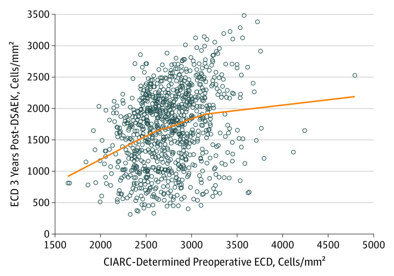
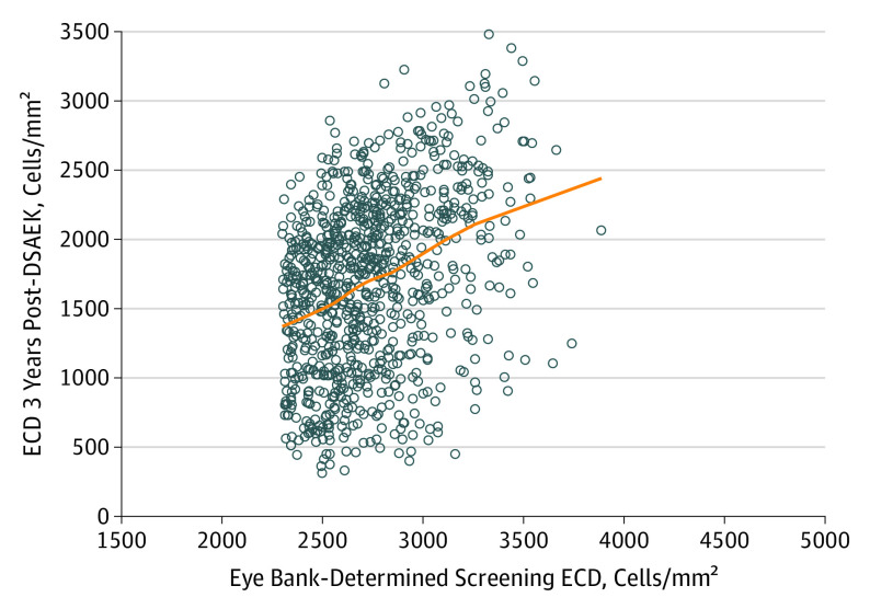
Main outcomes and measures: Endothelial cell density at 3 years as determined by a reading center from eye bank and clinical specular or confocal central endothelial images.
Results: The study included 1090 participants (median age, 70 years) with 1330 affected eyes (240 bilateral cases [22.0%]), who underwent DSAEK for Fuchs dystrophy (1255 eyes [94.4%]) or pseudophakic/aphakic corneal edema (PACE) (75 eyes [5.6%]). Of these, 801 eyes (60.2%) belonged to women and 1207 (90.8%) to white individuals. A total of 749 participants (913 eyes; 164 [21.9%] bilateral cases) had functioning grafts with acceptable endothelial images preoperatively and at 3 years postoperatively and were included in this analysis. Factors associated with a lower ECD at 3 years (estimated effect with 99% CI) in the final multivariable model included donors with diabetes (-103 [-196 to -9] cells/mm2), lower screening ECD (-234 [-331 to -137] per 500 cells/mm2), recipient diagnosis of PACE (-257 [-483 to -31] in cells/mm2), and operative complications (-324 [-516 to -133] in cells/mm2). Endothelial cell loss (ECL) from a preoperative measurement to a 3-year postoperative measurement was 47% (99% CI, 42%-52%) for participants receiving tissue from donors with diabetes vs 43% (99% CI, 39%-48%) without diabetes; it was 53% (99% CI, 44%-62%) for participants diagnosed with PACE vs 44% (99% CI, 39%-49%) for those diagnosed with Fuchs dystrophy, and 55% (99% CI, 48%-63%) in participants who experienced operative complications vs 44% (99% CI, 39%-48%) in those who did not. No other donor, recipient, or operative factors were significantly associated with 3-year ECD.
Conclusions and relevance: Donor diabetes, lower screening ECD, a PACE diagnosis in the recipient, and operative complications were associated with lower ECD at 3 years after DSAEK surgery and may be associated with long-term graft success. While causation cannot be inferred, further studies on the association of donor diabetes and PACE in recipients with lower 3-year ECD warrant further study.
Conflict of interest statement
Figures
References
-
- Lass JH, Gal RL, Dontchev M, et al. ; Cornea Donor Study Investigator Group . Donor age and corneal endothelial cell loss 5 years after successful corneal transplantation: specular microscopy ancillary study results. Ophthalmology. 2008;115(4):627-632.e8. doi:10.1016/j.ophtha.2008.01.004 - DOI - PMC - PubMed
-
- Lass JH, Benetz BA, Gal RL, et al. ; Writing Committee for the Cornea Donor Study Research Group . Donor age and factors related to endothelial cell loss 10 years after penetrating keratoplasty: Specular Microscopy Ancillary Study. Ophthalmology. 2013;120(12):2428-2435. doi:10.1016/j.ophtha.2013.08.044 - DOI - PMC - PubMed
Publication types
MeSH terms
LinkOut - more resources
Full Text Sources
Medical
