. 2019 May 15;199(10):1257-1266.
doi: 10.1164/rccm.201804-0628OC.

Tumor-derived Autoantibodies Identify Malignant Pulmonary Nodules

Affiliations

Tumor-derived Autoantibodies Identify Malignant Pulmonary Nodules

Kristin J Lastwika et al. Am J Respir Crit Care Med. .

Abstract

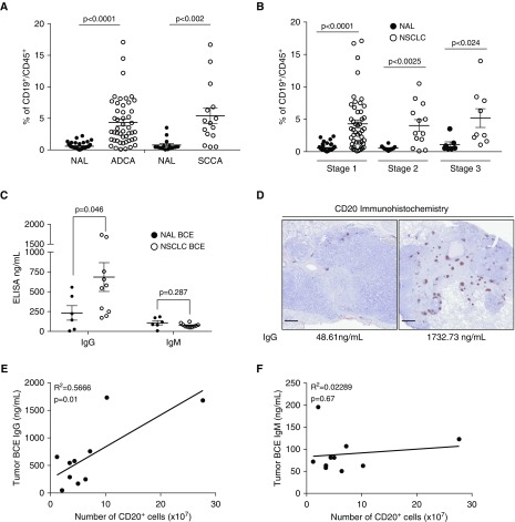
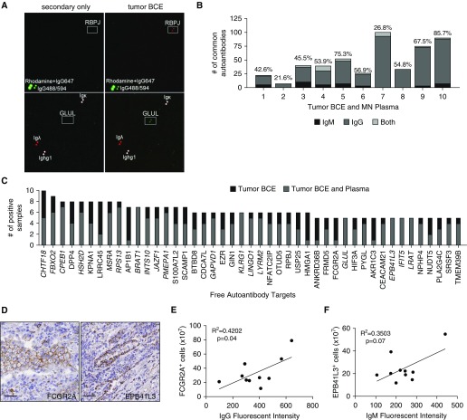
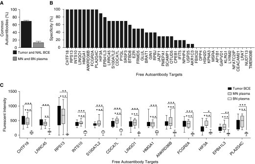
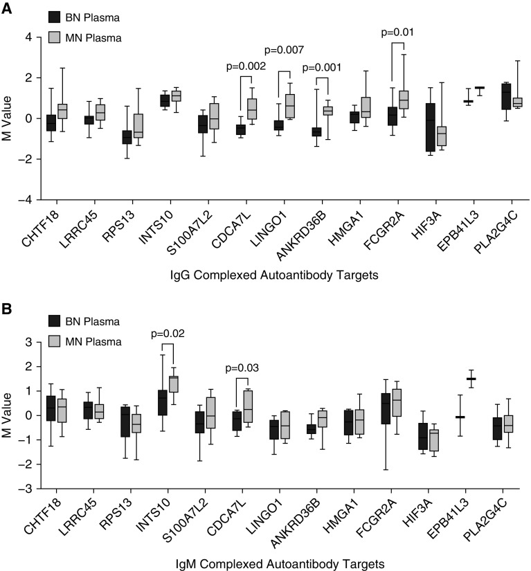
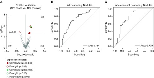
Rationale: Screening for non-small cell lung cancer is associated with earlier diagnosis and reduced mortality but also increased harm caused by invasive follow-up of benign pulmonary nodules. Lung tumorigenesis activates the immune system, components of which could serve as tumor-specific biomarkers. Objectives: To profile tumor-derived autoantibodies as peripheral biomarkers of malignant pulmonary nodules. Methods: High-density protein arrays were used to define the specificity of autoantibodies isolated from B cells of 10 resected lung tumors. These tumor-derived autoantibodies were also examined as free or complexed to antigen in the plasma of the same 10 patients and matched benign nodule control subjects. Promising autoantibodies were further analyzed in an independent cohort of 250 nodule-positive patients. Measurements and Main Results: Thirteen tumor B-cell-derived autoantibodies isolated ex vivo showed greater than or equal to 50% sensitivity and greater than or equal to 70% specificity for lung cancer. In plasma, 11 of 13 autoantibodies were present both complexed to and free from antigen. In the larger validation cohort, 5 of 13 tumor-derived autoantibodies remained significantly elevated in cancers. A combination of four of these autoantibodies could detect malignant nodules with an area under the curve of 0.74 and had an area under the curve of 0.78 in a subcohort of indeterminate (8-20 mm in the longest diameter) pulmonary nodules. Conclusions: Our novel pipeline identifies tumor-derived autoantibodies that could effectively serve as blood biomarkers for malignant pulmonary nodule diagnosis. This approach has future implications for both a cost-effective and noninvasive approach to determine nodule malignancy for widespread low-dose computed tomography screening.

Keywords: B cells; computed tomography imaging; early detection; indeterminate pulmonary nodules; lung cancer.


Figures

Figure 1.
Figure 1.
B-cell number and IgG concentration are significantly higher in tumors compared with normal adjacent lung. (A) Flow cytometry quantification of the proportion of CD19+ B cells in CD45+ leukocytes isolated from adenocarcinoma (ADCA), squamous cell carcinoma (SCCA), and normal adjacent lung (NAL). NAL n = 44 and ADCA n = 48; NAL n = 15 and SCCA n = 15. Data are shown as mean ± SEM. P values were determined via Welch’s t test. (B) Flow cytometry quantification of CD19+ B cells in CD45+ leukocytes by stage of non–small cell lung cancer (NSCLC). Tumors have significantly more CD19+ B cells in all stages of NSCLC than in NAL. Stage 1 n = 47, stage 2 n = 13, and stage 3 n = 9. Data are shown as mean ± SEM. P values were determined via Welch’s t test. (C) ELISA quantification of IgG and IgM antibodies isolated from tumor B-cell extracts (n = 10) and NAL (n = 6) (mean ± SEM). P values were determined via Welch’s t test. (D) Representative images of a tumor with low IgG quantification by ELISA and low CD20 staining by immunohistochemistry compared with a tumor with high CD20 staining and high IgG concentration. Scale bars = 1 mm. (E) Significant correlation of matched ELISA IgG and CD20 immunohistochemistry staining in 10 NSCLC tumors (n = 10, Pearson correlation). (F) ELISA IgM is not correlated with the number of B cells quantified by CD20+ immunohistochemical staining (n = 10, Pearson correlation). BCE = B-cell extract.
Figure 2.
Figure 2.
Common autoantibody targets can be identified in most lung tumors and plasma. (A) Representative images of IgG autoantibody target identification of RPBJ and IgM of GLUL (white boxes, proteins printed in duplicate) in tumor B-cell extracts (BCE) but not in a secondary-only control. Positive controls are rhodamine + IgG647, IgG-Alexa Fluor 488/594, and antibody fragments. (B) The number of autoantibodies found in tumor BCE and corresponding malignant nodule (MN) plasma. The percentage of identity is noted above the bar graph for each sample pair. (C) Targets of autoantibodies isolated from ≥50% tumor BCE identified using the HuProt array. Italicized autoantibodies were also present in ≥50% MN plasmas. (D) Representative images from tumor serial sections of immunohistochemical staining of EPB41L3 and FCGR2A (antigenic targets of lung-derived autoantibodies). Scale bars = 50 μm. (E) The number of FCGR2A-positive cells quantified by immunohistochemistry significantly correlates with the intensity of IgG autoantibody signal on the HuProt array (n = 10, Pearson correlation). (F) The number of EPB41L3-positive cells quantified by immunohistochemistry shows a trend toward correlation with the intensity of IgM autoantibody signal on the HuProt array (n = 10, Pearson correlation).
Figure 3.
Figure 3.
Autoantibodies specific for malignant nodules. (A) The percentage of common antibodies between tumor B-cell extracts (BCE) and normal adjacent lung BCE or malignant nodule plasma and benign nodules (BN) plasma (n = 2/group). (B) The specificity of free autoantibodies isolated from ≥50% tumor BCE (i.e., if none of the 10 BN plasmas had a given autoantibody, the specificity is indicated as 100%). (C) Quantification of fluorescent intensity of top autoantibodies with ≥50% sensitivity and ≥70% specificity (n = 10/group) (mean ± SEM; unpaired Student’s t test; *P < 0.02, **P < 0.0002, and ***P < 0.00001). MN = malignant nodule; NAL = normal adjacent lung.
Figure 4.
Figure 4.
Autoantibodies identified in lung tumors can be found in peripheral plasma complexed with antigen. Targeted antibody arrays were created by covalently linking human protein–specific antibodies to the top free autoantibody targets onto the slide surface. After hybridization with whole plasma, autoantibody–antigen complexes are detected via fluorescently labeled antihuman (A) IgG and (B) IgM secondary antibodies and quantified to compare complexed autoantibodies in plasma from malignant (n = 10) and benign (n = 10) nodule subjects (mean ± SEM; unpaired Student’s t test). BN = benign nodule; M value = log2(red channel or green channel signal) or the expression on the log2 scale after background correction; MN = malignant nodule.
Figure 5.
Figure 5.
Validated tumor-derived autoantibodies in plasma can detect malignant nodules. (A) Five autoantibodies are significantly higher in plasma from non–small cell lung cancer (NSCLC) nodule (n = 125) compared with benign nodule (n = 125) patients (unpaired Student’s t test). (B) Performance of a four-autoantibody panel by receiver operating characteristic curves in NSCLC cohort (n = 125 cases, 125 control subjects). (C) Performance of the same four-autoantibody panel in only indeterminant pulmonary nodules with the largest diameter 8–20 mm (n = 37 cases; n = 41 control subjects). AAb = autoantibody.

Comment in

References

    1. Humphrey L, Deffebach M, Pappas M, Baumann C, Artis K, Mitchell JP, et al. Screening for lung cancer: systematic review to update the US Preventive Services Task Force recommendation; US Preventive Services Task Force Evidence Syntheses, formerly Systematic Evidence Reviews. Rockville, MD: Agency for Healthcare Research and Quality; 2013. - PubMed
    1. Aberle DR, Adams AM, Berg CD, Black WC, Clapp JD, Fagerstrom RM, et al. National Lung Screening Trial Research Team. Reduced lung-cancer mortality with low-dose computed tomographic screening. N Engl J Med. 2011;365:395–409. - PMC - PubMed
    1. Smith RA, Andrews KS, Brooks D, Fedewa SA, Manassaram-Baptiste D, Saslow D, et al. Cancer screening in the United States, 2017: A review of current American Cancer Society guidelines and current issues in cancer screening. CA Cancer J Clin. 2017;67:100–121. - PubMed
    1. Freiman MR, Clark JA, Slatore CG, Gould MK, Woloshin S, Schwartz LM, et al. Patients’ knowledge, beliefs, and distress associated with detection and evaluation of incidental pulmonary nodules for cancer: results from a multicenter survey. J Thorac Oncol. 2016;11:700–708. - PMC - PubMed
    1. Kinsinger LS, Anderson C, Kim J, Larson M, Chan SH, King HA, et al. Implementation of lung cancer screening in the Veterans Health Administration. JAMA Intern Med. 2017;177:399–406. - PubMed

Publication types