Review
. 2018 Nov-Dec;38(7):2034-2050.
doi: 10.1148/rg.2018170173.

Deep Learning Electronic Cleansing for Single- and Dual-Energy CT Colonography

Affiliations
Review

Deep Learning Electronic Cleansing for Single- and Dual-Energy CT Colonography

Rie Tachibana et al. Radiographics. 2018 Nov-Dec.

Abstract

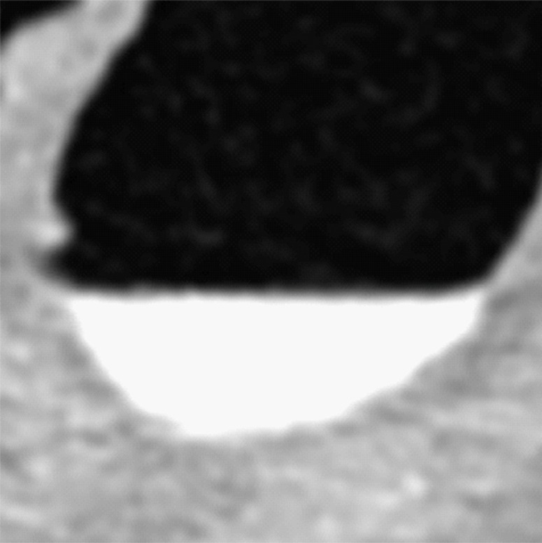
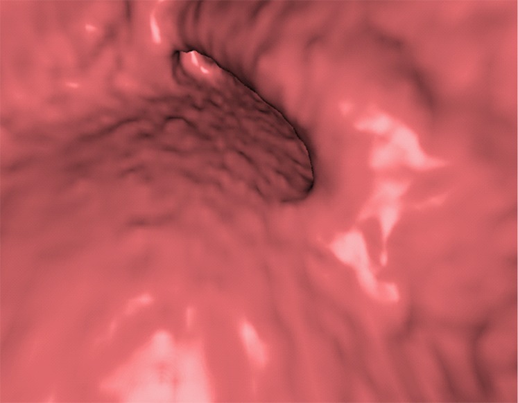
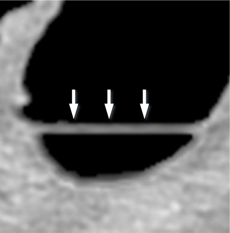
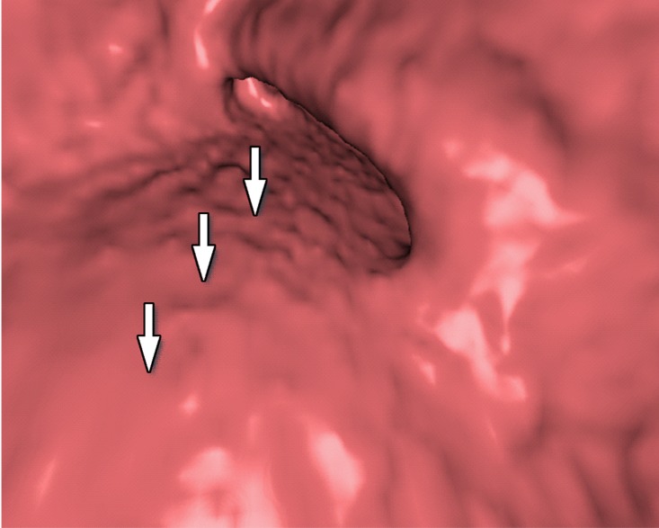
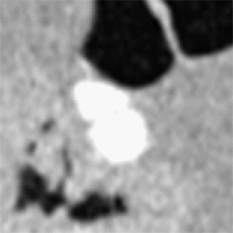
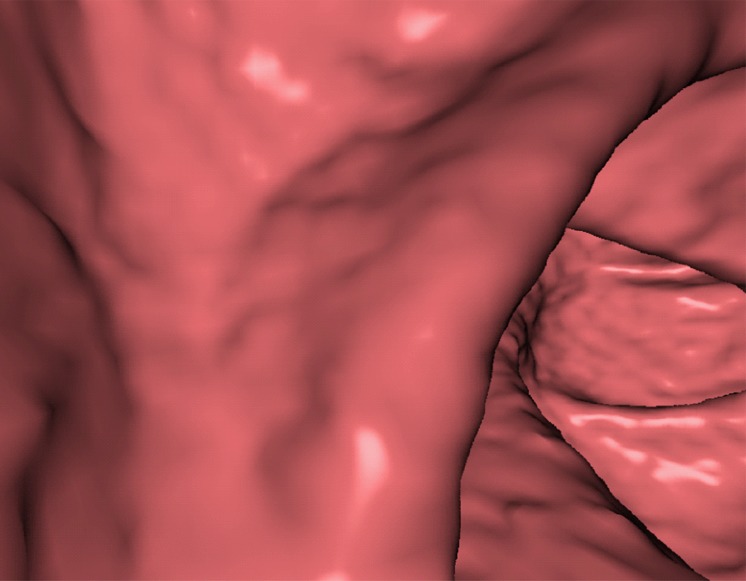
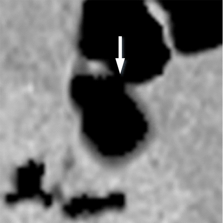
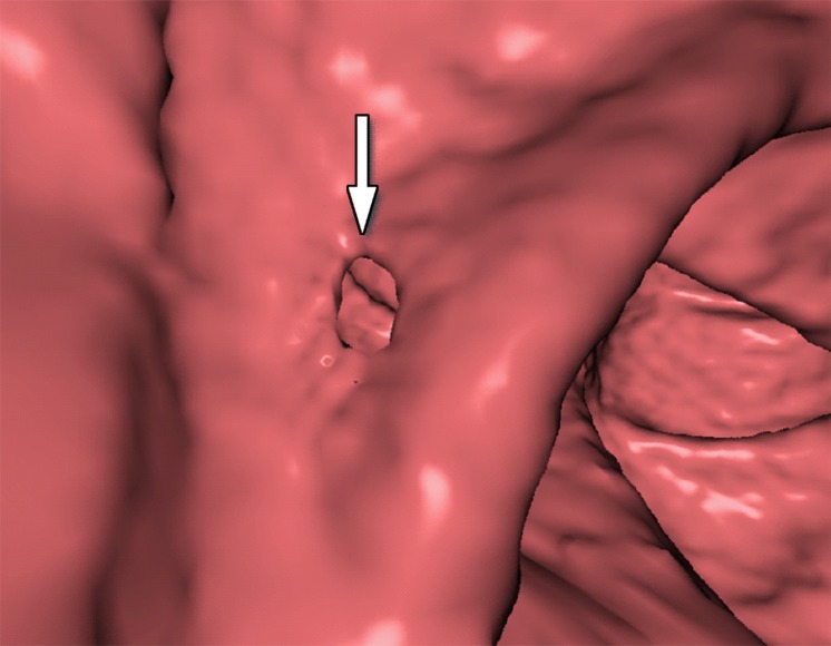
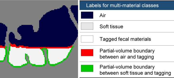
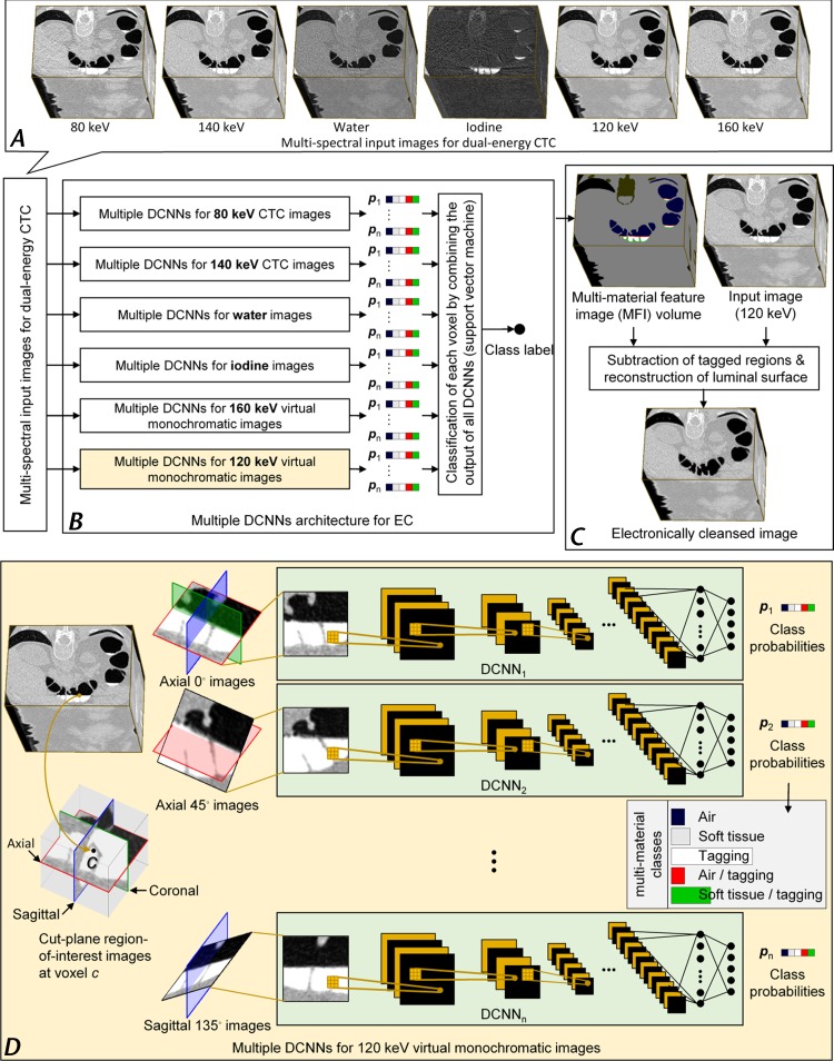
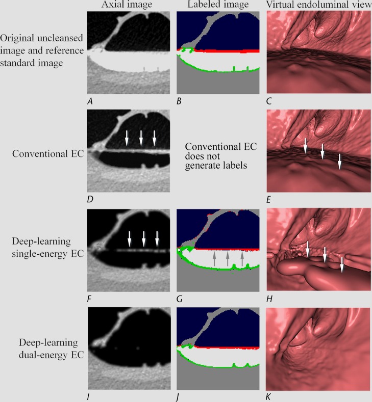
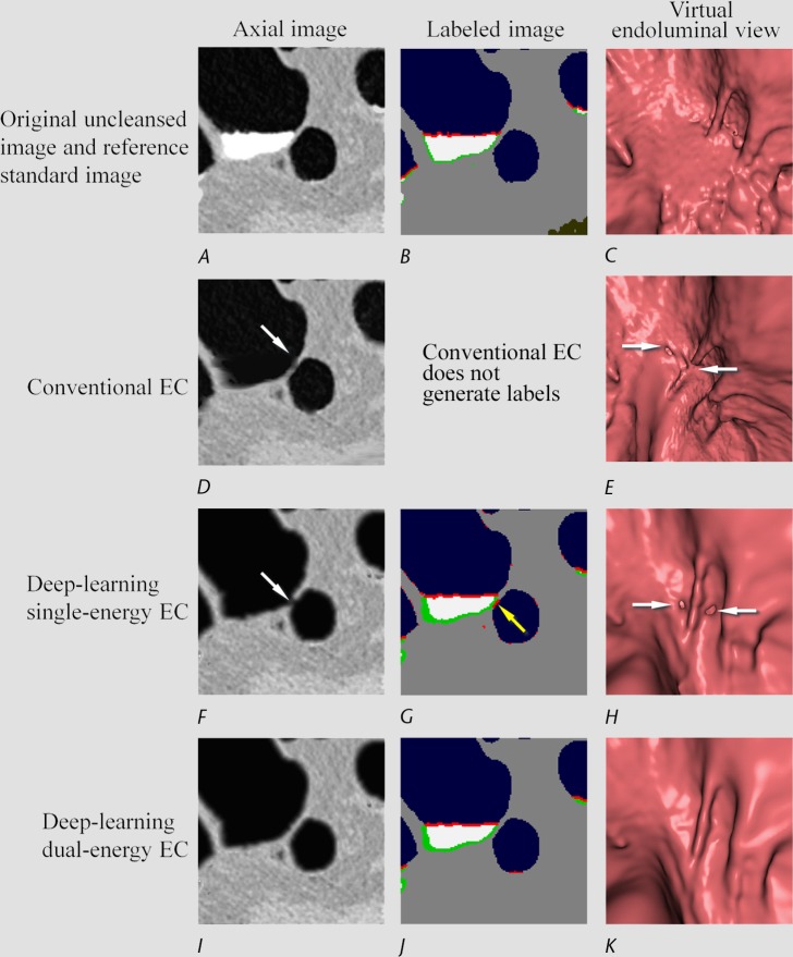
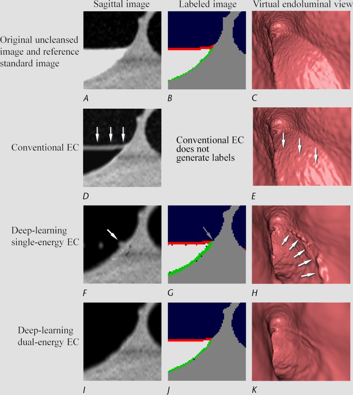
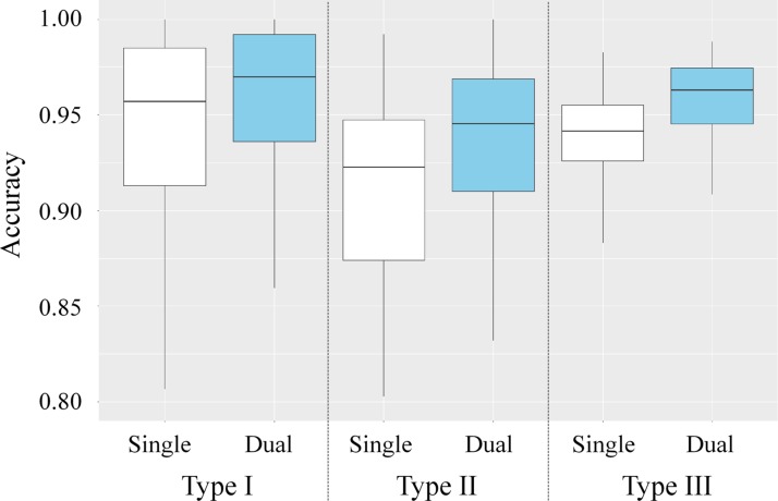
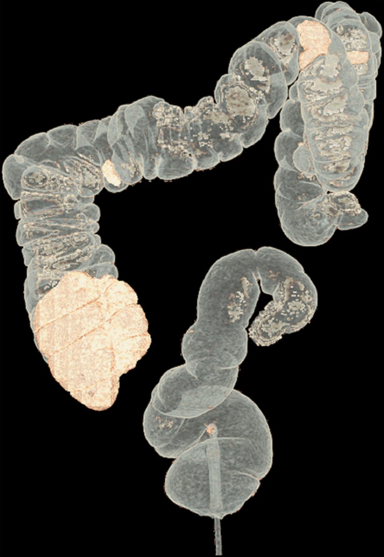
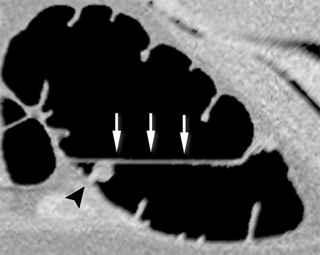
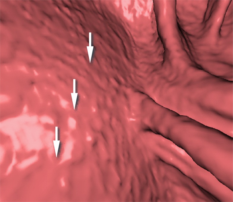
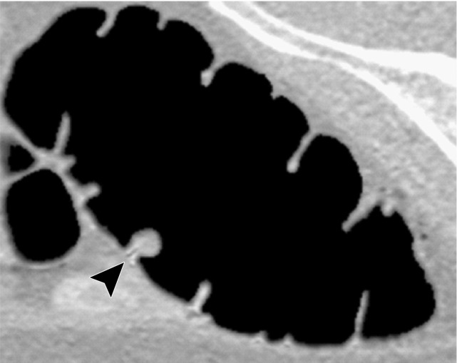
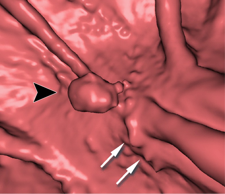
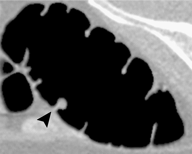
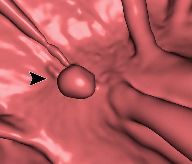
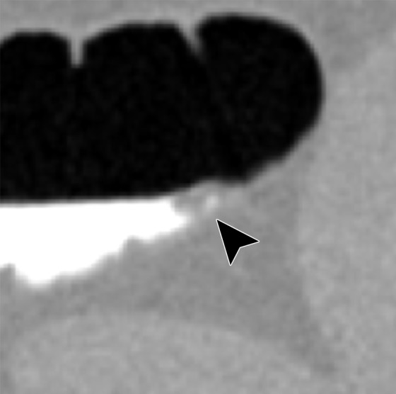
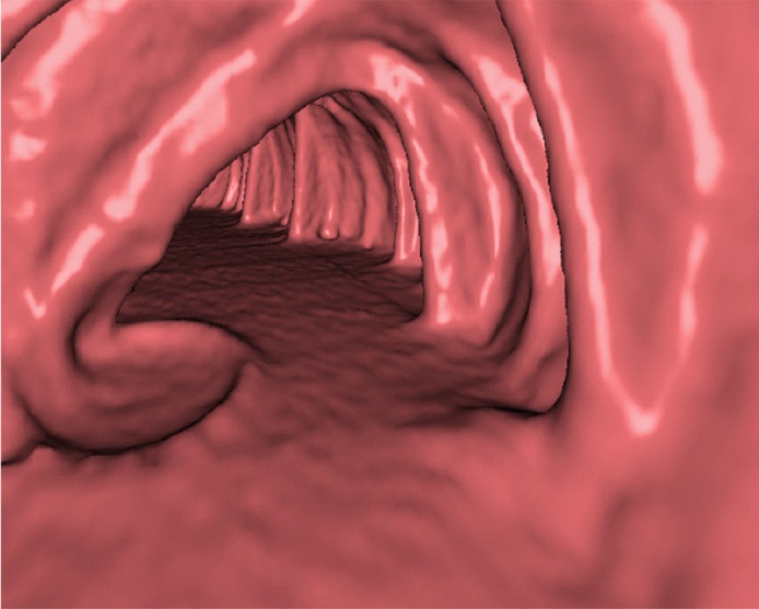
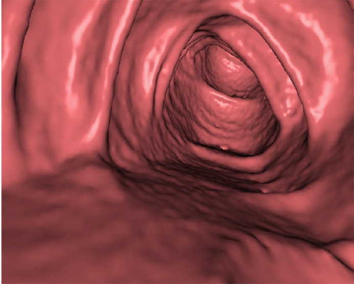
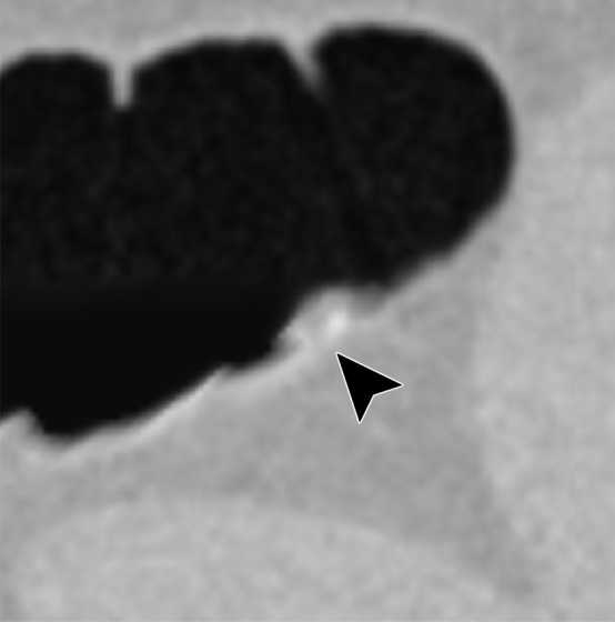
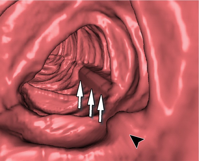
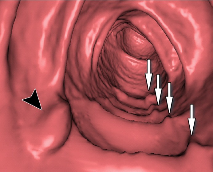
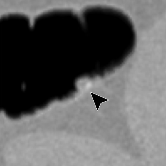
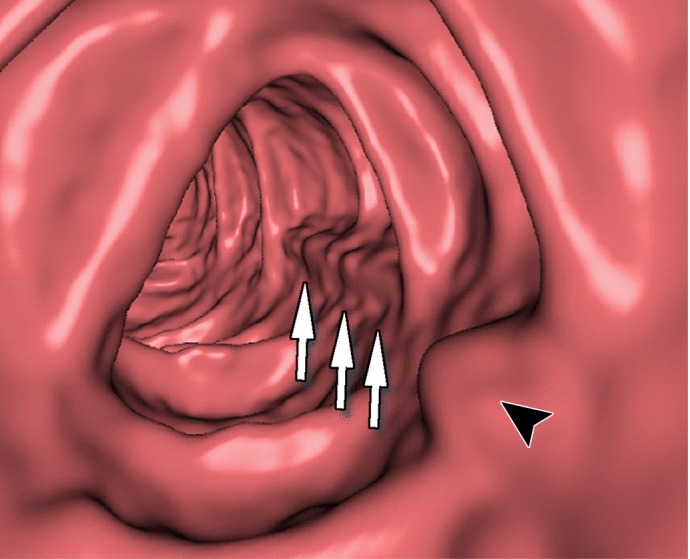
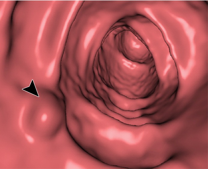
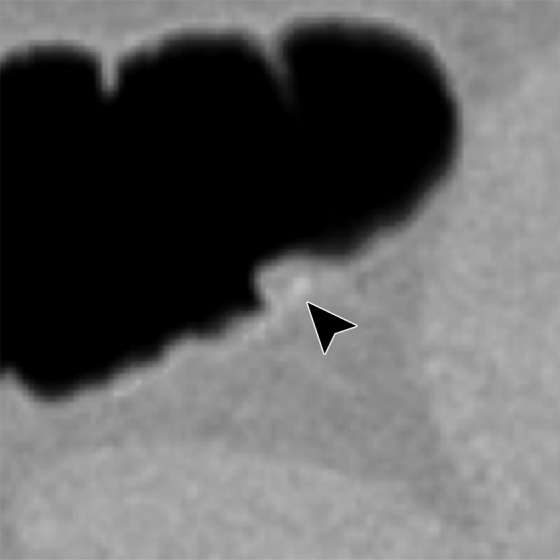
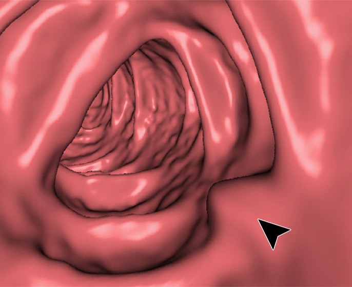
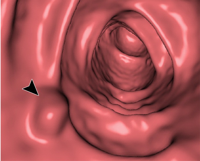
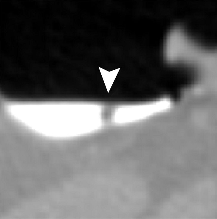
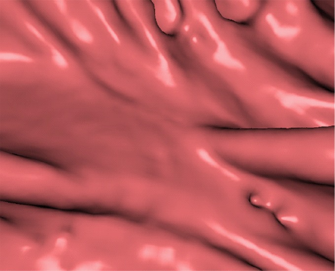
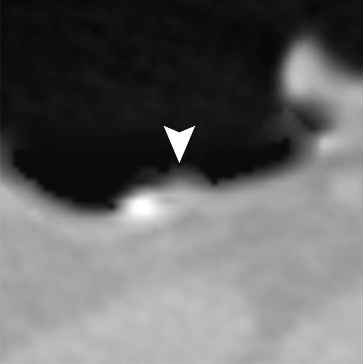
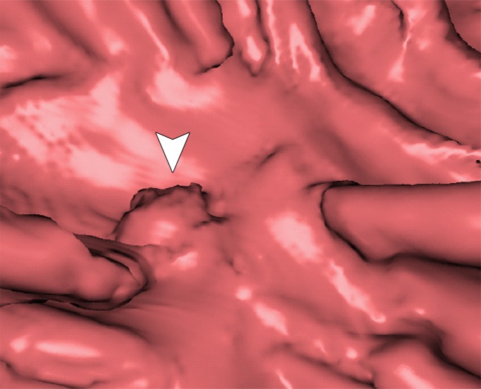
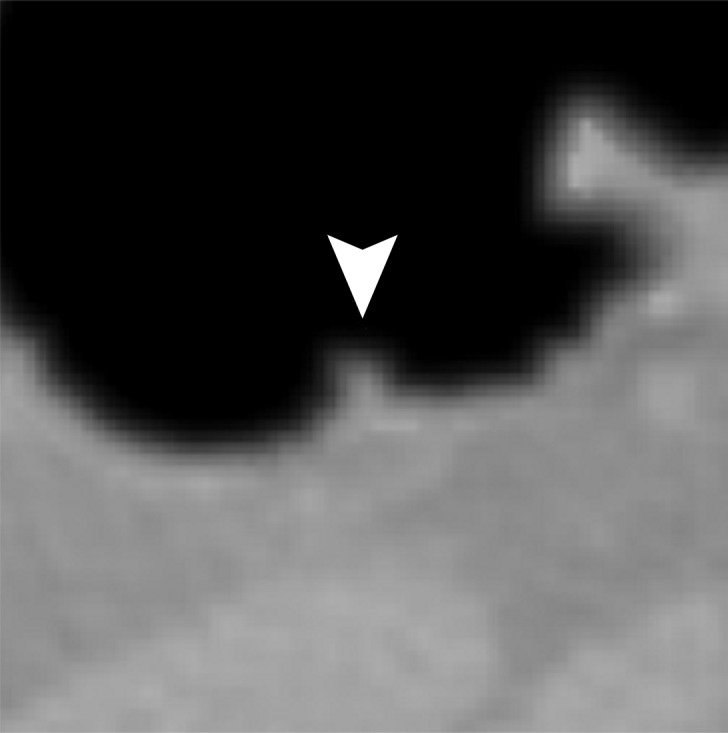
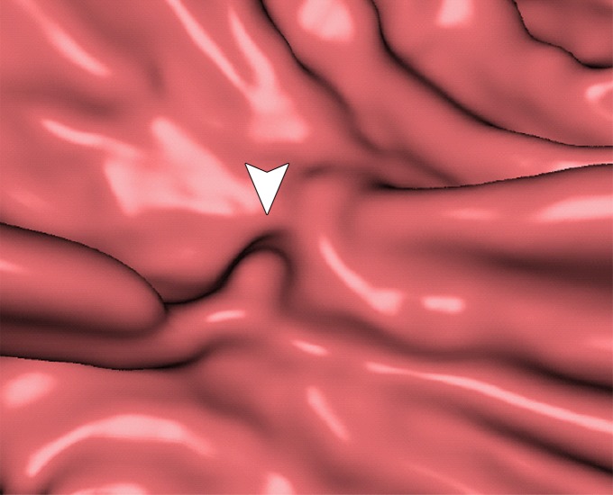
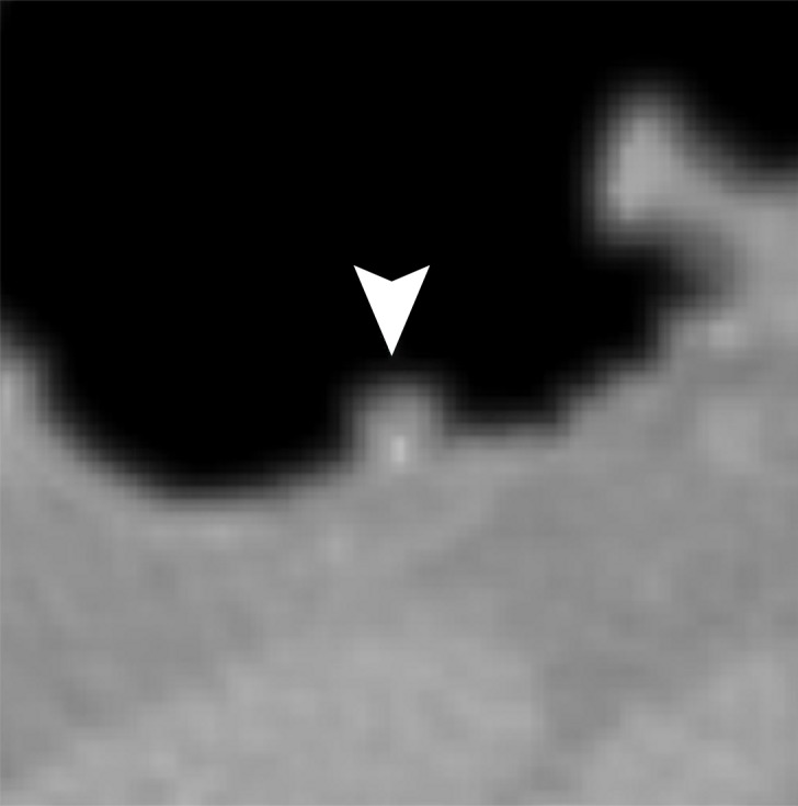
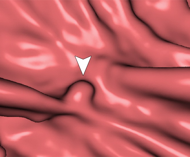
Electronic cleansing (EC) is used for computational removal of residual feces and fluid tagged with an orally administered contrast agent on CT colonographic images to improve the visibility of polyps during virtual endoscopic "fly-through" reading. A recent trend in CT colonography is to perform a low-dose CT scanning protocol with the patient having undergone reduced- or noncathartic bowel preparation. Although several EC schemes exist, they have been developed for use with cathartic bowel preparation and high-radiation-dose CT, and thus, at a low dose with noncathartic bowel preparation, they tend to generate cleansing artifacts that distract and mislead readers. Deep learning can be used for improvement of the image quality with EC at CT colonography. Deep learning EC can produce substantially fewer cleansing artifacts at dual-energy than at single-energy CT colonography, because the dual-energy information can be used to identify relevant material in the colon more precisely than is possible with the single x-ray attenuation value. Because the number of annotated training images is limited at CT colonography, transfer learning can be used for appropriate training of deep learning algorithms. The purposes of this article are to review the causes of cleansing artifacts that distract and mislead readers in conventional EC schemes, to describe the applications of deep learning and dual-energy CT colonography to EC of the colon, and to demonstrate the improvements in image quality with EC and deep learning at single-energy and dual-energy CT colonography with noncathartic bowel preparation. ©RSNA, 2018.


Figures

Figure 1a.
Figure 1a.
Type I cleansing artifact. (a, b) Original uncleansed axial CT colonographic image (a) and the corresponding virtual endoluminal view (b) show tagged fluid that obstructs the view of the bowel wall. (c, d) After cleansing with a conventional EC method, the corresponding axial image (c) and the virtual endoluminal view (d) show a Type I artifact of incomplete removal of the partial-volume boundary between air and tagged material (arrows on c and d).
Figure 1b.
Figure 1b.
Type I cleansing artifact. (a, b) Original uncleansed axial CT colonographic image (a) and the corresponding virtual endoluminal view (b) show tagged fluid that obstructs the view of the bowel wall. (c, d) After cleansing with a conventional EC method, the corresponding axial image (c) and the virtual endoluminal view (d) show a Type I artifact of incomplete removal of the partial-volume boundary between air and tagged material (arrows on c and d).
Figure 1c.
Figure 1c.
Type I cleansing artifact. (a, b) Original uncleansed axial CT colonographic image (a) and the corresponding virtual endoluminal view (b) show tagged fluid that obstructs the view of the bowel wall. (c, d) After cleansing with a conventional EC method, the corresponding axial image (c) and the virtual endoluminal view (d) show a Type I artifact of incomplete removal of the partial-volume boundary between air and tagged material (arrows on c and d).
Figure 1d.
Figure 1d.
Type I cleansing artifact. (a, b) Original uncleansed axial CT colonographic image (a) and the corresponding virtual endoluminal view (b) show tagged fluid that obstructs the view of the bowel wall. (c, d) After cleansing with a conventional EC method, the corresponding axial image (c) and the virtual endoluminal view (d) show a Type I artifact of incomplete removal of the partial-volume boundary between air and tagged material (arrows on c and d).
Figure 2a.
Figure 2a.
Type II cleansing artifact. (a, b) Original uncleansed axial CT colonographic image (a) shows tagged fecal material next to a thin fold, which appears complete on the corresponding virtual endoluminal view (b). (c, d) After cleansing with a conventional EC method, the corresponding coronal image (c) and the virtual endoluminal view (d) show a Type II artifact of an incorrectly removed thin soft-tissue surface of the fold (arrow in c and d).
Figure 2b.
Figure 2b.
Type II cleansing artifact. (a, b) Original uncleansed axial CT colonographic image (a) shows tagged fecal material next to a thin fold, which appears complete on the corresponding virtual endoluminal view (b). (c, d) After cleansing with a conventional EC method, the corresponding coronal image (c) and the virtual endoluminal view (d) show a Type II artifact of an incorrectly removed thin soft-tissue surface of the fold (arrow in c and d).
Figure 2c.
Figure 2c.
Type II cleansing artifact. (a, b) Original uncleansed axial CT colonographic image (a) shows tagged fecal material next to a thin fold, which appears complete on the corresponding virtual endoluminal view (b). (c, d) After cleansing with a conventional EC method, the corresponding coronal image (c) and the virtual endoluminal view (d) show a Type II artifact of an incorrectly removed thin soft-tissue surface of the fold (arrow in c and d).
Figure 2d.
Figure 2d.
Type II cleansing artifact. (a, b) Original uncleansed axial CT colonographic image (a) shows tagged fecal material next to a thin fold, which appears complete on the corresponding virtual endoluminal view (b). (c, d) After cleansing with a conventional EC method, the corresponding coronal image (c) and the virtual endoluminal view (d) show a Type II artifact of an incorrectly removed thin soft-tissue surface of the fold (arrow in c and d).
Figure 3a.
Figure 3a.
Type III cleansing artifact. (a, b) Original uncleansed sagittal CT colonographic image (a) shows tagged fecal material obstructing the bowel wall, as shown on the corresponding virtual endoluminal view (b). (c, d) After cleansing with a conventional EC method, the corresponding sagittal image (c) and the virtual endoluminal view (d) show a Type III artifact, an unsatisfactorily resolved mixture of air, soft tissue, and tagged material (arrows in c and d).
Figure 3b.
Figure 3b.
Type III cleansing artifact. (a, b) Original uncleansed sagittal CT colonographic image (a) shows tagged fecal material obstructing the bowel wall, as shown on the corresponding virtual endoluminal view (b). (c, d) After cleansing with a conventional EC method, the corresponding sagittal image (c) and the virtual endoluminal view (d) show a Type III artifact, an unsatisfactorily resolved mixture of air, soft tissue, and tagged material (arrows in c and d).
Figure 3c.
Figure 3c.
Type III cleansing artifact. (a, b) Original uncleansed sagittal CT colonographic image (a) shows tagged fecal material obstructing the bowel wall, as shown on the corresponding virtual endoluminal view (b). (c, d) After cleansing with a conventional EC method, the corresponding sagittal image (c) and the virtual endoluminal view (d) show a Type III artifact, an unsatisfactorily resolved mixture of air, soft tissue, and tagged material (arrows in c and d).
Figure 3d.
Figure 3d.
Type III cleansing artifact. (a, b) Original uncleansed sagittal CT colonographic image (a) shows tagged fecal material obstructing the bowel wall, as shown on the corresponding virtual endoluminal view (b). (c, d) After cleansing with a conventional EC method, the corresponding sagittal image (c) and the virtual endoluminal view (d) show a Type III artifact, an unsatisfactorily resolved mixture of air, soft tissue, and tagged material (arrows in c and d).
Figure 4a.
Figure 4a.
Multimaterial classes used with our EC method. (a) Original CT colonographic image contains the three base material classes of lumen air, soft tissue, and tagged fecal material. (b) The corresponding reference-standard labels are manually prepared labels delineating the five multimaterial classes indicated in the table on the right.
Figure 4b.
Figure 4b.
Multimaterial classes used with our EC method. (a) Original CT colonographic image contains the three base material classes of lumen air, soft tissue, and tagged fecal material. (b) The corresponding reference-standard labels are manually prepared labels delineating the five multimaterial classes indicated in the table on the right.
Figure 5.
Figure 5.
Schematic diagram of the deep learning EC method for dual-energy CT colonography (CTC). (A–B) An overview of the EC workflow shows that, A, six multispectral CT colonographic input volumetric images are reviewed by using, B, a DCNN ensemble classifier. The classifier consists of multiple DCNNs for reviewing each input volumetric image. With the use of output class probabilities of the individual DCNNs, a metaclassifier (support vector machine) determines the final multimaterial class labels for each voxel of an MFI volume. C, The virtually cleansed volumetric image is generated from a 120-keV CT colonographic volumetric image by converting its CT values that correspond to non–soft-tissue MFI volume labels to CT values of air, and by reconstructing cleansed soft-tissue surfaces within the MFI partial-volume boundary between soft tissue and tagged material. Each DCNN of the ensemble classifier in B has the same overall single-stream architecture. D, The regions of interest are represented by cut-plane images that are sampled at 0°, 45°, and 135° angles to the axial, coronal, and sagittal axes from the volumetric image. The use of multiple DCNNs allows calculation of class probabilities at each voxel from different input cut-plane region-of-interest images.
Figure 6.
Figure 6.
Electronic cleansing with Type I artifacts. (A–C) The first row shows, A, the original uncleansed axial CT colonographic image, B, the corresponding reference standard labels, and C, the uncleansed virtual endoluminal view. (D, E) The second row shows the outcome of a conventional EC method on, D, the axial CT colonographic image, and E, the virtual endoluminal view. White arrows on D and E indicate Type I artifacts. The corresponding space for the labeled image is kept blank, because the conventional EC method does not generate labels. (F–H) The third row shows the outcome of the deep learning single-energy EC on, F, the axial CT colonographic image, G, the corresponding automatically labeled image, and H, the corresponding virtual endoluminal view. The white arrows on F and H indicate Type I artifacts. The gray arrows on G indicate the portions of the partial-volume boundary between air and tagged material that were labeled incorrectly as soft tissue, and thus were causing the Type I artifacts. (I–K) The fourth row shows the outcome of the application of the deep learning dual-energy EC, I, on the axial CT colonographic image, J, the corresponding automatically labeled image, and K, the corresponding virtual endoluminal view. The entire partial-volume boundary between air and tagged material was correctly identified and removed, and thus no Type I artifacts were generated.
Figure 7.
Figure 7.
Electronic cleansing with Type II artifacts. (A–C) The first row shows, A, the original uncleansed axial CT colonographic image, B, the corresponding reference standard labeled image, and C, the corresponding uncleansed virtual endoluminal view. (D, E) The second row shows the outcome of a conventional EC method on, D, the axial image, and E, the corresponding virtual endoluminal view. The white arrows on D and E indicate Type II artifacts. The corresponding space for the labeled image is kept blank, because the conventional EC method does not generate labels. (FH) The third row shows the outcome of the deep learning single-energy EC on, F, the axial CT colonographic image, G, the corresponding automatically labeled image, and H, the corresponding virtual endoluminal view. The white arrows on F and H indicate Type II artifacts. The yellow arrow on G indicates a portion of the soft tissue that was labeled incorrectly as a partial-volume boundary between air and tagged material and thus was the cause of the Type II artifacts. (I–K) The fourth row shows the outcome of the application of the deep learning dual-energy EC on, I, the axial CT colonographic image, J, the corresponding automatically labeled image, and K, the corresponding virtual endoluminal view. The entire thin layer between the air and tagged material is identified correctly as soft tissue, and thus no Type II artifacts were generated.
Figure 8.
Figure 8.
Electronic cleansing with Type III artifacts. (A–C) The first row shows, A, the original uncleansed sagittal CT colonographic image, B, the corresponding reference standard labeled image, and C, the corresponding uncleansed virtual endoluminal view. (D, E) The second row shows the outcome of conventional EC on, D, the sagittal CT colonographic image and E, the virtual endoluminal view. The white arrows on D and E indicate Type I artifacts. The corresponding space for the labeled image is kept blank, because the conventional EC method does not generate labels. (F–H) The third row shows the outcome of the deep learning single-energy EC on, F, the sagittal CT colonographic image, G, the automatically labeled image, and H, the virtual endoluminal view. The gray arrow on G indicates that the junction of air, tagged material, and colonic wall is identified incorrectly as soft tissue, thereby generating the Type III artifacts indicated by white arrows on F and H. (IK) The fourth row shows the outcome of the application of the deep learning dual-energy EC scheme on, I, the sagittal CT colonographic image, J, the corresponding automatically labeled image, and K, the corresponding virtual endoluminal view. No type III artifacts were generated with this scheme.
Figure 9.
Figure 9.
Box plot shows a quantitative comparison of the accuracy of the MFI volume labels automatically calculated by deep learning single- and dual-energy EC schemes for cleansing of regions affected by the three types of artifacts and that of a conventional EC method. The metric for accuracy was the overlap ratio between the automatically calculated MFI volume labels and the manually labeled reference-standard volume labels. The box plot was calculated from 384 volumes of interest sampled from 18 patients, where each of the three types of artifacts was represented by 128 volumes of interest.
Figure 10a.
Figure 10a.
Virtual 3D reconstructions of a colon from virtual monochromatic CT colonographic images before and after virtual cleansing. (a) The uncleansed volume has residual materials (pink regions in a) distributed throughout the colon, and part of the descending colon is completely missing from the reconstruction due to a major obstruction of the lumen by the residual materials. (b) The corresponding volume that was virtually cleansed with a conventional EC method shows that some of the residual materials are still retained in the colon and that the reconstruction of the descending colon is still incomplete. (c) The corresponding volume that was virtually cleansed by our deep learning dual-energy EC scheme shows that all of the residual materials have been removed, and this also has enabled complete reconstruction of the descending colon for complete fly-through visualization.
Figure 10b.
Figure 10b.
Virtual 3D reconstructions of a colon from virtual monochromatic CT colonographic images before and after virtual cleansing. (a) The uncleansed volume has residual materials (pink regions in a) distributed throughout the colon, and part of the descending colon is completely missing from the reconstruction due to a major obstruction of the lumen by the residual materials. (b) The corresponding volume that was virtually cleansed with a conventional EC method shows that some of the residual materials are still retained in the colon and that the reconstruction of the descending colon is still incomplete. (c) The corresponding volume that was virtually cleansed by our deep learning dual-energy EC scheme shows that all of the residual materials have been removed, and this also has enabled complete reconstruction of the descending colon for complete fly-through visualization.
Figure 10c.
Figure 10c.
Virtual 3D reconstructions of a colon from virtual monochromatic CT colonographic images before and after virtual cleansing. (a) The uncleansed volume has residual materials (pink regions in a) distributed throughout the colon, and part of the descending colon is completely missing from the reconstruction due to a major obstruction of the lumen by the residual materials. (b) The corresponding volume that was virtually cleansed with a conventional EC method shows that some of the residual materials are still retained in the colon and that the reconstruction of the descending colon is still incomplete. (c) The corresponding volume that was virtually cleansed by our deep learning dual-energy EC scheme shows that all of the residual materials have been removed, and this also has enabled complete reconstruction of the descending colon for complete fly-through visualization.
Figure 11a.
Figure 11a.
Electronic cleansing of tagged fluid with a submerged 10-mm polyp (arrowhead in a, c, e–h). (a, b) Uncleansed sagittal CT colonographic image (a) and the corresponding uncleansed virtual endoluminal view (b) show how the fluid obstructs the view of the colonic wall, including the polyp. (c, d) After cleansing with a conventional EC method, the corresponding sagittal image (c) and virtual endoluminal image (d) show a Type I artifact (white arrows) that still prevents the submerged polyp and the colonic wall from being seen. (e, f) After cleansing with our deep learning single-energy EC scheme, the sagittal image (e) and virtual endoluminal view (f) show that the polyp is visible; however, the virtual endoluminal view is distorted by Type III artifacts (white arrows in f). (g, h) After cleansing with our deep learning dual-energy EC scheme, the sagittal image (g) and virtual endoluminal image (h) show that the polyp is visible and that there are no image artifacts.
Figure 11b.
Figure 11b.
Electronic cleansing of tagged fluid with a submerged 10-mm polyp (arrowhead in a, c, e–h). (a, b) Uncleansed sagittal CT colonographic image (a) and the corresponding uncleansed virtual endoluminal view (b) show how the fluid obstructs the view of the colonic wall, including the polyp. (c, d) After cleansing with a conventional EC method, the corresponding sagittal image (c) and virtual endoluminal image (d) show a Type I artifact (white arrows) that still prevents the submerged polyp and the colonic wall from being seen. (e, f) After cleansing with our deep learning single-energy EC scheme, the sagittal image (e) and virtual endoluminal view (f) show that the polyp is visible; however, the virtual endoluminal view is distorted by Type III artifacts (white arrows in f). (g, h) After cleansing with our deep learning dual-energy EC scheme, the sagittal image (g) and virtual endoluminal image (h) show that the polyp is visible and that there are no image artifacts.
Figure 11c.
Figure 11c.
Electronic cleansing of tagged fluid with a submerged 10-mm polyp (arrowhead in a, c, e–h). (a, b) Uncleansed sagittal CT colonographic image (a) and the corresponding uncleansed virtual endoluminal view (b) show how the fluid obstructs the view of the colonic wall, including the polyp. (c, d) After cleansing with a conventional EC method, the corresponding sagittal image (c) and virtual endoluminal image (d) show a Type I artifact (white arrows) that still prevents the submerged polyp and the colonic wall from being seen. (e, f) After cleansing with our deep learning single-energy EC scheme, the sagittal image (e) and virtual endoluminal view (f) show that the polyp is visible; however, the virtual endoluminal view is distorted by Type III artifacts (white arrows in f). (g, h) After cleansing with our deep learning dual-energy EC scheme, the sagittal image (g) and virtual endoluminal image (h) show that the polyp is visible and that there are no image artifacts.
Figure 11d.
Figure 11d.
Electronic cleansing of tagged fluid with a submerged 10-mm polyp (arrowhead in a, c, e–h). (a, b) Uncleansed sagittal CT colonographic image (a) and the corresponding uncleansed virtual endoluminal view (b) show how the fluid obstructs the view of the colonic wall, including the polyp. (c, d) After cleansing with a conventional EC method, the corresponding sagittal image (c) and virtual endoluminal image (d) show a Type I artifact (white arrows) that still prevents the submerged polyp and the colonic wall from being seen. (e, f) After cleansing with our deep learning single-energy EC scheme, the sagittal image (e) and virtual endoluminal view (f) show that the polyp is visible; however, the virtual endoluminal view is distorted by Type III artifacts (white arrows in f). (g, h) After cleansing with our deep learning dual-energy EC scheme, the sagittal image (g) and virtual endoluminal image (h) show that the polyp is visible and that there are no image artifacts.
Figure 11e.
Figure 11e.
Electronic cleansing of tagged fluid with a submerged 10-mm polyp (arrowhead in a, c, e–h). (a, b) Uncleansed sagittal CT colonographic image (a) and the corresponding uncleansed virtual endoluminal view (b) show how the fluid obstructs the view of the colonic wall, including the polyp. (c, d) After cleansing with a conventional EC method, the corresponding sagittal image (c) and virtual endoluminal image (d) show a Type I artifact (white arrows) that still prevents the submerged polyp and the colonic wall from being seen. (e, f) After cleansing with our deep learning single-energy EC scheme, the sagittal image (e) and virtual endoluminal view (f) show that the polyp is visible; however, the virtual endoluminal view is distorted by Type III artifacts (white arrows in f). (g, h) After cleansing with our deep learning dual-energy EC scheme, the sagittal image (g) and virtual endoluminal image (h) show that the polyp is visible and that there are no image artifacts.
Figure 11f.
Figure 11f.
Electronic cleansing of tagged fluid with a submerged 10-mm polyp (arrowhead in a, c, e–h). (a, b) Uncleansed sagittal CT colonographic image (a) and the corresponding uncleansed virtual endoluminal view (b) show how the fluid obstructs the view of the colonic wall, including the polyp. (c, d) After cleansing with a conventional EC method, the corresponding sagittal image (c) and virtual endoluminal image (d) show a Type I artifact (white arrows) that still prevents the submerged polyp and the colonic wall from being seen. (e, f) After cleansing with our deep learning single-energy EC scheme, the sagittal image (e) and virtual endoluminal view (f) show that the polyp is visible; however, the virtual endoluminal view is distorted by Type III artifacts (white arrows in f). (g, h) After cleansing with our deep learning dual-energy EC scheme, the sagittal image (g) and virtual endoluminal image (h) show that the polyp is visible and that there are no image artifacts.
Figure 11g.
Figure 11g.
Electronic cleansing of tagged fluid with a submerged 10-mm polyp (arrowhead in a, c, e–h). (a, b) Uncleansed sagittal CT colonographic image (a) and the corresponding uncleansed virtual endoluminal view (b) show how the fluid obstructs the view of the colonic wall, including the polyp. (c, d) After cleansing with a conventional EC method, the corresponding sagittal image (c) and virtual endoluminal image (d) show a Type I artifact (white arrows) that still prevents the submerged polyp and the colonic wall from being seen. (e, f) After cleansing with our deep learning single-energy EC scheme, the sagittal image (e) and virtual endoluminal view (f) show that the polyp is visible; however, the virtual endoluminal view is distorted by Type III artifacts (white arrows in f). (g, h) After cleansing with our deep learning dual-energy EC scheme, the sagittal image (g) and virtual endoluminal image (h) show that the polyp is visible and that there are no image artifacts.
Figure 11h.
Figure 11h.
Electronic cleansing of tagged fluid with a submerged 10-mm polyp (arrowhead in a, c, e–h). (a, b) Uncleansed sagittal CT colonographic image (a) and the corresponding uncleansed virtual endoluminal view (b) show how the fluid obstructs the view of the colonic wall, including the polyp. (c, d) After cleansing with a conventional EC method, the corresponding sagittal image (c) and virtual endoluminal image (d) show a Type I artifact (white arrows) that still prevents the submerged polyp and the colonic wall from being seen. (e, f) After cleansing with our deep learning single-energy EC scheme, the sagittal image (e) and virtual endoluminal view (f) show that the polyp is visible; however, the virtual endoluminal view is distorted by Type III artifacts (white arrows in f). (g, h) After cleansing with our deep learning dual-energy EC scheme, the sagittal image (g) and virtual endoluminal image (h) show that the polyp is visible and that there are no image artifacts.
Figure 12a.
Figure 12a.
Electronic cleansing of tagged fluid with a partially submerged 10-mm polyp located at a three-material junction of soft tissue, tagged fluid, and air (arrowhead in all images except b and c). (ac) Uncleansed sagittal CT colonographic image (a) clearly shows the polyp (arrowhead), but it is not as visible in the virtual endoluminal forward (b) and backward (c) views. (df) After cleansing with a conventional EC method, sagittal CT colonographic image (d) and virtual endoluminal views (e, f) show a distorted polyp with Type III artifacts (white arrows in e and f). (gi) After cleansing with deep learning single-energy EC, the sagittal CT colonographic image (g) and virtual endoluminal views (h, i) show that the shape of the polyp is preserved; however, some Type III artifacts remain (white arrows in h) and imitate polyps, thereby distracting and misleading readers. (j–l) After cleansing with deep learning dual-energy EC, the sagittal CT colonographic image (j) and virtual endoluminal images (k, l) show that the shape and size of the polyp are preserved without generation of cleansing artifacts.
Figure 12b.
Figure 12b.
Electronic cleansing of tagged fluid with a partially submerged 10-mm polyp located at a three-material junction of soft tissue, tagged fluid, and air (arrowhead in all images except b and c). (ac) Uncleansed sagittal CT colonographic image (a) clearly shows the polyp (arrowhead), but it is not as visible in the virtual endoluminal forward (b) and backward (c) views. (df) After cleansing with a conventional EC method, sagittal CT colonographic image (d) and virtual endoluminal views (e, f) show a distorted polyp with Type III artifacts (white arrows in e and f). (gi) After cleansing with deep learning single-energy EC, the sagittal CT colonographic image (g) and virtual endoluminal views (h, i) show that the shape of the polyp is preserved; however, some Type III artifacts remain (white arrows in h) and imitate polyps, thereby distracting and misleading readers. (j–l) After cleansing with deep learning dual-energy EC, the sagittal CT colonographic image (j) and virtual endoluminal images (k, l) show that the shape and size of the polyp are preserved without generation of cleansing artifacts.
Figure 12c.
Figure 12c.
Electronic cleansing of tagged fluid with a partially submerged 10-mm polyp located at a three-material junction of soft tissue, tagged fluid, and air (arrowhead in all images except b and c). (ac) Uncleansed sagittal CT colonographic image (a) clearly shows the polyp (arrowhead), but it is not as visible in the virtual endoluminal forward (b) and backward (c) views. (df) After cleansing with a conventional EC method, sagittal CT colonographic image (d) and virtual endoluminal views (e, f) show a distorted polyp with Type III artifacts (white arrows in e and f). (gi) After cleansing with deep learning single-energy EC, the sagittal CT colonographic image (g) and virtual endoluminal views (h, i) show that the shape of the polyp is preserved; however, some Type III artifacts remain (white arrows in h) and imitate polyps, thereby distracting and misleading readers. (j–l) After cleansing with deep learning dual-energy EC, the sagittal CT colonographic image (j) and virtual endoluminal images (k, l) show that the shape and size of the polyp are preserved without generation of cleansing artifacts.
Figure 12d.
Figure 12d.
Electronic cleansing of tagged fluid with a partially submerged 10-mm polyp located at a three-material junction of soft tissue, tagged fluid, and air (arrowhead in all images except b and c). (ac) Uncleansed sagittal CT colonographic image (a) clearly shows the polyp (arrowhead), but it is not as visible in the virtual endoluminal forward (b) and backward (c) views. (df) After cleansing with a conventional EC method, sagittal CT colonographic image (d) and virtual endoluminal views (e, f) show a distorted polyp with Type III artifacts (white arrows in e and f). (gi) After cleansing with deep learning single-energy EC, the sagittal CT colonographic image (g) and virtual endoluminal views (h, i) show that the shape of the polyp is preserved; however, some Type III artifacts remain (white arrows in h) and imitate polyps, thereby distracting and misleading readers. (j–l) After cleansing with deep learning dual-energy EC, the sagittal CT colonographic image (j) and virtual endoluminal images (k, l) show that the shape and size of the polyp are preserved without generation of cleansing artifacts.
Figure 12e.
Figure 12e.
Electronic cleansing of tagged fluid with a partially submerged 10-mm polyp located at a three-material junction of soft tissue, tagged fluid, and air (arrowhead in all images except b and c). (ac) Uncleansed sagittal CT colonographic image (a) clearly shows the polyp (arrowhead), but it is not as visible in the virtual endoluminal forward (b) and backward (c) views. (df) After cleansing with a conventional EC method, sagittal CT colonographic image (d) and virtual endoluminal views (e, f) show a distorted polyp with Type III artifacts (white arrows in e and f). (gi) After cleansing with deep learning single-energy EC, the sagittal CT colonographic image (g) and virtual endoluminal views (h, i) show that the shape of the polyp is preserved; however, some Type III artifacts remain (white arrows in h) and imitate polyps, thereby distracting and misleading readers. (j–l) After cleansing with deep learning dual-energy EC, the sagittal CT colonographic image (j) and virtual endoluminal images (k, l) show that the shape and size of the polyp are preserved without generation of cleansing artifacts.
Figure 12f.
Figure 12f.
Electronic cleansing of tagged fluid with a partially submerged 10-mm polyp located at a three-material junction of soft tissue, tagged fluid, and air (arrowhead in all images except b and c). (ac) Uncleansed sagittal CT colonographic image (a) clearly shows the polyp (arrowhead), but it is not as visible in the virtual endoluminal forward (b) and backward (c) views. (df) After cleansing with a conventional EC method, sagittal CT colonographic image (d) and virtual endoluminal views (e, f) show a distorted polyp with Type III artifacts (white arrows in e and f). (gi) After cleansing with deep learning single-energy EC, the sagittal CT colonographic image (g) and virtual endoluminal views (h, i) show that the shape of the polyp is preserved; however, some Type III artifacts remain (white arrows in h) and imitate polyps, thereby distracting and misleading readers. (j–l) After cleansing with deep learning dual-energy EC, the sagittal CT colonographic image (j) and virtual endoluminal images (k, l) show that the shape and size of the polyp are preserved without generation of cleansing artifacts.
Figure 12g.
Figure 12g.
Electronic cleansing of tagged fluid with a partially submerged 10-mm polyp located at a three-material junction of soft tissue, tagged fluid, and air (arrowhead in all images except b and c). (ac) Uncleansed sagittal CT colonographic image (a) clearly shows the polyp (arrowhead), but it is not as visible in the virtual endoluminal forward (b) and backward (c) views. (df) After cleansing with a conventional EC method, sagittal CT colonographic image (d) and virtual endoluminal views (e, f) show a distorted polyp with Type III artifacts (white arrows in e and f). (gi) After cleansing with deep learning single-energy EC, the sagittal CT colonographic image (g) and virtual endoluminal views (h, i) show that the shape of the polyp is preserved; however, some Type III artifacts remain (white arrows in h) and imitate polyps, thereby distracting and misleading readers. (j–l) After cleansing with deep learning dual-energy EC, the sagittal CT colonographic image (j) and virtual endoluminal images (k, l) show that the shape and size of the polyp are preserved without generation of cleansing artifacts.
Figure 12h.
Figure 12h.
Electronic cleansing of tagged fluid with a partially submerged 10-mm polyp located at a three-material junction of soft tissue, tagged fluid, and air (arrowhead in all images except b and c). (ac) Uncleansed sagittal CT colonographic image (a) clearly shows the polyp (arrowhead), but it is not as visible in the virtual endoluminal forward (b) and backward (c) views. (df) After cleansing with a conventional EC method, sagittal CT colonographic image (d) and virtual endoluminal views (e, f) show a distorted polyp with Type III artifacts (white arrows in e and f). (gi) After cleansing with deep learning single-energy EC, the sagittal CT colonographic image (g) and virtual endoluminal views (h, i) show that the shape of the polyp is preserved; however, some Type III artifacts remain (white arrows in h) and imitate polyps, thereby distracting and misleading readers. (j–l) After cleansing with deep learning dual-energy EC, the sagittal CT colonographic image (j) and virtual endoluminal images (k, l) show that the shape and size of the polyp are preserved without generation of cleansing artifacts.
Figure 12i.
Figure 12i.
Electronic cleansing of tagged fluid with a partially submerged 10-mm polyp located at a three-material junction of soft tissue, tagged fluid, and air (arrowhead in all images except b and c). (ac) Uncleansed sagittal CT colonographic image (a) clearly shows the polyp (arrowhead), but it is not as visible in the virtual endoluminal forward (b) and backward (c) views. (df) After cleansing with a conventional EC method, sagittal CT colonographic image (d) and virtual endoluminal views (e, f) show a distorted polyp with Type III artifacts (white arrows in e and f). (gi) After cleansing with deep learning single-energy EC, the sagittal CT colonographic image (g) and virtual endoluminal views (h, i) show that the shape of the polyp is preserved; however, some Type III artifacts remain (white arrows in h) and imitate polyps, thereby distracting and misleading readers. (j–l) After cleansing with deep learning dual-energy EC, the sagittal CT colonographic image (j) and virtual endoluminal images (k, l) show that the shape and size of the polyp are preserved without generation of cleansing artifacts.
Figure 12j.
Figure 12j.
Electronic cleansing of tagged fluid with a partially submerged 10-mm polyp located at a three-material junction of soft tissue, tagged fluid, and air (arrowhead in all images except b and c). (ac) Uncleansed sagittal CT colonographic image (a) clearly shows the polyp (arrowhead), but it is not as visible in the virtual endoluminal forward (b) and backward (c) views. (df) After cleansing with a conventional EC method, sagittal CT colonographic image (d) and virtual endoluminal views (e, f) show a distorted polyp with Type III artifacts (white arrows in e and f). (gi) After cleansing with deep learning single-energy EC, the sagittal CT colonographic image (g) and virtual endoluminal views (h, i) show that the shape of the polyp is preserved; however, some Type III artifacts remain (white arrows in h) and imitate polyps, thereby distracting and misleading readers. (j–l) After cleansing with deep learning dual-energy EC, the sagittal CT colonographic image (j) and virtual endoluminal images (k, l) show that the shape and size of the polyp are preserved without generation of cleansing artifacts.
Figure 12k.
Figure 12k.
Electronic cleansing of tagged fluid with a partially submerged 10-mm polyp located at a three-material junction of soft tissue, tagged fluid, and air (arrowhead in all images except b and c). (ac) Uncleansed sagittal CT colonographic image (a) clearly shows the polyp (arrowhead), but it is not as visible in the virtual endoluminal forward (b) and backward (c) views. (df) After cleansing with a conventional EC method, sagittal CT colonographic image (d) and virtual endoluminal views (e, f) show a distorted polyp with Type III artifacts (white arrows in e and f). (gi) After cleansing with deep learning single-energy EC, the sagittal CT colonographic image (g) and virtual endoluminal views (h, i) show that the shape of the polyp is preserved; however, some Type III artifacts remain (white arrows in h) and imitate polyps, thereby distracting and misleading readers. (j–l) After cleansing with deep learning dual-energy EC, the sagittal CT colonographic image (j) and virtual endoluminal images (k, l) show that the shape and size of the polyp are preserved without generation of cleansing artifacts.
Figure 12l.
Figure 12l.
Electronic cleansing of tagged fluid with a partially submerged 10-mm polyp located at a three-material junction of soft tissue, tagged fluid, and air (arrowhead in all images except b and c). (ac) Uncleansed sagittal CT colonographic image (a) clearly shows the polyp (arrowhead), but it is not as visible in the virtual endoluminal forward (b) and backward (c) views. (df) After cleansing with a conventional EC method, sagittal CT colonographic image (d) and virtual endoluminal views (e, f) show a distorted polyp with Type III artifacts (white arrows in e and f). (gi) After cleansing with deep learning single-energy EC, the sagittal CT colonographic image (g) and virtual endoluminal views (h, i) show that the shape of the polyp is preserved; however, some Type III artifacts remain (white arrows in h) and imitate polyps, thereby distracting and misleading readers. (j–l) After cleansing with deep learning dual-energy EC, the sagittal CT colonographic image (j) and virtual endoluminal images (k, l) show that the shape and size of the polyp are preserved without generation of cleansing artifacts.
Figure 13a.
Figure 13a.
Electronic cleansing of semi-solid tagged feces with a submerged 6-mm polyp. (a) The uncleansed axial CT colonographic image shows the polyp (arrowhead). (b) The virtual endoluminal view shows how the semisolid feces obstruct the view of the polyp. (c, d) After cleansing with a conventional EC method, the axial CT colonographic image (c) and virtual endoluminal view (d) show the distorted shape of the polyp (arrowhead). (e, f) After cleansing with the deep learning single-energy EC scheme, the axial CT colonographic image (e) and virtual endoluminal view (f) reveal the polyp (arrowhead), but the polyp appears inaccurately smaller. (g, h) After cleansing with the deep learning dual-energy EC scheme, axial CT colonographic image (g) and virtual endoluminal view (h) show that the size of the polyp (arrowhead) is preserved without producing cleansing artifacts.
Figure 13b.
Figure 13b.
Electronic cleansing of semi-solid tagged feces with a submerged 6-mm polyp. (a) The uncleansed axial CT colonographic image shows the polyp (arrowhead). (b) The virtual endoluminal view shows how the semisolid feces obstruct the view of the polyp. (c, d) After cleansing with a conventional EC method, the axial CT colonographic image (c) and virtual endoluminal view (d) show the distorted shape of the polyp (arrowhead). (e, f) After cleansing with the deep learning single-energy EC scheme, the axial CT colonographic image (e) and virtual endoluminal view (f) reveal the polyp (arrowhead), but the polyp appears inaccurately smaller. (g, h) After cleansing with the deep learning dual-energy EC scheme, axial CT colonographic image (g) and virtual endoluminal view (h) show that the size of the polyp (arrowhead) is preserved without producing cleansing artifacts.
Figure 13c.
Figure 13c.
Electronic cleansing of semi-solid tagged feces with a submerged 6-mm polyp. (a) The uncleansed axial CT colonographic image shows the polyp (arrowhead). (b) The virtual endoluminal view shows how the semisolid feces obstruct the view of the polyp. (c, d) After cleansing with a conventional EC method, the axial CT colonographic image (c) and virtual endoluminal view (d) show the distorted shape of the polyp (arrowhead). (e, f) After cleansing with the deep learning single-energy EC scheme, the axial CT colonographic image (e) and virtual endoluminal view (f) reveal the polyp (arrowhead), but the polyp appears inaccurately smaller. (g, h) After cleansing with the deep learning dual-energy EC scheme, axial CT colonographic image (g) and virtual endoluminal view (h) show that the size of the polyp (arrowhead) is preserved without producing cleansing artifacts.
Figure 13d.
Figure 13d.
Electronic cleansing of semi-solid tagged feces with a submerged 6-mm polyp. (a) The uncleansed axial CT colonographic image shows the polyp (arrowhead). (b) The virtual endoluminal view shows how the semisolid feces obstruct the view of the polyp. (c, d) After cleansing with a conventional EC method, the axial CT colonographic image (c) and virtual endoluminal view (d) show the distorted shape of the polyp (arrowhead). (e, f) After cleansing with the deep learning single-energy EC scheme, the axial CT colonographic image (e) and virtual endoluminal view (f) reveal the polyp (arrowhead), but the polyp appears inaccurately smaller. (g, h) After cleansing with the deep learning dual-energy EC scheme, axial CT colonographic image (g) and virtual endoluminal view (h) show that the size of the polyp (arrowhead) is preserved without producing cleansing artifacts.
Figure 13e.
Figure 13e.
Electronic cleansing of semi-solid tagged feces with a submerged 6-mm polyp. (a) The uncleansed axial CT colonographic image shows the polyp (arrowhead). (b) The virtual endoluminal view shows how the semisolid feces obstruct the view of the polyp. (c, d) After cleansing with a conventional EC method, the axial CT colonographic image (c) and virtual endoluminal view (d) show the distorted shape of the polyp (arrowhead). (e, f) After cleansing with the deep learning single-energy EC scheme, the axial CT colonographic image (e) and virtual endoluminal view (f) reveal the polyp (arrowhead), but the polyp appears inaccurately smaller. (g, h) After cleansing with the deep learning dual-energy EC scheme, axial CT colonographic image (g) and virtual endoluminal view (h) show that the size of the polyp (arrowhead) is preserved without producing cleansing artifacts.
Figure 13f.
Figure 13f.
Electronic cleansing of semi-solid tagged feces with a submerged 6-mm polyp. (a) The uncleansed axial CT colonographic image shows the polyp (arrowhead). (b) The virtual endoluminal view shows how the semisolid feces obstruct the view of the polyp. (c, d) After cleansing with a conventional EC method, the axial CT colonographic image (c) and virtual endoluminal view (d) show the distorted shape of the polyp (arrowhead). (e, f) After cleansing with the deep learning single-energy EC scheme, the axial CT colonographic image (e) and virtual endoluminal view (f) reveal the polyp (arrowhead), but the polyp appears inaccurately smaller. (g, h) After cleansing with the deep learning dual-energy EC scheme, axial CT colonographic image (g) and virtual endoluminal view (h) show that the size of the polyp (arrowhead) is preserved without producing cleansing artifacts.
Figure 13g.
Figure 13g.
Electronic cleansing of semi-solid tagged feces with a submerged 6-mm polyp. (a) The uncleansed axial CT colonographic image shows the polyp (arrowhead). (b) The virtual endoluminal view shows how the semisolid feces obstruct the view of the polyp. (c, d) After cleansing with a conventional EC method, the axial CT colonographic image (c) and virtual endoluminal view (d) show the distorted shape of the polyp (arrowhead). (e, f) After cleansing with the deep learning single-energy EC scheme, the axial CT colonographic image (e) and virtual endoluminal view (f) reveal the polyp (arrowhead), but the polyp appears inaccurately smaller. (g, h) After cleansing with the deep learning dual-energy EC scheme, axial CT colonographic image (g) and virtual endoluminal view (h) show that the size of the polyp (arrowhead) is preserved without producing cleansing artifacts.
Figure 13h.
Figure 13h.
Electronic cleansing of semi-solid tagged feces with a submerged 6-mm polyp. (a) The uncleansed axial CT colonographic image shows the polyp (arrowhead). (b) The virtual endoluminal view shows how the semisolid feces obstruct the view of the polyp. (c, d) After cleansing with a conventional EC method, the axial CT colonographic image (c) and virtual endoluminal view (d) show the distorted shape of the polyp (arrowhead). (e, f) After cleansing with the deep learning single-energy EC scheme, the axial CT colonographic image (e) and virtual endoluminal view (f) reveal the polyp (arrowhead), but the polyp appears inaccurately smaller. (g, h) After cleansing with the deep learning dual-energy EC scheme, axial CT colonographic image (g) and virtual endoluminal view (h) show that the size of the polyp (arrowhead) is preserved without producing cleansing artifacts.

References

    1. American Cancer Society . Cancer facts & figures 2018. Atlanta, Ga: American Cancer Society, 2018.
    1. Levin B, Lieberman DA, McFarland B, et al. . Screening and surveillance for the early detection of colorectal cancer and adenomatous polyps, 2008: a joint guideline from the American Cancer Society, the US Multi-Society Task Force on Colorectal Cancer, and the American College of Radiology. Gastroenterology 2008;134(5):1570–1595. - PubMed
    1. US Preventive Services Task Force , Bibbins-Domingo K, Grossman DC, et al. . Screening for colorectal cancer: US Preventive Services Task Force recommendation statement. JAMA 2016;315(23):2564–2575. - PubMed
    1. Rockey DC, Paulson E, Niedzwiecki D, et al. . Analysis of air contrast barium enema, computed tomographic colonography, and colonoscopy: prospective comparison. Lancet 2005;365(9456):305–311. - PubMed
    1. Pickhardt PJ. CT colonography for population screening: ready for prime time? Dig Dis Sci 2015;60(3):647–659. - PMC - PubMed