. 2019 Jan 1;30(1):115-123.
doi: 10.1093/annonc/mdy497.

Loss of function of NF1 is a mechanism of acquired resistance to endocrine therapy in lobular breast cancer

Affiliations

Loss of function of NF1 is a mechanism of acquired resistance to endocrine therapy in lobular breast cancer

E S Sokol et al. Ann Oncol. .

Abstract

Background: Invasive lobular carcinoma (ILC) as a disease entity distinct from invasive ductal carcinoma (IDC) has merited focused studies of the genomic landscape, but those to date are largely limited to the assessment of early-stage cancers. Given that genomic alterations develop as acquired resistance to endocrine therapy, studies on refractory ILC are needed.

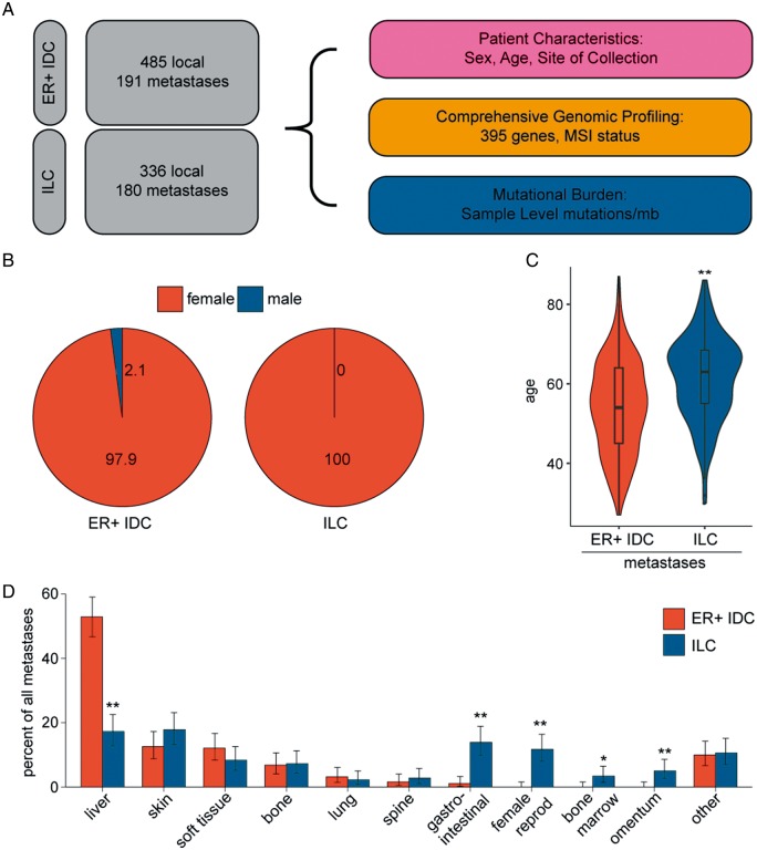
Patients and methods: Tissue from 336 primary-enriched, breast-biopsied ILC and 485 estrogen receptor (ER)-positive IDC and metastatic biopsy specimens from 180 ILC and 191 ER-positive IDC patients was assayed with hybrid-capture-based comprehensive genomic profiling for short variant, indel, copy number variants, and rearrangements in up to 395 cancer-related genes.

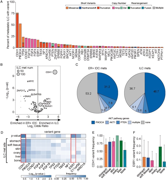
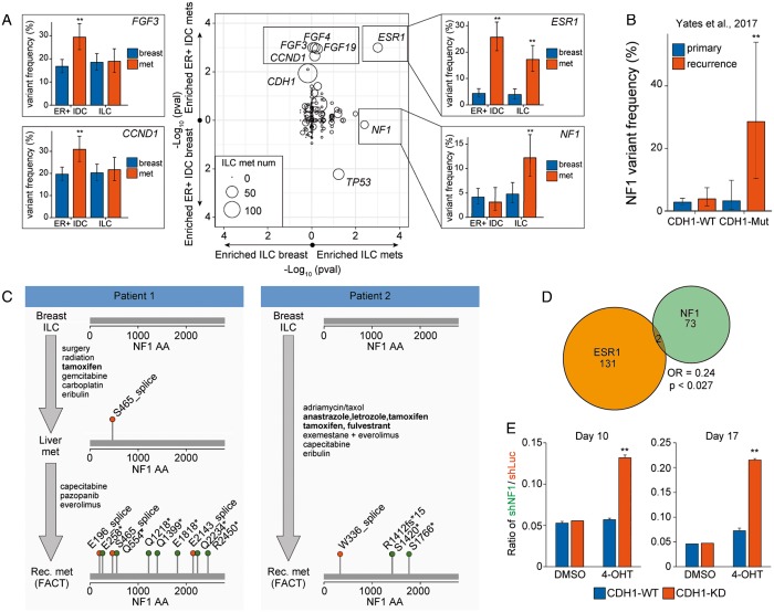
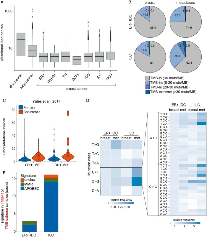
Results: Whereas ESR1 alterations are enriched in the metastases of both ILC and IDC compared with breast specimens, NF1 alterations are enriched only in ILC metastases (mILC). NF1 alterations are predominantly under loss of heterozygosity (11/14, 79%), are mutually exclusive with ESR1 mutations [odds ratio = 0.24, P < 0.027] and are frequently polyclonal in ctDNA assays. Assessment of paired specimens shows that NF1 alterations arise in the setting of acquired resistance. An in vitro model of CDH1 mutated ER-positive breast cancer demonstrates that NF1 knockdown confers a growth advantage in the presence of 4-hydroxy tamoxifen. Our study further identified a significant increase in tumor mutational burden (TMB) in mILCs relative to breast ILCs or metastatic IDCs (8.9% >20 mutations/mb; P < 0.001). Most TMB-high mILCs harbor an APOBEC trinucleotide signature (14/16; 88%).

Conclusions: This study identifies alteration of NF1 as enriched specifically in mILC. Mutual exclusivity with ESR1 alterations, polyclonality in relapsed ctDNA, and de novo acquisition suggest a role for NF1 loss in endocrine therapy resistance. Since NF1 loss leads to RAS/RAF kinase activation, patients may benefit from a matched inhibitor. Moreover, for an independent subset of mILC, TMB was elevated relative to breast ILC, suggesting possible benefit from immune checkpoint inhibitors.


Figures

Figure 1.
Figure 1.
Invasive lobular carcinoma (ILC) metastases are found at unique tissue sites. (A) Schematic of experimental design. In total, 676 estrogen receptor (ER)-positive breast invasive ductal carcinomas (IDCs) and 516 breast ILCs were profiled in the course of routine clinical care. Patient gender (B) and age (C) were compared in the metastatic ER-positive IDC and metastatic ILC cohorts. (D) Sites of metastatic biopsy were analyzed in the ER-positive IDC and ILC cohorts. P-values were calculated using the Mann–Whitney U test (C) and the Fisher’s exact test (D). *P <0.05; **P <0.01.
Figure 2.
Figure 2.
Metastatic invasive lobular carcinomas (ILCs) exhibit a unique genomic profile. (A) Waterfall plot showing the frequency of known/likely alterations in metastatic ILCs broken down by variant class and further subdivided by the specific alteration type in the variant class. The short variant category of alterations included missense mutations, nonframeshifting indels, and truncations (frameshifting indels, splice-affecting, nonsense, nonstart); the copy number variant category of alterations included amplifications and deletions; the rearrangement variant category included truncations and fusions; and the multiple variants category included samples with more than one variant in the sample. (B) Volcano plot comparing the frequency of gene alterations in metastatic ILCs and estrogen receptor (ER)-positive metastatic invasive ductal carcinoma (IDC) cohorts. P-values and odds ratios were calculated using the Fisher’s exact test. P values were capped at 1 × 10⁻¹⁰ and log2 odds ratios were capped at ±5. (C) Pie graph showing the frequency of known or likely AKT pathway alterations in the ER-positive metastatic IDC and metastatic ILC cohorts. Patients with multiple alterations are grouped in ‘multiple’. (D) The frequency of top-altered genes was compared across ILC metastatic sites with at least 10 samples and plotted as a heatmap. To determine whether alteration frequencies differed across sites, a Kruskal–Wallis test was applied. Genes that significantly differ across sites (CDH1 and ESR1; P <0.01) are boxed in red and plotted in panels (E) and (F), respectively. For panels (E) and (F), error bars represent the 90% binomial confidence interval.
Figure 3.
Figure 3.
NF1 alterations are frequently identified in metastatic invasive lobular carcinomas (ILCs) and may drive endocrine therapy resistance. (A) Gene variant frequencies were compared between local breast ILC and metastatic breast ILC (x-axis) and between local ER-positive breast invasive ductal carcinoma (IDC) and metastatic ER-positive breast IDC (y-axis). Plotted is the Fisher’s exact P-value (center). Alteration frequencies were plotted for a subset of significant genes (left and right panels) broken down by group (local versus met and ER-positive IDC versus ILC). Error bars represent the binomial 90% confidence interval. (B) NF1 alteration frequencies in the primary and recurrent breast cohorts from Yates et al. were calculated in CDH1-WT and CDH1-Mutant populations. Error bars represent the 90% binomial confidence interval. (C) Treatment and NF1 mutational status for two patients. Endocrine therapies are bolded. (D) Venn diagram examining the mutual exclusivity of ESR1 and NF1 in the entire breast cohort. Odds ratios and P-values were calculated using the Fisher’s exact test. (E) Quantification of CDH1-WT or CDH1-KD cells following T47D co-mixing experiments. For each background, 2.5 × 10⁴ T47D-shNF1-GFP cells and 4.75 × 10⁵ T47D-mCherry cells were mixed and treated with solvent control or 4-OHT for 10 days or 17 days, and fraction of cells with GFP or mCherry were measured by flow cytometry analysis. Plotted is the ratio of GFP-positive cells to mCherry-positive cells for each condition. **P <0.01 from the Fisher’s exact test (B) or Student’s t-test (E). FACT, FoundationACT.
Figure 4.
Figure 4.
Metastatic invasive lobular carcinomas (ILCs) exhibit a significantly elevated tumor mutational burden. (A) Box and whisker plots capturing the spread of mutational burden (mutations per megabase) in skin cancers, lung cancers, and breast cancers broken down by histological or diagnostic subtype. All relevant tumors from the foundation medicine database were included in this analysis. (B) Pie charts showing the mutational burden for ER-positive IDCs and ILCs, broken down by local/met status. (C) Violin plots capturing the distribution of mutational burden (mutations per megabase) in primary and recurrent breast cohorts from Yates et al. in CDH1-WT and CDH1-Mutant populations. (D) The relative frequency of nucleotide substitutions was compared between ER-positive breast IDCs and breast ILCs broken down by primary met status. The left panel examines the mononucleotide change (e.g. C to G transversion mutations) while the right panel examines the trinucleotide context of the C>T and C>G alterations. (E) TMB-H samples (mutational load ≥20 muts/mb) were examined for trinucleotide mutational signatures, as described by Zehir et al. Identified signatures included MMR (mismatch repair) and AID/APOBEC.

References

    1. Rakha EA, El-Sayed ME, Powe DG. et al. Invasive lobular carcinoma of the breast: response to hormonal therapy and outcomes. Eur J Cancer 2008; 44(1): 73–83. - PubMed
    1. Sikora MJ, Jankowitz RC, Dabbs DJ, Oesterreich S.. Invasive lobular carcinoma of the breast: patient response to systemic endocrine therapy and hormone response in model systems. Steroids 2013; 78(6): 568–575. - PubMed
    1. Metzger Filho O, Giobbie-Hurder A, Mallon E. et al. Relative effectiveness of letrozole compared with tamoxifen for patients with lobular carcinoma in the BIG 1-98 Trial. JCO 2015; 33(25): 2772–2779. - PMC - PubMed
    1. Robinson DR, Wu YM, Vats P. et al. Activating ESR1 mutations in hormone-resistant metastatic breast cancer. Nat Genet 2013; 45(12): 1446–1451. - PMC - PubMed
    1. Toy W, Shen Y, Won H. et al. ESR1 ligand-binding domain mutations in hormone-resistant breast cancer. Nat Genet 2013; 45(12): 1439–1445. - PMC - PubMed

MeSH terms