. 2019 Jan:20:442-450.
doi: 10.1016/j.redox.2018.11.001. Epub 2018 Nov 3.

Erythrocytic bioactivation of nitrite and its potentiation by far-red light

Affiliations

Erythrocytic bioactivation of nitrite and its potentiation by far-red light

Nadeem Wajih et al. Redox Biol. 2019 Jan.

Abstract

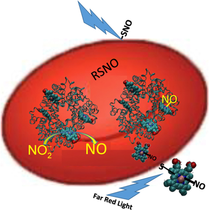
Background: Nitrite is reduced by heme-proteins and molybdenum-containing enzymes to form the important signaling molecule nitric oxide (NO), mediating NO signaling. Substantial evidence suggests that deoxygenated hemoglobin within red blood cells (RBCs) is the main erythrocytic protein responsible for mediating nitrite-dependent NO signaling. In other work, infrared and far red light have been shown to have therapeutic potential that some attribute to production of NO. Here we explore whether a combination of nitrite and far red light treatment has an additive effect in NO-dependent processes, and whether this effect is mediated by RBCs.

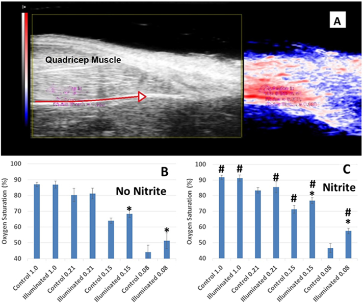
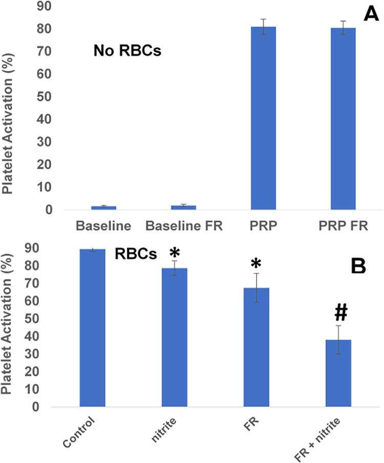
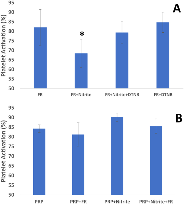
Methods and results: Using photoacoustic imaging in a rat model as a function of varying inspired oxygen, we found that far red light (660 nm, five min. exposure) and nitrite feeding (three weeks in drinking water at 100 mg/L) each separately increased tissue oxygenation and vessel diameter, and the combined treatment was additive. We also employed inhibition of human platelet activation measured by flow cytometry to assess RBC-dependent nitrite bioactivation and found that far red light dramatically potentiates platelet inhibition by nitrite. Blocking RBC-surface thiols abrogated these effects of nitrite and far-red light. RBC-dependent production of NO was also shown to be enhanced by far red light using a chemiluminescence-based nitric oxide analyzer. In addition, RBC-dependent bioactivation of nitrite led to prolonged lag times for clotting in platelet poor plasma that was enhanced by exposure to far red light.

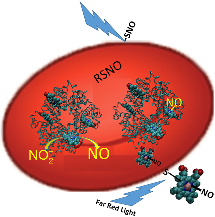
Conclusions: Our results suggest that nitrite leads to the formation of a photolabile RBC surface thiol-bound species such as an S-nitrosothiol or heme-nitrosyl (NO-bound heme) for which far red light enhances NO signaling. These findings expand our understanding of RBC-mediated NO production from nitrite. This pathway of NO production may have therapeutic potential in several applications including thrombosis, and, thus, warrants further study.

Keywords: Hemoglobin; Light therapy; Nitric oxide; Nitrite; Photobiomodulation; Red blood cells.


Figures

fx1
Graphical abstract
Fig. 1
Fig. 1
Microvascular oxygen saturation measurements obtained from photoacoustic imaging in rats. Oxygen saturation was measured using photoacoustic imaging while rats inhaled different fractions of inspired oxygen (FiO2). (A) Representative images. Left: standard 2D B-mode ultrasound image. Right: co-registered photoacoustic image with setting optimized to the depth of that vessel and tissues above it. The red arrow in the 2D B-mode indicates the course of the femoral artery. Intense red indicates high oxygen saturation; blue indicates low saturation. (B) Effects of far red light (660 nm, 150 mW, 1 cm spot size, 5 min) in control-fed rats. Tissue oxygenation is shown without illumination (blue bars) by far red light adjacent to that with far red light (red bars) for each FiO2. n = 8, *p < 0.05 illuminated vs not illuminated. Data are plotted as mean ± one standard deviation. (C) Effects of far red light in nitrite-fed rats. n = 8, *p < 0.05 illuminated vs not illuminated, #P < 0.05 nitrite vs no nitrite. In two-way Anova, P < 0.0001 nitrite vs no nitrite. Data are plotted as mean ± one standard deviation.
Fig. 2
Fig. 2
Red blood cell bioactivation of nitrite is enhanced by far red light. (A) Platelet activation in the absence of RBCs. Platelets in platelet rich plasma (PRP) were activated with 1 μM ADP compared to baseline. Exposure to far red light (FR, 660 nm, 560 mW, 5 min) had no effect on baseline activation or PRP with ADP. n = 9. (B) Platelet activation in the presence of deoxygenated RBCs. Nitrite (10 μM) reduced platelet activation in the presence of deoxygenated RBCs (15% Hct, *P < 0.02, paired t-test, n = 9). Illumination with far red light also reduced platelet activation compared to control (*P < 0.02, paired t-test, n = 9). Data are plotted as mean ± one standard error. The combination of light and nitrite treatments further reduced platelet activation (#P < 0.001, paired t-test FR + nitrite vs FR, n = 9). Data are plotted as mean ± one standard error.
Fig. 3
Fig. 3
Mechanistic aspects of effects of nitrite and light. (A) Effect of blocking surface thiols on effects of nitrite and far red light. Platelets were activated with 1 μM ADP yielding 84.2 ± 6.8% activation (bar labeled PRP). Addition of deoxygenated RBCs (deoxyRBC) and five minutes of exposure to far red light (FR, 660 nm, 150 mW) did not significantly affect activation. However, combination of nitrite (10 μM) and light reduced platelet activation. When the RBCs were pre-incubated with DTNB (2.5 mM, one hour), inhibition of platelet activation was abrogated (n = 5, *P < 0.01 for FR+nitrite vs any other condition shown). Data are plotted as mean ± one standard deviation. (B) Effects of light and nitrite on platelet activation in the absence of RBCs. Platelets were activated by 1 μM ADP (PRP). Illumination with far red light (FR, 660 nm, 5 min), 10 μM nitrite, or the combination of the two had no significant effect on platelet activation (n = 3). Data are plotted as mean ± one standard deviation.
Fig. 4
Fig. 4
Nitric oxide production by nitrite and red blood cells. Deoxygenated RBCs (50% Hct, 100 µL) were injected into 20 ml of PBS (pH 6.8) containing 20 mM nitrite in a three neck round bottom flask that was connected directly to a chemiluminescence-based nitric oxide analyzer with and without illumination by far red light (660 nm, 65 mW). Data are plotted as mean ± one standard error. (A) Representative raw data from the nitric oxide analyzer. RBCs were injected at 5 min and the signal increases more with light than without (indicated by arrow). (B) Average areas of the signals showing greater NO production in the presence of light (P = 0.04, n = 8).
Fig. 5
Fig. 5
Effects of nitrite and far red light on clotting. Platelet poor plasma containing fibrinogen and an activation solution containing thrombin were prepared separately and, in some cases, incubated with deoxygenated RBCs (20% Hct), nitrite (10 μM), and/or treated with far red light (660 nm, 10 min, 107 mW). All experiments were performed pair-wise; that is two types of samples were performed side by side per day and the results compared. Data are plotted as mean ± one standard error. (A) Representative plots of optical density (OD) vs time after initiation of clotting, shown here for the case of a control (CNT, RBCs but no other treatment) vs a sample administered nitrite and light treatment (Nit + Lit). Except for the nitrite and light treatment, both samples were treated identically. (B) Average lag times and standard errors for repeated measures. In the absence of RBCs, nitrite had no effect on the lag time (control (CNT) vs Nit, P = 0.78, n = 5). When mixtures were pre-incubated with RBCs, nitrite treatment resulted in prolonged lag times compared to no nitrite treatment (CNT vs NIT, *P < 0.01, n = 8). Illumination with far red light (660 nm, 10 min) along with the nitrite treatment during pre-incubation with deoxygenated RBCs also prolonged lag time compared to when the nitrite and light treatments were both absent (CNT vs Nit+ FR, *P < 0.01, n = 5). Inclusion of the illumination treatment along with the nitrite treatment prolonged lag times compared to nitrite treatment alone (Nit vs Nit +FR, *P < 0.05, n = 10).
Fig. 6
Fig. 6
Potential mechanism of RBC-mediated nitrite bioactivation and its potentiation by far red light. Nitrite reacts with deoxygenated Hb to make NO which then binds other vacant hemes or forms a nitrosothiol (RSNO). The nitrosyl-heme or nitrosothiol is exported and binds a surface thiol. Another potential NO species that may form is a DNIC (not shown). The NO congener can then be transported in plasma and interact with platelets and other blood components and this action is potentiated by photolysis using far red light.

References

    1. Ignarro L.J., Byrns R.E., Buga G.M., Wood K.S. Endothelium-derived relaxing factor from pulmonary-artery and vein possesses pharmacological and chemical-properties identical to those of nitric-oxide radical. Circ. Res. 1987;61(6):866–879. - PubMed
    1. Palmer R.M.J., Ferrige A.G., Moncada S. Nitric-oxide release accounts for the biological-activity of endothelium-derived relaxing factor. Nature. 1987;327(6122):524–526. - PubMed
    1. Furchgott R.F. Studies on relaxation of rabbit aorta by sodium nitrite: the basis for the proposal that the acid- activatable factor from bovine retractor penis is inorganic nitrite and the endothelium-derived relaxing factor is nitric oxide. In: Vanhoutte P.M., editor. Vasodilatation: Vascular Smooth Muscle, Peptides, Autonomic Nerves, and Endothelium. Raven Press; New York: 1988. pp. 401–414.
    1. Ignarro L.J., Byrns R.E., Wood K.S. Biochemical and pharmacological properties of endothelium-derived relaxing factor and its similarity to nitric oxide radical. In: Vanhoutte P.M., editor. Vasodilatation: Vascular Smooth Muscle, Peptides, Autonomic Nerves, and Endothelium. Raven Press; New York: 1988. pp. 427–435.
    1. Loscalzo J. Nitric oxide insufficiency, platelet activation, and arterial thrombosis. Circ. Res. 2001;88(8):756–762. - PubMed

Publication types

LinkOut - more resources