

Gold is for the mistress, silver for the maid: Enhanced mechanical properties, osteoinduction and antibacterial activity due to iron doping of tricalcium phosphate bone cements
- PMID: 30423766
- PMCID: PMC6366449
- DOI: 10.1016/j.msec.2018.10.028
Gold is for the mistress, silver for the maid: Enhanced mechanical properties, osteoinduction and antibacterial activity due to iron doping of tricalcium phosphate bone cements
Abstract
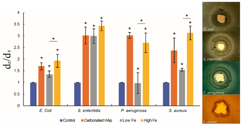
Self-hardening calcium phosphate cements present ideal bone tissue substitutes from the standpoints of bioactivity and biocompatibility, yet they suffer from (a) weak mechanical properties, (b) negligible osteoinduction without the use of exogenous growth factors, and (c) a lack of intrinsic antibacterial activity. Here we attempt to improve on these deficiencies by studying the properties of self-setting Fe-doped bone-integrative cements containing two different concentrations of the dopant: 0.49 and 1.09 wt% Fe. The hardening process, which involved the transformation of Fe-doped β-tricalcium phosphate (Fe-TCP) to nanocrystalline brushite, was investigated in situ by continuously monitoring the cements using the Energy Dispersive X-Ray Diffraction technique. The setting time was 20 min and the hardening time 2 h, but it took 50 h for the cement to completely stabilize compositionally and mechanically. Still, compared to other similar systems, the phase transformation during hardening was relatively fast and it also followed a relatively simple reaction path, virtually free of complex intermediates and noisy background. Mössbauer spectrometry demonstrated that 57Fe atoms in Fe-TCP were located in two non-equivalent crystallographic sites and distributed over positions with a strong crystal distortion. The pronounced presence of ultrafine crystals in the final, brushite phase contributed to the reduction of the porosity and thereby to the enhancement of the mechanical properties. The compressive strength of the hardened TCP cements increased by more than twofold when Fe was added as a dopant, i.e., from 11.5 ± 0.5 to 24.5 ± 2.0 MPa. The amount of iron released from the cements in physiological media steadied after 10 days and was by an order of magnitude lower than the clinical threshold that triggers the toxic response. The cements exhibited osteoinductive activity, as observed from the elevated levels of expression of genes encoding for osteocalcin and Runx2 in both undifferentiated and differentiated MC3T3-E1 cells challenged with the cements. The osteoinductive effect was inversely proportional to the content of Fe ions in the cements, indicating that an excessive amount of iron can have a detrimental effect on the induction of bone growth by osteoblasts in contact with the cement. In contrast, the antibacterial activity of the cement in the agar assay increased against all four bacterial species analysed (E. coli, S. enteritidis, P. aeruginosa, S. aureus) in direct proportion with the concentration of Fe ions in it, indicating their key effect on the promotion of the antibacterial effect in this material. This effect was less pronounced in broth assays. Experiments involving co-incubation of cements with cells in an alternate magnetic radiofrequency field for 30 min demonstrated a good potential for the use of these magnetic cements in hyperthermia cancer therapies. Specifically, the population of human glioblastoma cells decreased six-fold at the 24 h time point following the end of the magnetic field treatment, while the population of the bone cancer cells dropped approximately twofold. The analysis of the MC3T3-E1 cell/cement interaction reiterated the effects of iron in the cement on the bone growth marker expression by showing signs of adverse effects on the cell morphology and proliferation only for the cement containing the higher concentration of Fe ions (1.09 wt%). Biological testing concluded that the effects of iron are beneficial from the perspective of a magnetic hyperthermia therapy and antibacterial prophylaxis, but its concentration in the material must be carefully optimized to avoid the adverse effects induced above a certain level of iron concentrations.
Keywords: Bone cement; Escherichia coli; Hardening behaviour; Iron-doped tricalcium phosphate; Osteoblastic MC3T3-E1; Pseudomonas aeruginosa; Salmonella enteritidis; Staphylococcus aureus.
Copyright © 2018 Elsevier B.V. All rights reserved.
Conflict of interest statement
The authors declare no competing interests.
Figures


References
-
- Ambard AJ, Mueninghoff L, Calcium phosphate cement: review of mechanical and biological properties, J Prosthodont. 15 (2006) 321–328. - PubMed
-
- Zhang J, Liu W, Schnitzler V, Tancret F, Bouler JM, Calcium phosphate cements for bone substitution: chemistry, handling and mechanical properties, Acta Biomater. 10 (2014) 1035–49. - PubMed
-
- Luo J, Ajaxon I, Ginebra MP, Engqvist H, Persson C, Compressive, diametral tensile and biaxial flexural strength of cutting-edge calcium phosphate cements, Journal of the mechanical behavior of biomedical materials 60 (2016) 617–627. - PubMed
-
- Charriére E, Terrazzoni S, Pittet C, Mordasini Ph., Dutoit M, Lemaître J, Zysset Ph., Mechanical characterization of brushite and hydroxyapatite cements, Biomaterials 22 (2001) 2937–2945. - PubMed
MeSH terms
Substances
Grants and funding
LinkOut - more resources
Full Text Sources
Medical
