

Ursolic Acid Attenuates Hepatic Steatosis, Fibrosis, and Insulin Resistance by Modulating the Circadian Rhythm Pathway in Diet-Induced Obese Mice
- PMID: 30423963
- PMCID: PMC6266464
- DOI: 10.3390/nu10111719
Ursolic Acid Attenuates Hepatic Steatosis, Fibrosis, and Insulin Resistance by Modulating the Circadian Rhythm Pathway in Diet-Induced Obese Mice
Abstract
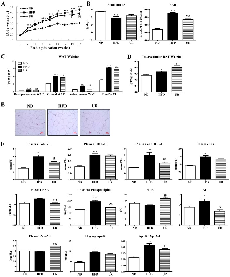
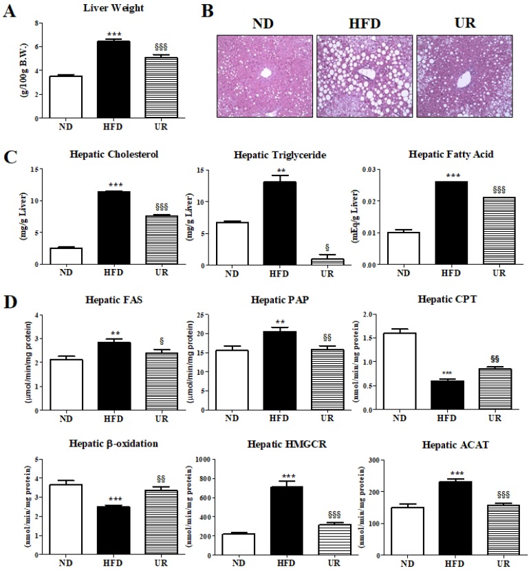
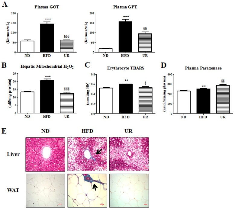
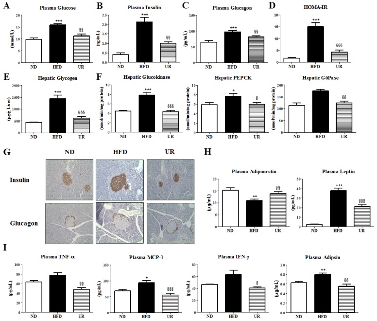
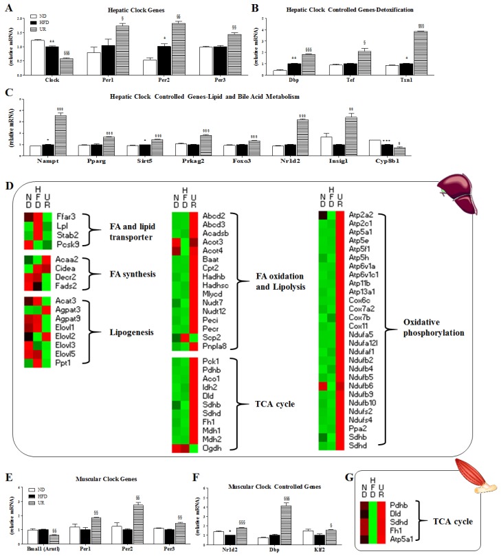
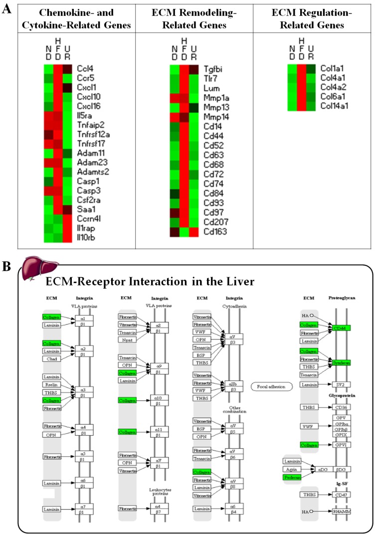
The aim of the current study was to elucidate the effects of long-term supplementation with dietary ursolic acid (UR) on obesity and associated comorbidities by analyzing transcriptional and metabolic responses, focusing on the role of UR in the modulation of the circadian rhythm pathway in particular. C57BL/6J mice were divided into three groups and fed a normal diet, high-fat diet, or high-fat + 0.05% (w/w) UR diet for 16 weeks. Oligonucleotide microarray profiling revealed that UR is an effective regulator of the liver transcriptome, and canonical pathways associated with the "circadian rhythm" and "extracellular matrix (ECM)⁻receptor interactions" were effectively regulated by UR in the liver. UR altered the expression of various clock and clock-controlled genes (CCGs), which may be linked to the improvement of hepatic steatosis and fibrosis via lipid metabolism control and detoxification enhancement. UR reduced excessive reactive oxygen species production, adipokine/cytokine dysregulation, and ECM accumulation in the liver, which also contributed to improve hepatic lipotoxicity and fibrosis. Moreover, UR improved pancreatic islet dysfunction, and suppressed hepatic gluconeogenesis, thereby reducing obesity-associated insulin resistance. Therapeutic approaches targeting hepatic circadian clock and CCGs using UR may ameliorate the deleterious effects of diet-induced obesity and associated complications such as hepatic fibrosis.
Keywords: circadian rhythm; extracellular matrix; fibrosis; liver-specific; ursolic acid.
Conflict of interest statement
The authors declare no conflict of interest.
Figures
Similar articles
-
Dietary Eriodictyol Alleviates Adiposity, Hepatic Steatosis, Insulin Resistance, and Inflammation in Diet-Induced Obese Mice.Int J Mol Sci. 2019 Mar 11;20(5):1227. doi: 10.3390/ijms20051227. Int J Mol Sci. 2019. PMID: 30862092 Free PMC article.
-
Phlorizin Supplementation Attenuates Obesity, Inflammation, and Hyperglycemia in Diet-Induced Obese Mice Fed a High-Fat Diet.Nutrients. 2016 Feb 16;8(2):92. doi: 10.3390/nu8020092. Nutrients. 2016. PMID: 26891322 Free PMC article.
-
Effect of Green Tea Extract on Systemic Metabolic Homeostasis in Diet-Induced Obese Mice Determined via RNA-Seq Transcriptome Profiles.Nutrients. 2016 Oct 14;8(10):640. doi: 10.3390/nu8100640. Nutrients. 2016. PMID: 27754422 Free PMC article.
-
Adipocytokines and hepatic fibrosis.Trends Endocrinol Metab. 2015 Mar;26(3):153-61. doi: 10.1016/j.tem.2015.01.002. Epub 2015 Feb 2. Trends Endocrinol Metab. 2015. PMID: 25656826 Free PMC article. Review.
-
Circadian Rhythms in the Pathogenesis and Treatment of Fatty Liver Disease.Gastroenterology. 2020 May;158(7):1948-1966.e1. doi: 10.1053/j.gastro.2020.01.050. Epub 2020 Feb 13. Gastroenterology. 2020. PMID: 32061597 Free PMC article. Review.
Cited by
-
Current Perspective on the Role of the Circadian Clock and Extracellular Matrix in Chronic Lung Diseases.Int J Environ Res Public Health. 2023 Jan 30;20(3):2455. doi: 10.3390/ijerph20032455. Int J Environ Res Public Health. 2023. PMID: 36767821 Free PMC article. Review.
-
Therapeutic potential of bioactive phytoconstituents found in fruits in the treatment of non-alcoholic fatty liver disease: A comprehensive review.Heliyon. 2023 Apr 10;9(4):e15347. doi: 10.1016/j.heliyon.2023.e15347. eCollection 2023 Apr. Heliyon. 2023. PMID: 37101636 Free PMC article. Review.
-
Network Pharmacology and Molecular Docking Reveal the Mechanism of Isodon ternifolius (D. Don) Kudo Against Liver Fibrosis.Drug Des Devel Ther. 2023 Aug 7;17:2335-2351. doi: 10.2147/DDDT.S412818. eCollection 2023. Drug Des Devel Ther. 2023. PMID: 37576085 Free PMC article.
-
Underlying Molecular Mechanism and Construction of a miRNA-Gene Network in Idiopathic Pulmonary Fibrosis by Bioinformatics.Int J Mol Sci. 2023 Aug 27;24(17):13305. doi: 10.3390/ijms241713305. Int J Mol Sci. 2023. PMID: 37686108 Free PMC article.
-
Ursolic Acid Inhibits Collagen Production and Promotes Collagen Degradation in Skin Dermal Fibroblasts: Potential Antifibrotic Effects.Biomolecules. 2025 Mar 3;15(3):365. doi: 10.3390/biom15030365. Biomolecules. 2025. PMID: 40149901 Free PMC article.
References
MeSH terms
Substances
Grants and funding
LinkOut - more resources
Full Text Sources
Medical
Molecular Biology Databases
