Review
. 2018 Nov 9;19(11):3534.
doi: 10.3390/ijms19113534.

Structure and Physiological Regulation of AMPK

Affiliations
Review

Structure and Physiological Regulation of AMPK

Yan Yan et al. Int J Mol Sci. .

Abstract

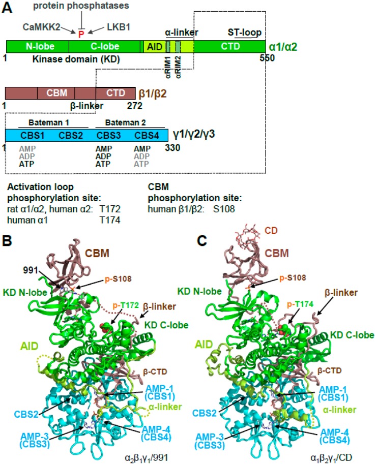
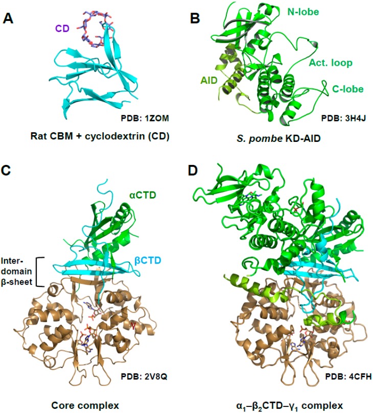
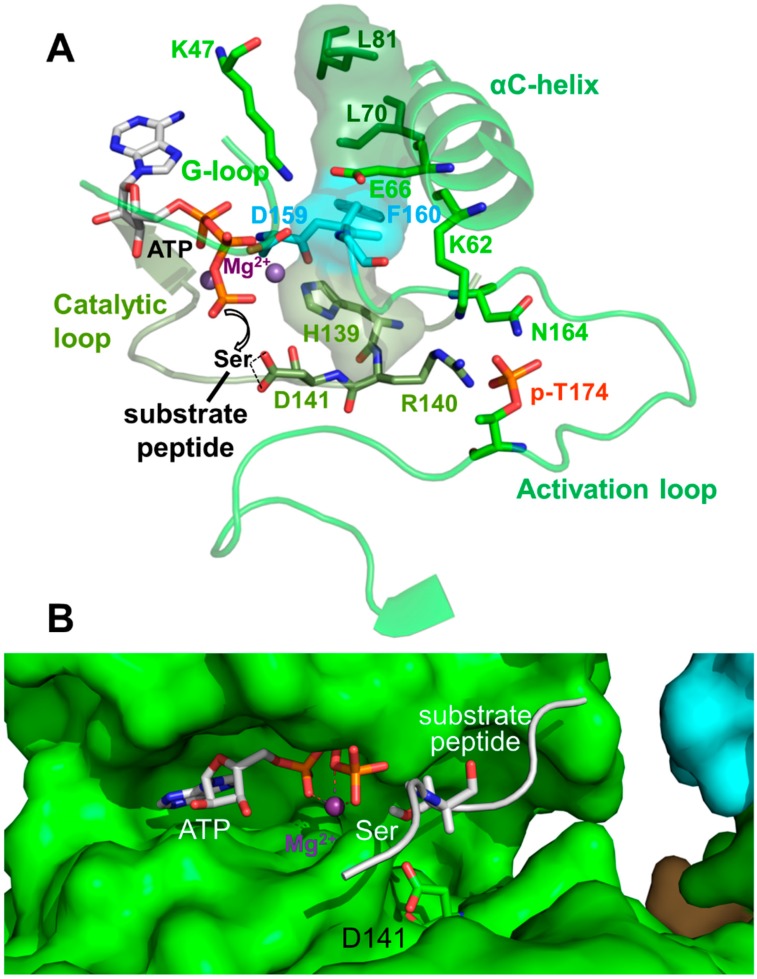
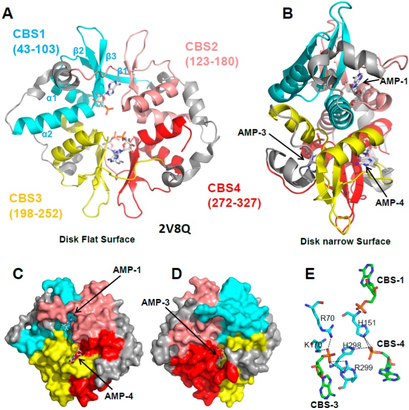
Adenosine monophosphate (AMP)-activated protein kinase (AMPK) is a heterotrimeric αβγ complex that functions as a central regulator of energy homeostasis. Energy stress manifests as a drop in the ratio of adenosine triphosphate (ATP) to AMP/ADP, which activates AMPK's kinase activity, allowing it to upregulate ATP-generating catabolic pathways and to reduce energy-consuming catabolic pathways and cellular programs. AMPK senses the cellular energy state by competitive binding of the three adenine nucleotides AMP, ADP, and ATP to three sites in its γ subunit, each, which in turn modulates the activity of AMPK's kinase domain in its α subunit. Our current understanding of adenine nucleotide binding and the mechanisms by which differential adenine nucleotide occupancies activate or inhibit AMPK activity has been largely informed by crystal structures of AMPK in different activity states. Here we provide an overview of AMPK structures, and how these structures, in combination with biochemical, biophysical, and mutational analyses provide insights into the mechanisms of adenine nucleotide binding and AMPK activity modulation.

Keywords: AID; AMPK; CBS; CaMKK2; LKB1; activation loop; energy metabolism; α-linker; αRIM; β-linker.


Conflict of interest statement

The authors declare no conflict of interest. The funders had no role in the design of the study; in the collection, analyses, or interpretation of data; in the writing of the manuscript, or in the decision to publish the results.

Figures

Figure 1
Figure 1
Overall structure of human adenosine monophosphate (AMP)-activated protein kinase (AMPK). (A). Domain structure and AMPK isoforms. Activation loop and carbohydrate-binding module (CBM) phosphorylation sites of different isoforms are indicated below the domain map (B,C). Crystal structures of phosphorylated, AMP-bound AMPK α2β1γ1/991 ((B); PDB: 4CFE) and α1β2γ1/cyclodextrin (CD) ((C); PDB: 4RER).
Figure 2
Figure 2
Structure of AMPK domains and subcomplexes. (A) Rat CBM bound to cyclodextrin; (B) Fission yeast kinase domain–autoinhibitory domain (KD‒AID) complex; (C) AMP-bound, phosphorylated mammalian AMPK core complex (rat α1-human β2-rat γ1); (D) AMP-bound, phosphorylated rat α1—human β2CTD—rat γ1 complex.
Figure 3
Figure 3
Active protein kinase catalytic cleft. (A) Key residues and structural elements of phosphorylated AMP-bound α1β2γ1 AMPK (4RER). Active kinase structures are characterized by a precisely positioned set of motifs for substrate- and adenosine triphosphate (ATP)-binding, in which four residues (L70, L81, H139, F160; shown in stick plus translucent surface presentation) are stacked against each other to form a regulatory spine. In this conformation, the activation loop p-T174 (p-T172 in human α2) positions R140 and D141 from the catalytic loop for peptide substrate binding, and K62 from the αC-helix for aligning the ATP-binding K47 and the Mg2+-binding DFG loop. The AMPK active protein kinase cleft resembles the canonical protein kinase A (PKA) site. To better visualize the active structure, we modeled the serine residue of a substrate peptide and the co-substrate ATP from the structure of PKA (PDB: 1ATP) in the catalytic cleft. Spheres: Mg2+ ions. (B) Surface presentation of the AMPK catalytic cleft (4RER) overlaid with a stick model of the aligned substrate peptide and ATP from the structure of substrate-bound CDK2 (PDB: 1QMZ). The Ser hydroxyl-positioning AMPK D141 is shown in green stick representation.
Figure 4
Figure 4
AMP binds three of the four CBS sites of the γ subunit. (A,B) Cartoon representation of the γ subunit in two different orientations. AMP molecules are shown in stick representation. The four CBS sites are shown in different colors with the secondary structure elements of CBS1 labeled. (C,D) Surface representation of the front and back sides of the disk flat surfaces illustrating the AMP-occupied binding pockets 1, 3, and 4, and the empty CBS2 pocket. (E) The phosphate groups (orange) of the three AMP molecules (cyan C atoms) coordinately interact with a set of polar γ subunit residues (green C atoms); O: red, N: blue.
Figure 5
Figure 5
The AID is in equilibrium between KD- and γ-bound conformations. (A) Cartoon structure of the human α1 KD-AID complex. (B) Overlay of the inactive KD-AID structure with the structure of active holo-AMPK (α subunit: green; β- and γ-subunits: grey). The arrow indicates the repositioning of the AID in the active structure. (C,D) Catalytic center of the inactive (C) and active (D) AMPK conformation. Stick plus translucent surface presentations indicate the regulatory spine residues L70, L81, H139, and F160. Mg2+-ATP was modeled into both structures for orientation, even though it cannot bind to the inactive structure shown in panel C. Spheres: Mg2+ ions.
Figure 6
Figure 6
αRIM2/CBS3 and AID-αRIM1/CBS2 interactions are linked. Structure of human AMP-bound AMPK α1β2γ1 (4RER) with key residues shown in a stick presentation; the α-linker is shown in magenta, the γ subunit in cyan, and the AID in light green. AMP bound at CBS3 and αRIM2 E364 directly interact with γ1 K170, which positions the αRIM1-binding residues R171, and indirectly through K174 and F175, F179, thus stabilizing the AID‒γ subunit interaction. Consistently, mutations of the αRIM1/γ subunit (and αRIM2/CBS3) interface residues highlighted by oval outlines (human α1: F342D/Y343D, I335D/M336D, E364, R365; γ1: R171A, F179D) are constitutively AMP-non-responsive. Dashed lines indicate hydrogen bonds.
Figure 7
Figure 7
The β-CTD binds and stabilizes the activation loop. Structure of AMP-bound, phosphorylated AMPK α1–β2CTD–γ1 (PDB: 4CFH). The activation loop is highlighted in orange, and p-T172 is shown in sphere presentation.

References

    1. Yuan H.X., Xiong Y., Guan K.L. Nutrient sensing, metabolism, and cell growth control. Mol. Cell. 2013;49:379–387. doi: 10.1016/j.molcel.2013.01.019. - DOI - PMC - PubMed
    1. Garcia D., Shaw R.J. AMPK: Mechanisms of Cellular Energy Sensing and Restoration of Metabolic Balance. Mol. Cell. 2017;66:789–800. doi: 10.1016/j.molcel.2017.05.032. - DOI - PMC - PubMed
    1. Hardie D.G. AMP-activated protein kinase: An energy sensor that regulates all aspects of cell function. Genes Dev. 2011;25:1895–1908. doi: 10.1101/gad.17420111. - DOI - PMC - PubMed
    1. Hardie D.G. Keeping the home fires burning: AMP-activated protein kinase. J. R. Soc. Interface. 2018;15:20170774. doi: 10.1098/rsif.2017.0774. - DOI - PMC - PubMed
    1. Steinberg G.R., Kemp B.E. AMPK in Health and Disease. Physiol. Rev. 2009;89:1025–1078. doi: 10.1152/physrev.00011.2008. - DOI - PubMed

Substances

LinkOut - more resources