Review
. 2018 Oct 18;9(10):529.
doi: 10.3390/mi9100529.

Tunable Adhesion for Bio-Integrated Devices

Affiliations
Review

Tunable Adhesion for Bio-Integrated Devices

Zhaozheng Yu et al. Micromachines (Basel). .

Abstract

With the rapid development of bio-integrated devices and tissue adhesives, tunable adhesion to soft biological tissues started gaining momentum. Strong adhesion is desirable when used to efficiently transfer vital signals or as wound dressing and tissue repair, whereas weak adhesion is needed for easy removal, and it is also the essential step for enabling repeatable use. Both the physical and chemical properties (e.g., moisture level, surface roughness, compliance, and surface chemistry) vary drastically from the skin to internal organ surfaces. Therefore, it is important to strategically design the adhesive for specific applications. Inspired largely by the remarkable adhesion properties found in several animal species, effective strategies such as structural design and novel material synthesis were explored to yield adhesives to match or even outperform their natural counterparts. In this mini-review, we provide a brief overview of the recent development of tunable adhesives, with a focus on their applications toward bio-integrated devices and tissue adhesives.

Keywords: bio-integrated devices; dry/wet conditions; soft biological tissue; tissue adhesives; tunable adhesion.


Conflict of interest statement

The authors declare no competing financial interests.

Figures
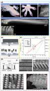
Figure 1
Figure 1
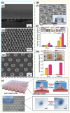
Gecko-inspired dry adhesive. (A) (i) A close-up view of a gecko climbing a glass wall and (ii) mesostructure of gecko toe pad with (iii) microscale array of high-aspect-ratio setae structure. (iv) Cryo-SEM view of a single seta (ca. 110 µm in length and 4.2 µm in diameter) that branches at the tips into 100–1000 more structures known as spatulas. Reproduced with permission from Reference [56]; Copyright 2006, The Company of Biologists. (B) Schematic illustrations of adhesives with (i) a relatively flat surface and (ii) an array of fibrils in contact with a rough surface. Reproduced with permission from Reference [49]; Copyright 2010, John Wiley and Sons. (C) Dependence of the terminal element density (NA) of the attachment pads on the body mass (m) in hairy-pad systems of diverse animal groups. The red line that fits all data corresponds to the self-similarity criterion with a slope of ~0.67. The green lines (slope of ~0.33) correspond to the model with a curvature invariance of contacts with radius R. The blue line shows the approximate limit of maximum contact for such attachment devices. Reproduced with permission from Reference [47]; Copyright 2003, United States National Academy of Sciences. (D) (i) A schematic showing the mechanism of a plasma sheath with a Faraday cage, where ions are incident on the substrate surface in a direction normal to the grid plane for an angled etching in the silicon substrate. (ii) SEM images of polySi etch profiles with angles of 30°/45°/60°, and (iii) polyurethane acrylate (PUA) nano-hairs formed on a poly(ethylene terephthalate) (PET) film substrate with a slanted angle of 60°. (iv) SEM image of two-level hierarchical PUA hairs with well-defined high aspect ratio over a large area using two-step ultraviolet (UV)-assisted capillary force lithography; the inset shows angled nano-hairs on the tip. Reproduced with permission from Reference [9]; Copyright 2009, United States National Academy of Sciences.
Figure 2
Figure 2
Octopus-inspired adhesive. (A) SEM images of (i) mushroom-like structures consisting of silica caps and polymer stems on a silicon wafer prepared by plasma etching; (ii) a polyvinyl alcohol (PVA) composite film embedding silica nanoparticles; (iii) a polydimethylsiloxane (PDMS) nano-sucker array obtained from the PVA composite mold. Reproduced with permission from Reference [84]; Copyright 2017, American Chemical Society. (B) (i) SEM image and cross-sectional optical image of micro-suckers (100 µm in diameter and 75 µm in height); (ii) dry/wet adhesion strength increases as the curvature of the cross-sectional profile increases; (iii) pull-off strength for PDMS-based micro-suckers and a non-patterned PDMS patch against a rough pigskin, with dry and moist pigskins shown in the inset. Reproduced with permission from Reference [85]; Copyright 2018, John Wiley and Sons. (C) Schematic representation and SEM image (scale bar: 10 μm) of micro-cavity arrays within a smart adhesive pad and its corresponding switchable adhesion mechanism (i.e., temperature-dependent hydrogel actuation in response to the environmental temperature). The inset in the bottom right shows SEM images of a single hole on the smart adhesive pad at a low temperature (left, T ≈ 3 °C) and a high temperature (right, T ≈ 40 °C) (scale bar: 500 nm). Reproduced with permission from Reference [85]; Copyright 2018, John Wiley and Sons.
Figure 3
Figure 3
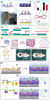
Mussel-inspired wet adhesives. (A) (i) PDMS casting onto the master is followed by curing, and lift-off results in gecko-foot-mimetic nano-pillar arrays. Next, the fabricated nanopillars are coated with a mussel-adhesive-protein-mimetic polymer that contains catechols, a key component of wet adhesive proteins found in mussel holdfasts. (ii) Adhesion force per pillar for gecko and geckel in water (red) and air (black). (iii) Performance of geckel adhesive during multiple contact cycles in water (red) and air (black) with error bars to represent the standard deviation. Reproduced with permission from Reference [7]; Copyright 2007, Nature Publishing Group. (B) Schematic illustrations of alkanethiol monolayer and poly(ethylene glycol) (PEG) polymer grafting, as well as the glycosaminoglycan hyaluronic acid (HA) conjugation to polydopamine (PDA)-coated surfaces. Reproduced with permission from Reference [7]; Copyright 2007, American Association for the Advancement of Science. (C) Polydopamine decorating carbon nanotubes (PDA-CNTs) in water results in PDA-CNT dispersion. Copolymerizing acrylamide (AM) and acrylic acid (AA) monomers in the PDA-CNT dispersion forms a glycerol–water (GW) hydrogel in a glycerol–water binary solvent. Frostbite and burn models on rats’ back skin demonstrate anti-freezing and anti-burning properties of the adhesive GW-hydrogel. Reproduced with permission from Reference [102]; Copyright 2018, John Wiley and Sons. (D) (i) Schematic diagram showing the specific procedure to prepare the wet adhesive, where dip-coating an adhesive guest copolymer poly(3,4-dihydroxy-l-phenylalanine) (pDOPA)/adamantine (AD)/methoxyethyl acrylate (MEA) on a clean Si substrate is followed by the self-assembly of host copolymer poly(N-isopropylacrylamide) (pNIPAM)/cyclodextrin (CD) using host–guest molecular recognition. (ii) Schematic drawing showing the tunable wet adhesion that responds to a local temperature trigger. When the local temperature of the adhesive is below lower critical solution temperature (LCST), the pNIPAM easily forms intermolecular hydrogen bonding with adjacent water molecules, and the infused water layer transforms the pNIPAM side chains to a swelling layer, which spatially stabilizes and confines the underlying adhesive moiety, DOPA. On the other hand, heating above the LCST leads to a phase transition and the collapse of pNIPAM-CD chains to form numerous agglomerates, exposing the adhesive group. Reproduced with permission from Reference [10]; Copyright 2017, Nature Publishing Group.
Figure 4
Figure 4
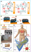
Adhesives on soft tissues. (A) Chondroitin sulfate N-succinimidyl succinate (CS–NHS) reacts with primary amines of both six-arm polyethylene glycol amine PEG–(NH2)6 and proteins of the tissue to form a covalently bonded hydrogel to the tissue. Reproduced with permission from Reference [115]; Copyright 2010, Elsevier. (B) When the buckypaper (BP) comes into contact with the wet biological tissue, water suction leads to deformation of the soft tissue and conformal contact of the BP to the tissue. The resulting water bridge formation and BP–tissue mechanical interlocking help enhance the adhesion during peeling and shearing tests. Reproduced with permission from Reference [116]; Copyright 2013, American Chemical Society. (C) (i) Inking vinylsiloxane (VS) precursor onto uniformly shaped cylindrical microfibers followed by curing vs. directly onto the skin surface. The inset shows the cross-sectional SEM image of an adhesive film attached to an artificial skin replica, where the red dashed line indicates the interface between the skin-adhesive film and artificial skin. (ii) Optical image of a skin-adhesive film attached to the human forearm during the retraction cycle of the adhesion experiment (dashed red line indicates the interfacing border between the adhesive film and the skin; scale bar: 1 cm). (iii) The output signal from a microfibrillar strain sensor mounted onto the radial artery of the wrist; the inset shows a photograph of the strain sensor attached to the wrist through the inking and printing process. Reproduced with permission from Reference [26]; Copyright 2017, John Wiley and Sons. (D) Schematic illustration of the multifunctional electronic patch integrated onto an octopus-inspired adhesive layer with miniaturized suction cups (mSCs). The multifunctional electronic patch is capable of monitoring vital signs from electrodes, strain gauges, and temperature sensors, as well as transdermally delivering drugs loaded in mesoporous silica nanoparticles via iontophoresis. The smart band (connected to the mSC patch) is used to provide wireless functionalities and a power source. Reproduced with permission from Reference [83]; Copyright 2015, John Wiley and Sons.
Figure 5
Figure 5
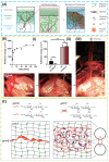
Tough and topological adhesives. (A) Tough adhesives (TAs) consist of a dissipative matrix (light-blue square), made of a hydrogel containing both ionically (calcium; red circles) cross-linked and covalently cross-linked polymers (black and blue lines), and an adhesive surface that contains a bridging polymer with primary amines (green lines). The bridging polymer can penetrate into the TA and the substrate (light-green region) to facilitate covalent-bond formation. In the presence of a crack, the process zone (orange area) dissipates significant amounts of energy as ionic bonds between alginate chains and calcium ions break. (B) (i) Tough adhesives exhibit a rapid increase in adhesion energy to porcine skin over time. (ii) Comparing with cyanoacrylate (CA), TAs showed strong adhesion even when the porcine skin was exposed to blood in the in vitro experiment. n = 4–6. (iii) The TAs were further tested in an in vivo experiment on a beating porcine heart with blood exposure. (A,B) Reproduced with permission from References [105]; Copyright 2017, American Association for the Advancement of Science. (C) Chitosan chains dissolve in water at pH 5 and form a network in water at pH 7. Placing an aqueous solution of chitosan of pH 5 between two hydrogels (or biological tissues) of pH 7 is followed by the diffusion of chitosan chains into the two hydrogels, forming a network that topologically entangles with the networks of both hydrogels. Reproduced with permission from Reference [125]; Copyright 2018, John Wiley and Sons.

Similar articles

Cited by

References

    1. Shull K.R. Contact mechanics and the adhesion of soft solids. Mater. Sci. Eng. R Rep. 2002;36:1–45. doi: 10.1016/S0927-796X(01)00039-0. - DOI
    1. Zhu J., Dexheimer M., Cheng H. Reconfigurable systems for multifunctional electronics. npj Flex. Electron. 2017;1:8. doi: 10.1038/s41528-017-0009-6. - DOI
    1. Choi S., Lee H., Ghaffari R., Hyeon T., Kim D.H. Recent Advances in Flexible and Stretchable Bio-Electronic Devices Integrated with Nanomaterials. Adv. Mater. 2016;28:4203–4218. doi: 10.1002/adma.201504150. - DOI - PubMed
    1. Rogers J.A., Someya T., Huang Y. Materials and mechanics for stretchable electronics. Science. 2010;327:1603–1607. doi: 10.1126/science.1182383. - DOI - PubMed
    1. Kwak M.K., Jeong H.E., Suh K.Y. Rational design and enhanced biocompatibility of a dry adhesive medical skin patch. Adv. Mater. 2011;23:3949–3953. doi: 10.1002/adma.201101694. - DOI - PubMed

LinkOut - more resources