Review
. 2018 Nov 12;6(4):106.
doi: 10.3390/biomedicines6040106.

Alcoholic Liver Disease: Alcohol Metabolism, Cascade of Molecular Mechanisms, Cellular Targets, and Clinical Aspects

Affiliations
Review

Alcoholic Liver Disease: Alcohol Metabolism, Cascade of Molecular Mechanisms, Cellular Targets, and Clinical Aspects

Rolf Teschke. Biomedicines. .

Abstract

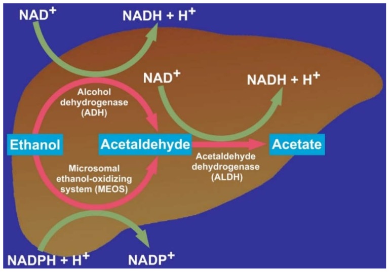
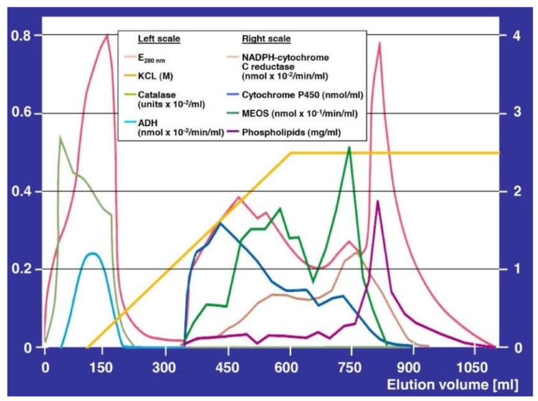
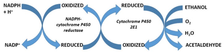
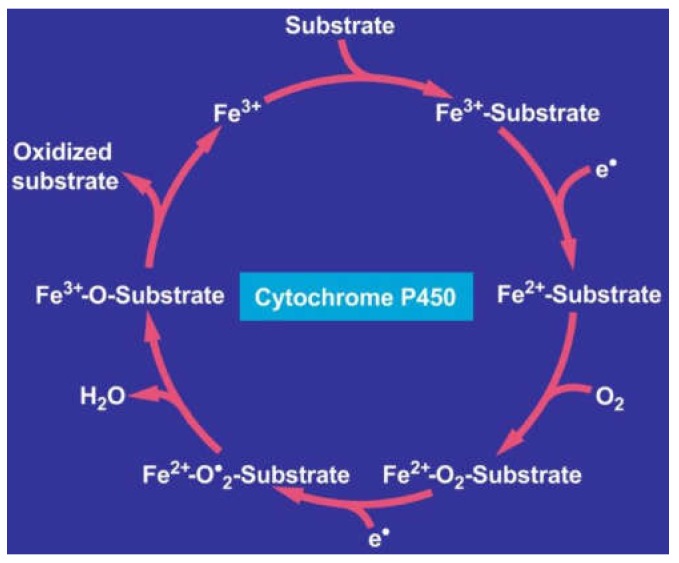
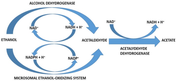
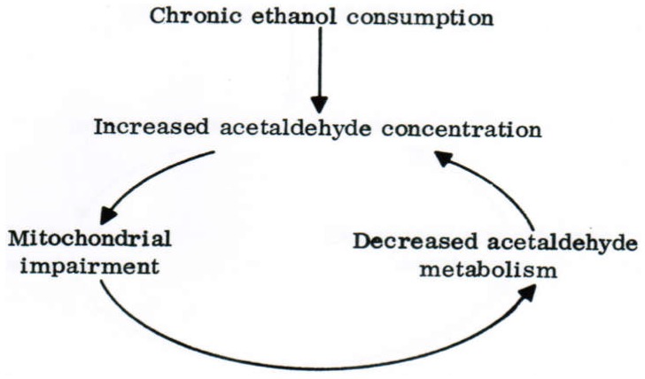
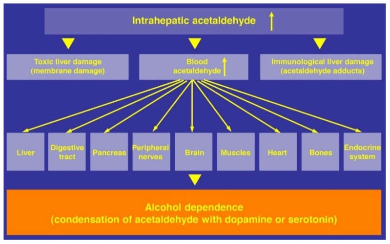
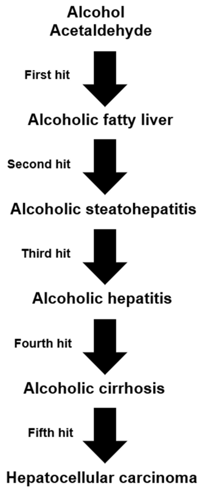
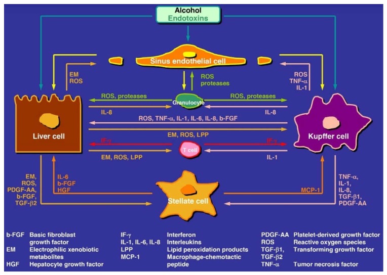
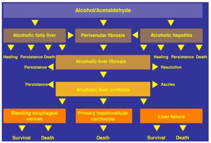
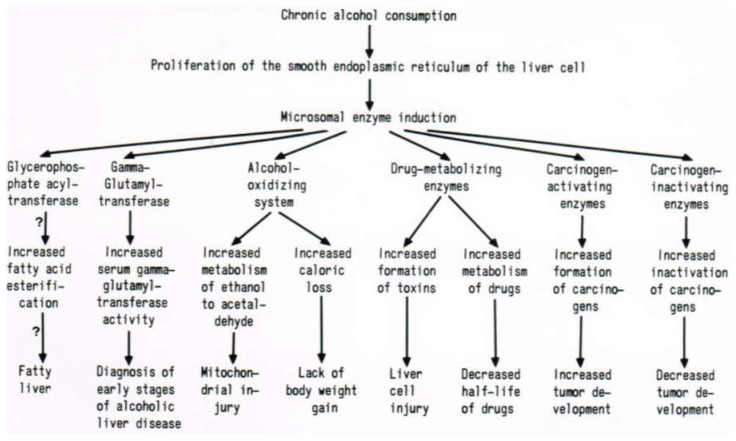
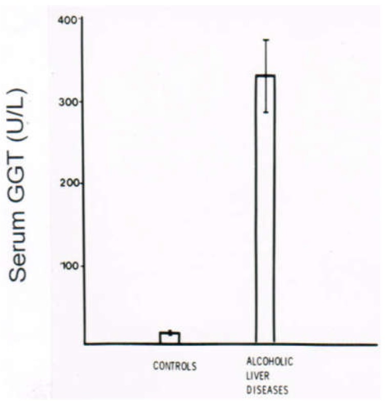
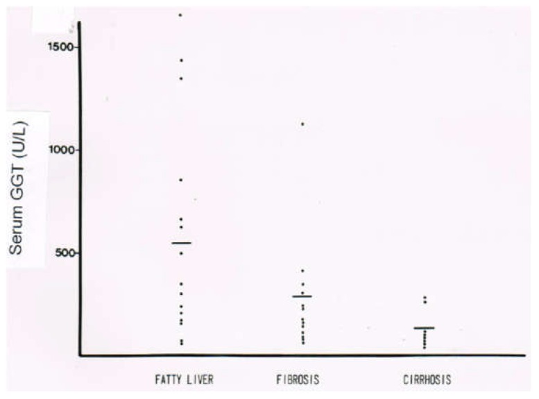
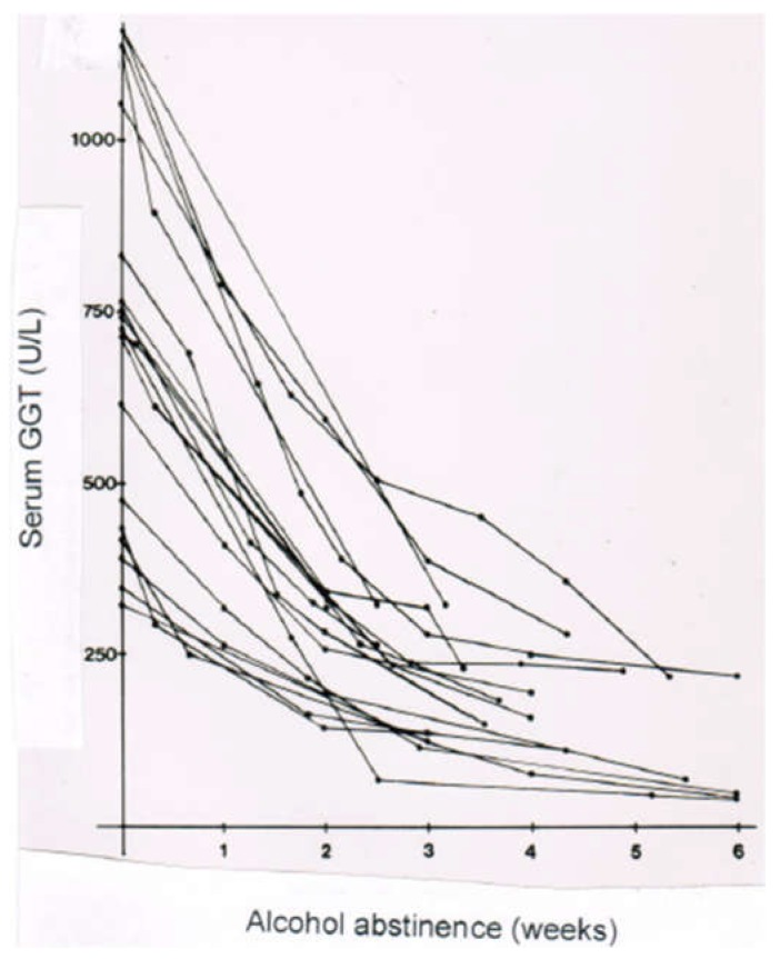
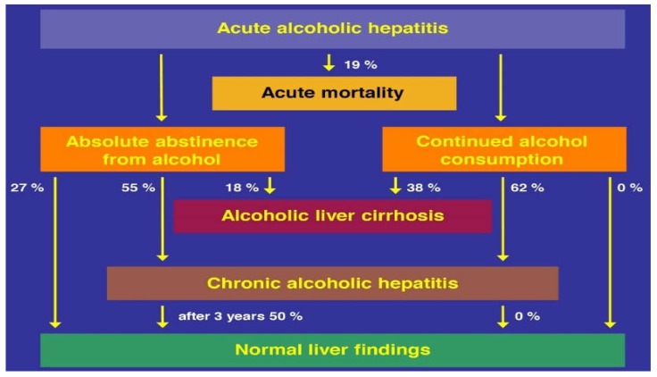
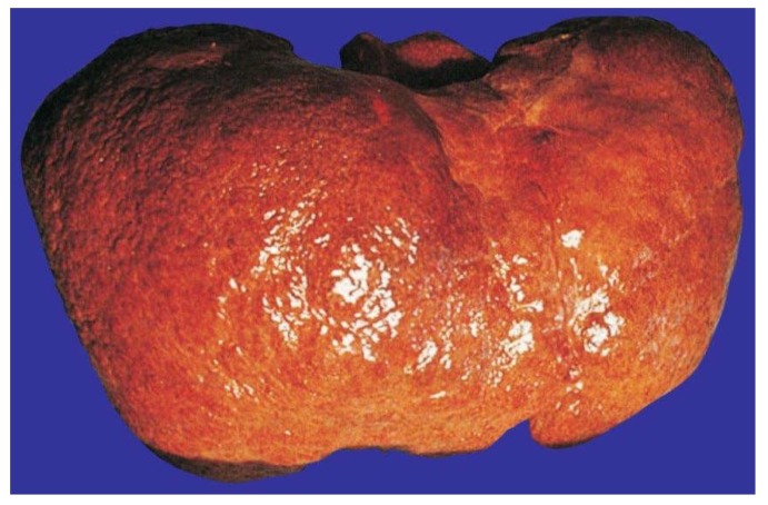
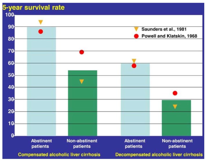
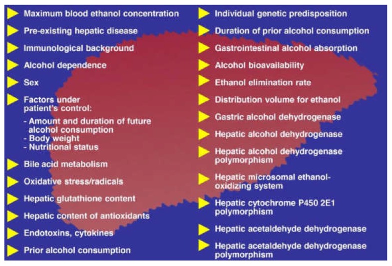
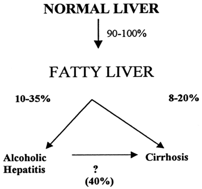
Alcoholic liver disease is the result of cascade events, which clinically first lead to alcoholic fatty liver, and then mostly via alcoholic steatohepatitis or alcoholic hepatitis potentially to cirrhosis and hepatocellular carcinoma. Pathogenetic events are linked to the metabolism of ethanol and acetaldehyde as its first oxidation product generated via hepatic alcohol dehydrogenase (ADH) and the microsomal ethanol-oxidizing system (MEOS), which depends on cytochrome P450 2E1 (CYP 2E1), and is inducible by chronic alcohol use. MEOS induction accelerates the metabolism of ethanol to acetaldehyde that facilitates organ injury including the liver, and it produces via CYP 2E1 many reactive oxygen species (ROS) such as ethoxy radical, hydroxyethyl radical, acetyl radical, singlet radical, superoxide radical, hydrogen peroxide, hydroxyl radical, alkoxyl radical, and peroxyl radical. These attack hepatocytes, Kupffer cells, stellate cells, and liver sinusoidal endothelial cells, and their signaling mediators such as interleukins, interferons, and growth factors, help to initiate liver injury including fibrosis and cirrhosis in susceptible individuals with specific risk factors. Through CYP 2E1-dependent ROS, more evidence is emerging that alcohol generates lipid peroxides and modifies the intestinal microbiome, thereby stimulating actions of endotoxins produced by intestinal bacteria; lipid peroxides and endotoxins are potential causes that are involved in alcoholic liver injury. Alcohol modifies SIRT1 (Sirtuin-1; derived from Silent mating type Information Regulation) and SIRT2, and most importantly, the innate and adapted immune systems, which may explain the individual differences of injury susceptibility. Metabolic pathways are also influenced by circadian rhythms, specific conditions known from living organisms including plants. Open for discussion is a 5-hit working hypothesis, attempting to define key elements involved in injury progression. In essence, although abundant biochemical mechanisms are proposed for the initiation and perpetuation of liver injury, patients with an alcohol problem benefit from permanent alcohol abstinence alone.

Keywords: CYP 2E1; MEOS; ROS; SIRT; acetaldehyde; alcohol dehydrogenase; alcohol metabolism; alcoholic cirrhosis; alcoholic fatty liver; alcoholic hepatitis; alcoholic liver disease; alcoholic steatohepatitis; circadian rhythms; endotoxins; ethanol; hepatocellular carcinoma; intestinal microbiome; microsomal ethanol-oxidizing system.


Conflict of interest statement

The author has no conflict of interest to declare, with respect to this invited review article.

Figures

Figure 1
Figure 1
Significant pathways of hepatic alcohol and acetaldehyde metabolism. For alcohol metabolism, presented are cytosolic alcohol dehydrogenase (ADH) and the microsomal ethanol-oxidizing system (MEOS); both pathways metabolize ethanol to acetaldehyde. Reproduced from a previous report [25], with permission of the Publisher Taylor & Francis (Didcot, UK).
Figure 2
Figure 2
Purification of the microsomal ethanol-oxidizing system (MEOS) and its separation from catalase and alcohol dehydrogenase (ADH) activities. Separation was achieved by DEAE (Diethyl-Amino-Ethyl) cellulose ion exchange column chromatography after solubilization of liver microsomes obtained from rats fed an ethanol containing liquid diet for three weeks. In the void volume eluted up to around 220 mL, the highest peak represents the protein curve assessed as E280 nm, and the peak below that is the catalase peak, whereas ADH presents as the lowest peak. Starting with an elution volume of around 330 mL, microsomal components begin to appear. The first peak represents cytochrome P450, the second peak represents E280 nm, followed by a third peak with two shoulders and by a fourth peak representing MEOS. At around 770 mL, the reductase peak emerges, followed by the phospholipid peak at around 790 mL elution volume. Overall, this experimental approach was challenging, putting active MEOS on the top of the column and expecting active MEOS in the effluents. There was a high risk of inactivation of MEOS, not only during the solubilization procedure using ultrasonication and deoxycholate that disintegrated MEOS out of the intact microsomal membranes, but also during the chromatography procedure itself that could lead to the inactive cytochrome P420 from the active P450. The original figure was published in a previous report [30] and is reproduced with permission of the Publisher Elsevier (Amsterdam, The Netherlands).
Figure 3
Figure 3
Constituents of MEOS. A key role is attributed to the hepatic microsomal cytochrome P450 2E1, but NADPH-cytochrome P450 reductase plays also an obligatory role; the metabolic reaction of MEOS requires phospholipids, the site of their reaction is unknown. Reproduced from a previous report [25], with permission of the Publisher Taylor & Francis (Didcot, UK).
Figure 4
Figure 4
Hepatic microsomal cytochrome P450 and its interaction with substrates. Cytochrome P450 catalyzes the oxidation of substrates such as drugs and ethanol, which bind to the ferric (3+) iron of the cytochrome P450 as the initial metabolic step leading finally to the oxidized substrate. The original figure was published in a recent article [257].
Figure 5
Figure 5
Interconnected action of hepatic alcohol dehydrogenase (ADH) and the microsomal ethanol-oxidizing system (MEOS). ADH produces reducing equivalents that are used by MEOS, showing that both enzymes depend on each other. The original figure was published in an earlier report [25], reproduced with permission of the Publisher Taylor & Francis (Didcot, UK).
Figure 6
Figure 6
Hypothesis of a vicious circle of acetaldehyde in the liver. Acetaldehyde is increasingly generated from ethanol through MEOS, which is adaptively induced in activity following chronic ethanol consumption. Increased acetaldehyde levels in the liver in turn impair mitochondrial functions, including the activity of mitochondrial acetaldehyde dehydrogenase, which again likely enhances hepatic acetaldehyde concentrations at least temporarily, representing a vicious circle. Discussed and presented as a figure in a previous report [46], and reproduced with permission of the Publisher American Association for the Advancement of Science (AAAS, Washington, DC, USA).
Figure 7
Figure 7
Actions of acetaldehyde. The increasingly generated acetaldehyde in the liver spills over in the blood and reaches many organs, which are injured by direct toxic attacks or through condensation products. Alcohol dependence is considered to be triggered by the condensation of acetaldehyde with dopamine or serotonin. Symbol ↑: Increase.
Figure 8
Figure 8
The 5-hit working hypothesis in alcoholic liver disease. The 5-hit hypothesis presents various possible steps leading from alcoholic fatty liver, eventually to hepatocellular carcinoma. In clinical practice, some patients with alcoholic hepatitis do not have steatosis/steatohepatitis as a precursor, with additional details provided in Table 4. The original figure was published in an earlier report [25] and is reproduced with permission of the Publisher Taylor & Francis (Didcot, UK).
Figure 9
Figure 9
Hypothetical steps leading to alcoholic hepatitis. The pathogenesis of alcoholic hepatitis involves various mediators and cell types of the liver, some of the steps need confirmation and are therefore hypothetical. The original figure was published in a recent report [25] and is reproduced with permission of the Publisher Taylor & Francis (Didcot, UK).
Figure 10
Figure 10
Stages of alcoholic liver diseases with potential clinical outcomes. The clinical outcome is variable among the different stages. Clinical deterioration is most commonly associated with continuation of alcohol use.
Figure 11
Figure 11
Differential diagnosis of alcoholic liver disease. Patients with a history of alcohol abuse presenting with increased liver values, require a careful diagnosis to exclude liver diseases that are unrelated to alcohol abuse.
Figure 12
Figure 12
Hypothesis of events, leading to increased serum gamma-glutamyltransferase (GGT) activities, following chronic alcohol consumption. Mechanisms leading to increased GGT in the serum following alcohol abuse include microsomal GGT induction and enzyme solubilization via ethanol and bile acids. Symbol: ?, process under discussion. Abbreviation: GGT, gamma-glutamyltransferase.
Figure 13
Figure 13
Proliferation of the smooth endoplasmic reticulum associated with microsomal induction of gamma-glutamyltransferase (GGT) due to alcohol abuse. Chronic alcohol consumption induces also various other microsomal functions, which are of potential clinical relevance. In addition, increased GGT activities of the plasma membranes may contribute to increases in the serum [296,297]. Symbol: ?, under discussion. The original figure was published in an earlier report [23] and is reproduced with the permission of the Publisher Wiley (Hoboken, NJ, USA).
Figure 14
Figure 14
Serum gamma-glutamyltransferase (GGT) activity in alcoholic liver diseases. Patients with an alcoholic liver disease show increased serum GGT activities as compared to a control group lacking a previous history of alcohol abuse and with normal liver tests.
Figure 15
Figure 15
Serum gamma-glutamyltransferase (GGT) activities in patients with different stages of alcoholic liver diseases. Highest GGT activities were found in patients with alcoholic fatty liver, with decreasing values along with increasing fibrosis. Relative low values are found in patients with alcoholic cirrhosis, possibly due to reduced GGT enzyme induction because of impaired liver function.
Figure 16
Figure 16
Decline of serum gamma-glutamyltransferase (GGT) activities due to alcohol abstinence. Alcohol abstinence leads to a reduction of serum activities of GGT in patients with alcoholic liver disease of all stages including alcoholic fatty liver, alcoholic steatohepatitis, alcoholic hepatitis, and alcoholic cirrhosis. This approach is extremely valuable in any clinical setting for checking whether a patient has followed the professional advice to stop alcohol use. The original figure was published in a previous report [301] and is reproduced with the permission of the Publisher Springer (Berlin, Germany).
Figure 17
Figure 17
Risk factors of exogenous substrates for alcoholic fatty liver. At the stage of alcoholic fatty liver, and due to microsomal induction of cytochrome P450, various exogenous substrates are increasingly metabolized, leading to additional liver injury or to decreased blood drug levels.
Figure 18
Figure 18
Natural course of alcoholic hepatitis under absolute alcohol abstinence or continued alcohol use. Data are compiled from results published in a previous report [336].
Figure 19
Figure 19
Macroscopic picture of alcoholic cirrhosis. The surface of alcoholic cirrhosis is granular, reflecting the regenerative nodules, which can be seen upon histological evaluation.
Figure 20
Figure 20
Prognosis of alcoholic cirrhosis. Shown is the 5-year survival rate, prognosis is better in abstinent patients with compensated cirrhosis as compared to abstinent patients with decompensated cirrhosis. In both cohorts, continued alcohol use deteriorates the survival rate. The original figure was published in a previous report [341] and is reproduced with permission of the Publisher Wiley (Hoboken, NJ, USA).
Figure 21
Figure 21
Hypothesis of risk factors of alcoholic cirrhosis.
Figure 22
Figure 22
Development of alcoholic cirrhosis Good evidence exists that alcoholic hepatitis is responsible for most cases of alcoholic cirrhosis, but it may emerge also from alcoholic fatty liver with its perivenular and perisinusoidal fibrosis. Symbol: ?, pathway under discussion. The figure was published in a previous report [345], and is reproduced with permission of the Publisher Springer (Berlin, Germany).

References

    1. Ingólfsson H.I., Anderson O.S. Alcohol’s effects on lipid bilayer properties. Biophys. J. 2011;101:847–855. doi: 10.1016/j.bpj.2011.07.013. - DOI - PMC - PubMed
    1. Sergent O., Djoudi-Aliche F., Lagadic-Gossmann D. Up-to date insight about membrane remodeling as a mechanism of action for ethanol-induced liver toxicity. In: Shimizu I., editor. Trends in Alcoholic Liver Disease–Clinical and Scientific Aspects. InTech; London, UK: [(accessed on 26 October 2018)]. Available online: https://cdn.intechopen.com/pdfs-wm/25884.pdf.
    1. Dopico A.M., Lovinger D.M. Acute alcohol action and desensibilization of ligandgated ion channels. Pharmacol. Rev. 2009;61:98–114. doi: 10.1124/pr.108.000430. - DOI - PMC - PubMed
    1. Lieber C.S. Alcoholic fatty liver: Its pathogenesis and mechanism of progression to inflammation and fibrosis. Alcohol. 2004;34:9–19. doi: 10.1016/j.alcohol.2004.07.008. - DOI - PubMed
    1. Gao B., Bataller R. Alcoholic liver disease: Pathogenesis and new therapeutic targets. Gastroenterology. 2011;141:1572–1585. doi: 10.1053/j.gastro.2011.09.002. - DOI - PMC - PubMed

LinkOut - more resources