. 2019 Jan;7(1):50-61.
doi: 10.1158/2326-6066.CIR-18-0395. Epub 2018 Nov 13.

High-Throughput Stability Screening of Neoantigen/HLA Complexes Improves Immunogenicity Predictions

Affiliations

High-Throughput Stability Screening of Neoantigen/HLA Complexes Improves Immunogenicity Predictions

Dylan T Blaha et al. Cancer Immunol Res. 2019 Jan.

Abstract

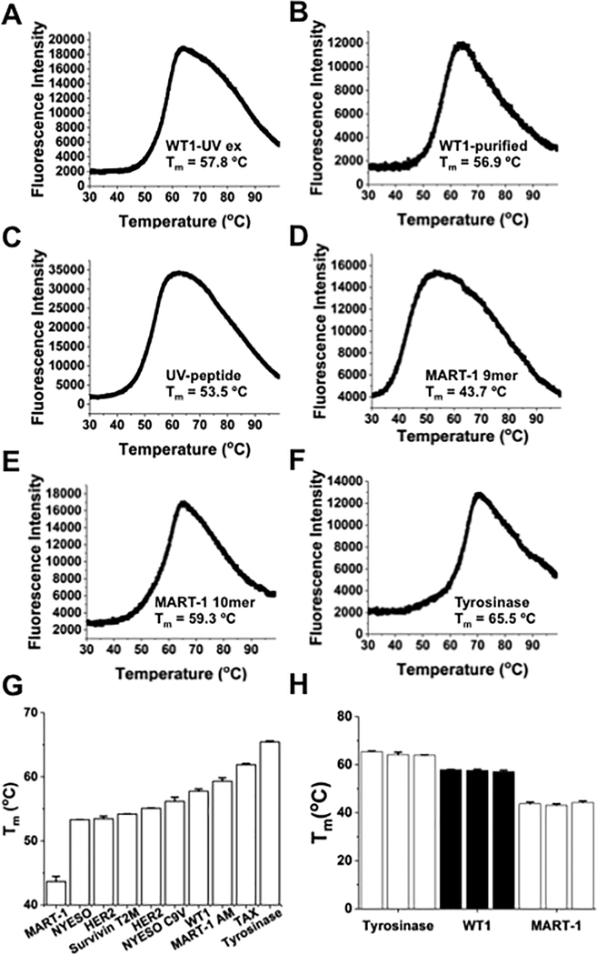
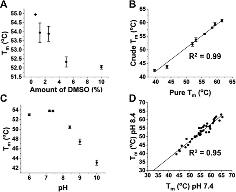
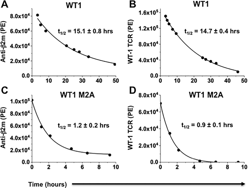
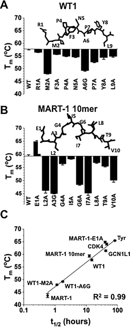
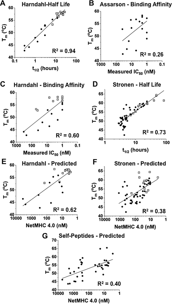
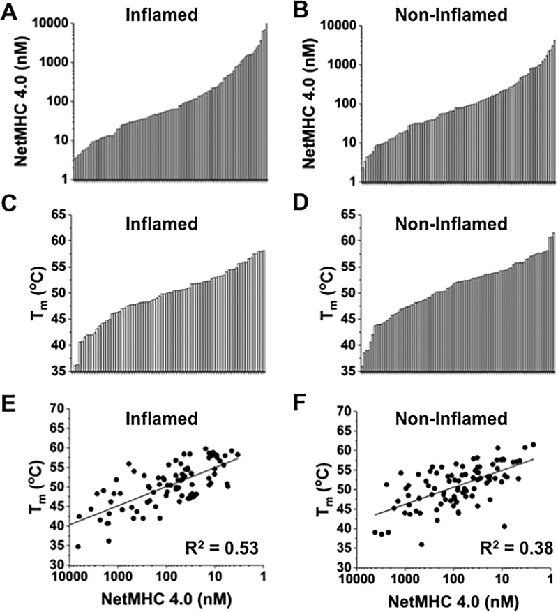
Mutated peptides (neoantigens) from a patient's cancer genome can serve as targets for T-cell immunity, but identifying which peptides can be presented by an MHC molecule and elicit T cells has been difficult. Although algorithms that predict MHC binding exist, they are not yet able to distinguish experimental differences in half-lives of the complexes (an immunologically relevant parameter, referred to here as kinetic stability). Improvement in determining actual neoantigen peptide/MHC stability could be important, as only a small fraction of peptides in most current vaccines are capable of eliciting CD8+ T-cell responses. Here, we used a rapid, high-throughput method to experimentally determine peptide/HLA thermal stability on a scale that will be necessary for analysis of neoantigens from thousands of patients. The method combined the use of UV-cleavable peptide/HLA class I complexes and differential scanning fluorimetry to determine the Tm values of neoantigen complexes. Measured Tm values were accurate and reproducible and were directly proportional to the half-lives of the complexes. Analysis of known HLA-A2-restricted immunogenic peptides showed that Tm values better correlated with immunogenicity than algorithm-predicted binding affinities. We propose that temperature stability information can be used as a guide for the selection of neoantigens in cancer vaccines in order to focus attention on those mutated peptides with the highest probability of being expressed on the cell surface.


Conflict of interest statement

Conflict of Interest: The authors declare that they have no conflicts of interest with this article.

Figures

Figure 1.
Figure 1.. Thermal denaturation curves generated from differential scanning fluorimetry (DSF) of self-peptide/HLA-A2 complexes.
(A) DSF denaturation profile of WT-1 (RMFPNAPYL)/HLA-A2 complex prepared through UV-mediated ligand exchange at 4 uM. For all measurements, the temperature ramp rate was 1°C/minute from 25°C to 99°C. The curve was created using OriginPro 2017 software to plot all 820 time points generated from QuantStudio™ Real-Time PCR. (B) Thermal denaturation curve of refolded WT-1/HLA-A2 monomer at 4 uM, used for comparison with UV-generated complex. (C) DSF profile of refolded UV-peptide/HLA-A2. The UV-peptide (KILGFVFJV) contains 3-amino-3-(2-nitro)phenyl-propionic acid, denoted with a J in the peptide sequence. (D-F) DSF profiles for UV-exchanged self-peptide/HLA-A2 complexes with MART-1 9-mer (AAGIGILTV), MART-1 anchor-modified 10-mer (ELAGIGILTV), and Tyrosinase (YMDGTMSQV). For (A-F), curves are representative of 3 independent experiments, with three replicates per experiment. (G) Comparison of Tm values from several well-characterized self-peptides, including NYESO-1 (SLLMWITNC) and HER2 (KIFGSLAFL). (H) Comparison of Tm values from three separate DSF experiments with UV-exchanged self-peptide/HLA-A2 complexes. UV-exchanged complexes with Tyrosinase (YMDGTMSQV), WT-1 (RMFPNAPYL), and MART-1 (AAGIGILTV) were analyzed in three separate experiments of three replicates each. Different UV-exchanged preparations were used for each trial. Error bars represent the standard deviation between replicates of individual DSF runs.
Figure 2.
Figure 2.. Effects of DMSO, peptide purity, and pH on thermal denaturation of pep/HLA-A2 complexes.
(A) Tm values of UV-peptide/HLA-A2 in PBS and various concentrations of dimethyl sulfoxide (DMSO). The UV-exchange method uses peptides dissolved in DMSO, yielding final concentrations of DMSO present at 0.5% in a typical DSF experiment. (B) Comparison of Tm values from DSF of HLA-A2 complexes generated with either pure (90%) or crude (as low as 30%) peptide preparations. Correlation co-efficient (R2=0.99) was calculated using OriginPro 2017. (C) Tm values were determined by DSF at various pH values, using assay buffers containing HEPES, boric acid, or acetic acid. Unless otherwise noted, all subsequent DSF experiments were performed at pH 7.4 using HEPES as the assay buffer. (D) Comparison of Tm values (R2 = 0.95) from various self-peptide/HLA-A2 and neoantigen/HLA-A2 complexes at pH 7.4 and pH 8.4. Melting temperatures were increased at 7.4 (compared to pH 8.4) by an average of 2.5°C. Error bars represent the average and standard deviation of three replicates in a single experiment.
Figure 3.
Figure 3.. Dissociation measurements of WT-1/HLA-A2 complexes using two different probes.
(A) Dissociation curve of WT-1/HLA-A2 complexes using anti-β2m as a probe (t1/2=15.7). (B) Dissociation curve of WT-1/HLA-A2 complexes using soluble high-affinity T-cell receptor (TCR) as a probe (t1/2 =14.4). (C) Dissociation curve of WT-1–M2A complexes, a reduced stability variant (RAFPNAPYL), using anti-β2m (t1/2=1.3). (D) Dissociation curve of WT-1–M2A/HLA-A2 using soluble TCR (t1/2=1.0). In these experiments, refolded WT-1/HLA-A2 or WT-1–M2A/HLA-A2 were biotinylated with BirA ligase, purified by HPLC ion exchange and gel filtration chromatography, and immobilized on SA-coated 5 μm microspheres. Curves show data from a single experiment, representative of two independent experiments, with one replicate each. Half-life values are the averages of two independent experiments, with one replicate each.
Figure 4.
Figure 4.. DSF analysis of self-peptides and single peptide variants.
(A) Comparison of Tm values for self-peptide WT-1 (RMFPNAPYL) and its alanine variants. In positions where an alanine was present in the wild-type sequence, it was replaced with a glycine (WT1-A6G). The structure of the WT-1 peptide from the WT-1/HLA-A2 structure is shown (PDB: 3HPJ). (B) Tm values for MART-1 anchor-modified 10-mer (ELAGIGILTV) and its alanine variants. In positions where an alanine was present in the wild-type sequence, it was replaced with a glycine (MART1-A3G). The structure of the MART-1 10mer peptide from the MART-1/HLA-A2 structure is shown (PDB: 1JF1). (C) Correlation (R2=0.99) between half-lives measured experimentally using the anti-β2m probe and Tm values obtained from DSF. Error bars represent the average and standard deviation of two independent experiments, with three replicates per experiment.
Figure 5.
Figure 5.. Correlation of Tm values with half-lives, predicted binding affinities, or measured binding affinities.
(A) Plot of Tm values, determined by DSF, versus half-lives for 23 T-cell epitopes restricted by HLA-A2 (9). Epitopes reported as immunogenic are shown in open squares (R2=0.94). (B, C) Peptides with known binding affinity measurements analyzed by Assarson et al. (31) and Harndahl et al. (9) were examined by DSF and Tm values were plotted versus IC50 measurements from Assarson et al. (R2=0.26) or Harndahl et al. (R2=0.60). Epitopes that they defined as immunogenic are shown in open squares. (D) Plot of Tm values, determined by DSF, versus half-lives of 53 HLA-A2 restricted neoantigens derived from three melanoma patients (10). The correlation (R2=0.73) between published half-lives and measured Tm values was increased (R2=0.81) if data for those peptides that reached the shortest measurable half-life (10). Immunogenic neoantigens (12 of 53) from the latter study are shown in open squares. (E) Correlation (R2=0.62) between binding affinity, predicted in silico with NetMHC 4.0, and experimentally measured Tm values for the 23 unique T-cell epitopes (9). Immunogenic epitopes are shown in open squares. (F) Correlation (R2=0.39) between binding affinity, predicted in silico with NetMHC 4.0, and experimentally measured Tm values for 53 melanoma-specific, HLA-A2–restricted neoantigens (10). The 12 immunogenic neoantigens are shown in open squares. (G) Correlation (R2=0.40) between binding affinity, predicted in silico with NetMHC 4.0, and experimentally measured Tm values for HLA-A2–restricted self-peptides or their single peptide variants (Supplementary Table S2).
Figure 6.
Figure 6.. Analysis of neoantigen/HLA-A2 complexes identified from TCGA.
(A, B) Distribution of NetMHC 4.0 values for HLA-A2-restricted neoantigens from TCGA-indicated inflamed and non-inflamed tumor microenvironments, respectively. (C, D) Distribution of Tm values, obtained from DSF, for HLA-A2–restricted neoantigens from inflamed and non-inflamed tumor microenvironments, respectively. (E) Relationship (R2=0.53) between predicted binding affinity and Tm for 85 neoantigens found in inflamed tumor microenvironments. (F) Correlation (R2=0.38) of predicted binding affinity and Tm for 91 neoantigens found in non-inflamed tumor microenvironments. Analysis revealed no statistically significant difference between Tm values measured for peptides from the two sources (inflamed and non-inflamed). P=0.4 assessed from a single experiment with three replicates of each neoantigen in the inflamed and non-inflamed cohorts, using a two-tailed Student t test (t=0.85, two degrees of freedom) with significance accepted at p<0.05).

References

    1. Gubin MM, Zhang X, Schuster H, Caron E, Ward JP, Noguchi T, et al. Checkpoint blockade cancer immunotherapy targets tumour-specific mutant antigens. Nature 2014;515(7528):577–81 doi 10.1038/nature13988. - DOI - PMC - PubMed
    1. Ribas A, Wolchok JD. Cancer immunotherapy using checkpoint blockade. Science 2018;359(6382):1350–5 doi 10.1126/science.aar4060. - DOI - PMC - PubMed
    1. Schumacher TN, Schreiber RD. Neoantigens in cancer immunotherapy. Science 2015;348(6230):69–74 doi 10.1126/science.aaa4971. - DOI - PubMed
    1. Blank CU, Haanen JB, Ribas A, Schumacher TN. CANCER IMMUNOLOGY. The “cancer immunogram”. Science 2016;352(6286):658–60 doi 10.1126/science.aaf2834. - DOI - PubMed
    1. Schumacher TN, Hacohen N. Neoantigens encoded in the cancer genome. Curr Opin Immunol 2016;41:98–103 doi 10.1016/j.coi.2016.07.005. - DOI - PubMed

Publication types