Review
. 2018 Nov 13;9(11):1126.
doi: 10.1038/s41419-018-1157-x.

Renal tubular epithelial cells: the neglected mediator of tubulointerstitial fibrosis after injury

Affiliations
Review

Renal tubular epithelial cells: the neglected mediator of tubulointerstitial fibrosis after injury

Ruochen Qi et al. Cell Death Dis. .

Abstract

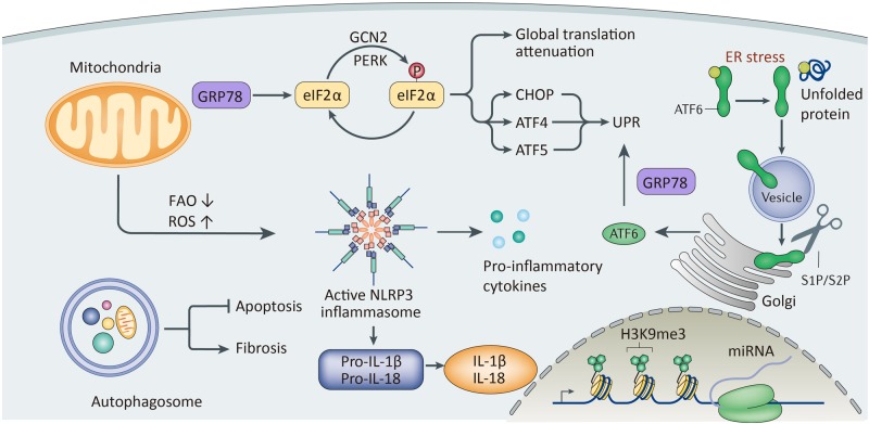
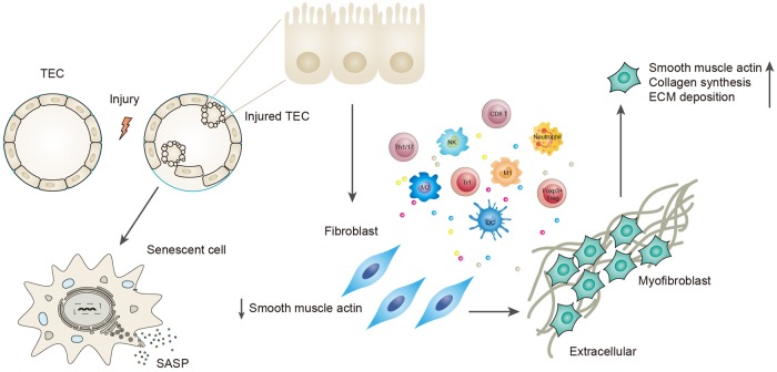
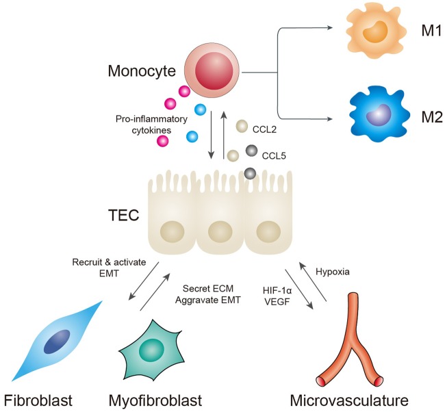
Renal fibrosis, especially tubulointerstitial fibrosis, is the inevitable outcome of all progressive chronic kidney diseases (CKDs) and exerts a great health burden worldwide. For a long time, interests in renal fibrosis have been concentrated on fibroblasts and myofibroblasts. However, in recent years, growing numbers of studies have focused on the role of tubular epithelial cells (TECs). TECs, rather than a victim or bystander, are probably a neglected mediator in renal fibrosis, responding to a variety of injuries. The maladaptive repair mechanisms of TECs may be the key point in this process. In this review, we will focus on the role of TECs in tubulointerstitial fibrosis. We will follow the fate of a tubular cell and depict the intracellular changes after injury. We will then discuss how the repair mechanism of tubular cells becomes maladaptive, and we will finally discuss the intercellular crosstalk in the interstitium that ultimately proceeds tubulointerstitial fibrosis.


Conflict of interest statement

The authors declare that they have no conflict of interest.

Figures

Fig. 1
Fig. 1. A schematic view of intracellular changes in tubular cells upon injury.
Mitochondria undergo metabolic disorders, manifested by decreased fatty acid oxidation. Mitochondria can also produce ROS and further activate the NLRP3/inflammasome. Production of mitochondrial ROS is also related to the expression of proinflammatory factors. ER stress caused by misfolded proteins can start the unfolded protein reaction (UPR), including activation of GRP78 and downstream signaling, including the PERK–eIF2–ATF4 pathway and ATF6 pathway. However, persistent activation of UPR can result in apoptosis. Autophagy plays a dual role. This process can either avoid apoptosis or aggravate renal fibrosis, depending on different situations. Injuries can also lead to epigenetic changes and changes in mRNA expression
Fig. 2
Fig. 2. Description of maladaptive repair.
It is the severity and frequency of the injury that determine whether the repair mechanism adopted by tubular cells is beneficial or maladaptive. Severe and persistent injury exceeds normal repair mechanisms, and these cells become maladaptive to survive the injury. Maladaptive repair is manifested by cell cycle arrested at the G2/M phase and a senescence-associated secretory phenotype. The former is characterized by the expression of proteins that include p53, p21, and p16INK4a. The latter includes secretion of proinflammatory and profibrotic factors, including TGF-beta1, CTGF, CXCL1, IL-6, IL-8, etc. Tubular cells can also undergo EMT to avoid apoptosis, with loss of some epithelial markers (E-cadherin, ZO-1) and acquisition of partial myofibroblast markers (alpha-SMA, vimentin, FSP-1). This process involves changes in the immune microenvironment. Proinflammatory factors secreted by TECs recruit and activate different inflammatory cells, and these recruited cells can further produce cytokines that drive TECs to undergo EMT. TECs finally obtain a myofibroblast phenotype, express alpha-SMA, and are responsible for collagen synthesis and ECM deposition
Fig. 3
Fig. 3. Interactions between TECs and other cells in the interstitium.
TECs can secrete CCL2 and CCL5 to recruit monocytes. Monocytes/macrophages can further produce proinflammatory factors, but their functions depend on their polarization phenotypes. Monocytes can also induce EMT by producing MMP-9. Capillary rarefaction causes hypoxia in tubular cells. Injured TECs thus secrete HIF-1alpha and VEGF to stimulate new capillary formation to meet their oxygen demand. However, these new capillaries are often leaky and are incapable of performing normal functions, thus forming a vicious cycle. Maladaptive TECs can produce a microenvironment suitable for fibroblast recruitment and activation. Activated myofibroblasts can mediate ECM deposition and execute the final process of renal fibrosis. Increased matrix rigidity can also aggravate tubular hypoxia and the progression of EMT

References

    1. Webster AC, Nagler EV, Morton RL, Masson P. Chronic kidney disease. Lancet. 2017;389:1238–1252. doi: 10.1016/S0140-6736(16)32064-5. - DOI - PubMed
    1. Liu Y. Cellular and molecular mechanisms of renal fibrosis. Nat. Rev. Nephrol. 2011;7:684–696. doi: 10.1038/nrneph.2011.149. - DOI - PMC - PubMed
    1. Tanaka T. A mechanistic link between renal ischemia and fibrosis. Med. Mol. Morphol. 2017;50:1–8. doi: 10.1007/s00795-016-0146-3. - DOI - PubMed
    1. Gewin LS. Renal fibrosis: primacy of the proximal tubule. Matrix Biol. 2018;68-69:248–262. doi: 10.1016/j.matbio.2018.02.006. - DOI - PMC - PubMed
    1. Liu BC, Tang TT, Lv LL, Lan HY. Renal tubule injury: a driving force toward chronic kidney disease. Kidney Int. 2018;93:568–579. doi: 10.1016/j.kint.2017.09.033. - DOI - PubMed

Publication types

MeSH terms