. 2018 Nov 13;9(11):1130.
doi: 10.1038/s41419-018-1107-7.

TGFβ1 signaling sustains aryl hydrocarbon receptor (AHR) expression and restrains the pathogenic potential of TH17 cells by an AHR-independent mechanism

Affiliations

TGFβ1 signaling sustains aryl hydrocarbon receptor (AHR) expression and restrains the pathogenic potential of TH17 cells by an AHR-independent mechanism

Kalil Alves de Lima et al. Cell Death Dis. .

Abstract

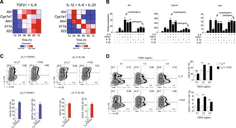
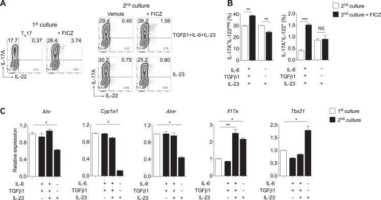
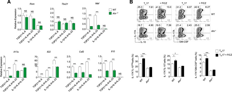
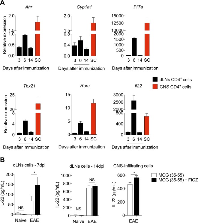
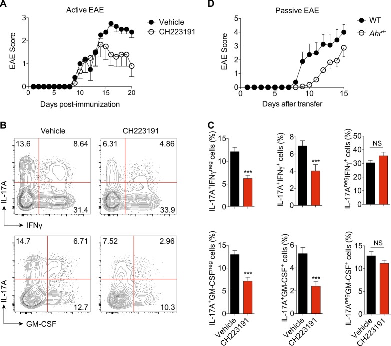
The aryl hydrocarbon receptor (AHR) is a transcription factor activated by ligand highly expressed on TH17 cells, and AHR-deficient CD4+ T cells have impaired production of IL-17A and IL-22. Although AHR activation can exacerbate in vivo TH17 cell-mediated autoimmunity, accumulating data indicate that AHR is a nonpathogenic TH17 marker. Thus it remains unclear how AHR activation is regulated and impacts on the generation of TH17 subsets. Here we demonstrated that AHR pathway is activated during in vitro pathogenic TH17 polarization, but it is quickly downregulated. Under these conditions, additional AHR activation promoted IL-22 but not IL-17A. Interestingly, AHR high sustained expression and IL-17A promotion were only achieved when TGFβ1 was present in the culture. In addition to the effect on AHR regulation, TGFβ1 presented a dual role by simultaneously suppressing the TH17 pathogenic phenotype acquisition. This latter effect was independent of AHR stimulation, since its activation did not confer a TH17 anti-inflammatory profile and Ahr-/- cells did not upregulate any TH17 pathogenic marker. Through the use of EAE model, we demonstrated that AHR is still functional in encephalitogenic CD4+ T cells and the adoptive transfer of Ahr-/- TH17 cells to recipient mice resulted in milder EAE development when compared to their WT counterparts. Altogether, our data demonstrated that although AHR is highly expressed on in vitro-generated nonpathogenic TH17 cells, its ligation does not shift TH17 cells to an anti-inflammatory phenotype. Further studies investigating the role of AHR beyond TH17 differentiation may provide a useful understanding of the physiopathology of autoimmune diseases.


Conflict of interest statement

The authors declare that they have no conflict of interest.

Figures

Fig. 1
Fig. 1. AHR is activated during in vitro pathogenic TH17 cell differentiation, and it regulates IL-17A production in a TGFβ1-dependent manner.
a Heatmap of Ahr, Cyp1a1, Ahrr, Il22, and Il17a mRNA expression in CD4+CD44loCD62Lhi naive T cells differentiated for 12, 24, 36, 48, 60, and 72 h under nonpathogenic (TGFβ1 plus IL-6) and pathogenic (IL-1β, IL-6, and IL-23) TH17 conditions. b qPCR analysis of Ahr, Cyp1a1, and Ahrr mRNA expression of naive CD4+ T cells (white bars) activated 24 h under different combinations of IL-6, TGFβ1, IL-1β, and IL-23 (as indicated). c Frequency of IL-17A+ and IL-22+ cells from pathogenic TH17 cells differentiated with TGFβ1, IL-6 and IL-23 (pTH17 (TGFβ1)) or IL-1β, IL-6 plus IL-23 (pTH17 (IL-1β)) in the presence of FICZ. d Frequency of IL-17A+ and IL-22+ cells from pTH17 cells (TGFβ1, IL-6, and IL-23) differentiated under different concentrations of TGFβ1. b P < 0.05 when compared to amedium; bIL-6; cTGFβ1, and dTGFβ1+IL-6 groups (two-way ANOVA). NS not significant; *P < 0.05, *P< 0.01, and ***P< 0.001 (c, unpaired, two-tailed Student’s t test and d two-way ANOVA). Data are representative of more than three independent experiments with similar results
Fig. 2
Fig. 2. TGFβ1 signaling maintains Ahr expression in TH17 cells, while it opposes IL-23-driven conversion into pathogenic TH17 cells.
a Naive CD4+ T cells were cultured 3 days under nonpathogenic TH17-polarizing conditions (TGFβ1+IL-6) in the absence or presence of FICZ and then stained intracellularly for IL-17A and IL-22 (first culture). A fraction of recovered cells cultured without FICZ was re-stimulated for additional 3 days with TGFβ1, IL-6 plus IL-23, or only IL-23, with or without FICZ stimulation (second culture). b Frequencies of IL-17A+ and IL-22+ cells after the second culture shown in a. c qPCR analysis of Ahr, Cyp1a1, Ahrr, Il17a, and Tbx21 of naive CD4+ T cells cultured under nonpathogenic TH17-polarizing conditions (TGFβ1+IL-6) for 3 days (first culture, white bars) and re-stimulated with TGFβ1, IL-6, and IL-23 combinations for 24 h (second culture). NS not significant; *P < 0.05, **P < 0.01, and ***P < 0.001 (two-way ANOVA)
Fig. 3
Fig. 3. AHR signaling does not drive anti-inflammatory properties to TH17 cells.
Quantitative RT-PCR analysis of Rorc, Tbx21, Maf, Il17a, Il22, Csf2, and l10 from WT (white bars) or Ahr-deficient (green bars) cells differentiated for 72 h under nonpathogenic (TGFβ1 and IL-6) and pathogenic TH17 cell conditions (TGFβ1, IL-6 plus IL-23 or IL-1β, IL-6 and IL-23). b Frequency of IL-17A+, IL-10+, and GM-CSF+ cells from WT and Ahr/− naive CD4+ T cells differentiated for 72 h with TGFβ1+IL-6+IL-23 under FICZ stimulation. NS not significant; *P < 0.05 and ***P < 0.001 (two-way ANOVA)
Fig. 4
Fig. 4. AHR is still highly expressed and functional on CNS-infiltrating pathogenic CD4+ T cells.
a qPCR analysis of Ahr, Cyp1a1, Il17a, Tbx21, Rorc, and Il22 transcripts of CD4+ T cells isolated from draining lymph nodes (dLNs) after 3, 6, or 14 days after EAE induction. Spinal cord (SC) CD4+ T cells were isolated 14 days after MOG immunization. b IL-22 production of purified dLNs cells from WT mice 7 and 14 days after EAE induction and CNS-infiltrating cells 14 days after MOG-CFA immunization re-stimulated for 72 h with MOG (50 µg/mL) with or without FICZ (300 nM). NS not significant; *P< 0.05 (unpaired, two-tailed Student’s t test)
Fig. 5
Fig. 5. AHR activation promotes effector functions to encephalitogenic TH17 cells.
a EAE clinical score of WT mice treated with AHR antagonist for 10 consecutive days (CH223191, i.p. 10 mg/kg). b Representative plots of IL-17A+, IFN-γ, and GM-CSF+ CNS-derived CD4+ T cells from vehicle and CH223191-treated EAE animals. c Frequency of cytokine-producer cells shown in b. d EAE clinical score of Rag1/− mice recipients of WT or Ahr/− MOG-specific TH17 cells. NS not significant; ***P < 0.001 (unpaired, two-tailed Student’s t test)

References

    1. Korn T, Bettelli E, Oukka M, Kuchroo VK. IL-17 and Th17 Cells. Annu. Rev. Immunol. 2009;27:485–517. doi: 10.1146/annurev.immunol.021908.132710. - DOI - PubMed
    1. Gaffen SL, Jain R, Garg AV, Cua DJ. The IL-23-IL-17 immune axis: from mechanisms to therapeutic testing. Nat. Rev. Immunol. 2014;14:585–600. doi: 10.1038/nri3707. - DOI - PMC - PubMed
    1. Bettelli E, et al. Reciprocal developmental pathways for the generation of pathogenic effector TH17 and regulatory T cells. Nature. 2006;441:235–238. doi: 10.1038/nature04753. - DOI - PubMed
    1. Mangan PR, et al. Transforming growth factor-beta induces development of the T(H)17 lineage. Nature. 2006;441:231–234. doi: 10.1038/nature04754. - DOI - PubMed
    1. Veldhoen M, Hocking RJ, Atkins CJ, Locksley RM, Stockinger B. TGFbeta in the context of an inflammatory cytokine milieu supports de novo differentiation of IL-17-producing T cells. Immunity. 2006;24:179–189. doi: 10.1016/j.immuni.2006.01.001. - DOI - PubMed

Publication types

MeSH terms

Substances