. 2018 Nov 13;8(1):16766.
doi: 10.1038/s41598-018-35176-y.

Comprehensive molecular and cellular studies suggest avian scutate scales are secondarily derived from feathers, and more distant from reptilian scales

Affiliations

Comprehensive molecular and cellular studies suggest avian scutate scales are secondarily derived from feathers, and more distant from reptilian scales

Ping Wu et al. Sci Rep. .

Abstract

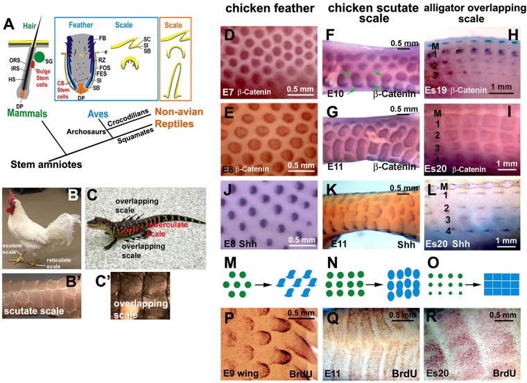
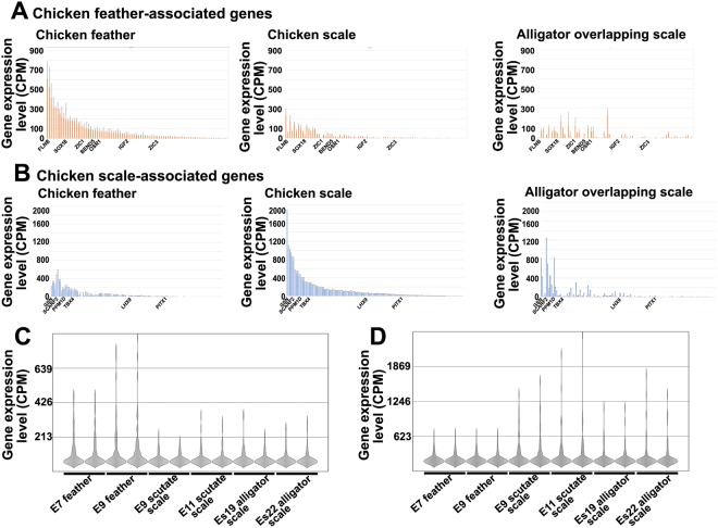
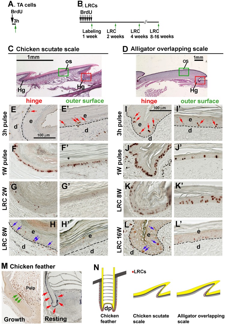
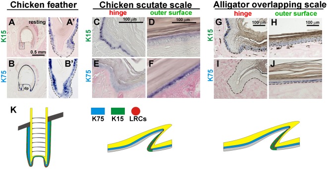
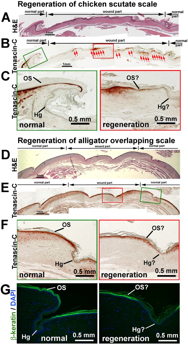
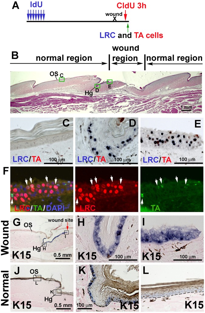
Amniote skin appendages such as feathers, hairs and scales, provide thermoregulation, physical protection and display different color patterns to attract a mate or frighten an adversary. A long-standing question is whether "reptile scale" and "avian leg scales" are of the same origin. Understanding the relation between avian feathers, avian scales and reptilian scales will enhance our understanding of skin appendage evolution. We compared the molecular and cellular profiles in chicken feather, chicken scales and alligator scales and found that chicken scutate scales are similar to chicken feathers in morphogenesis at the early placode stage. When we compared the expression of the recently identified feather-specific genes and scale-specific genes in these skin appendages, we found that at the molecular level alligator scales are significantly different from both chicken feathers and chicken scales. Furthermore, we identified a similarly diffuse putative stem cell niche in morphologically similar chicken and alligator scales. These putative stem cells participate in alligator scale regeneration. In contrast, avian feathers have a more condensed stem cell niche, which may be responsible for cycling. Thus, our results suggest that chicken and alligator scales formed independently through convergent evolution.


Conflict of interest statement

The authors declare no competing interests.

Figures

Figure 1
Figure 1
Development of avian and reptilian scales. (A) Schematic drawing of the stem cell niche in mammalian hairs and avian feathers. (B) Adult chicken showing feathers and scales. (B’) Scutate scales. (C) Juvenile alligator showing different types of scales. (C’) Overlapping scale. D-I, β-catenin whole mount in situ hybridization. (D) E7 chicken dorsal feather tract (placode stage). (E) E8 chicken dorsal feather tract (short bud stage). (F) E10 chicken scutate scale (placode stage). Green arrows indicate the fusion of scutate scale placodes. (G) E11 chicken scutate scale (short bud stage). (H) Es19 alligator overlapping scale (placode stage). (I) Es20 alligator overlapping scale (short bud stage). (J–L) Shh whole mount in situ hybridization. J, E8 chicken dorsal feather tract. (K) E11 chicken scutate scale. (L) Es20 alligator overlapping scale. (M–O) Schematic drawing of skin appendage development. (M) Chicken feather, (N) chicken scutate scale, (O) alligator overlapping scale. (PR) Whole mount BrdU staining. (P) Feather buds in an E9 chicken wing showed different feather developmental stages, from short buds to long buds. (Q) E11 chicken scutate scale. (R) Es20 alligator overlapping scale. Note the feathers have a broader localized growth zone than scales. CB, collar bulge; DP, dermal papilla; e, epidermis; FB; feather barb ridge; FES, feather sheath; FOS, feather follicle sheath; HS, hair shaft; IRS, inner root sheath; M, dorsal middle line of alligator embryo; ORS, outer root sheath; RZ, ramogenic zone; SG, sebaceous gland; SB, stratum basal; SC, stratum corneum; SI, stratum intermedium; 1, 2, 3, 4 indicate the row number with 1 closest to the middle of the dorsal region.
Figure 2
Figure 2
RNA-seq analysis of embryonic chicken feathers, chicken scutate scales and alligator overlapping scales. (A) Comparing the chicken feather-associated gene expression profile among chicken feathers (E7 and E9), chicken scutate scales (E9 and E11) and alligator overlapping scales (Es19 and Es22). The X-axis is the average CPM (count per million) value of feather-associated genes (Y-axis) from high to low in feather samples (left panel). Chicken scale (middle panel) and alligator scale (right panel) expression levels of feather-associated gene are shown. (B) Comparing the chicken scale-associated gene expression profile among chicken feathers (E7 and E9), chicken scutate scales (E9 and E11) and alligator overlapping scales (Es19 and Es22). The X-axis is the average CPM (count per million) value of scale-associated genes (Y-axis) from high to low in chicken scutate scale samples (middle panel). Chicken feather (left panel) and alligator scale (right panel) expression levels of scale-associated gene are shown. (C) Violin plots showing the chicken feather-associated genes are down-regulated in chicken scales and alligator scales. (D) Violin plot analysis showing the chicken scale-associated genes are down-regulated in both chicken feathers and alligator scales.
Figure 3
Figure 3
The topology of putative stem cells in chicken scutate scales and alligator overlapping scales. (A) Strategy of TA cell labeling. (B) Strategy of LRC labeling. (C) H&E staining of a chicken scutate scale. (D) H&E staining of an alligator overlapping scale. The red and green rectangular boxes in panel C and D indicate the hinge region and outer surface region in chicken and alligator scales. (E–H’) TA and LRCs in chicken scutate scales. (E) BrdU 3-hour pulse labeling. Red arrows indicate BrdU positive cells. (F) BrdU 1-week labeling. (G) A 2-week chase period after 1-week of labeling. (H) An 8-week chase period after 1-week of labeling. Blue arrows indicate the LRCs in the hinge region. (I–L’) TA and LRCs in alligator overlapping scales. I. 3-hour BrdU pulse labeling. Red arrows indicate BrdU positive cells. (J) BrdU labeling for 1-week. (K) An 8-week chase period after 1-week of labeling. (L) A 16-week chase period after 1-week of labeling. Blue arrows indicate the LRCs in the hinge region. Note that LRCs exist in the hinge regions but are negative in the outer surface in both chicken scutate and alligator overlapping scales. (M) LRCs in chicken feathers. Left panel, growth phase, green arrows indicate the LRCs in the feather bulge region. Right panel, resting phase, red arrows indicate the LRCs in epidermis surrounding the dermal papilla (papilla ectoderm) and in the follicle sheath. (N) Schematic drawing showing the distribution of LRCs (red dots). The dotted lines separate epidermis and dermis. d, dermis; e, epidermis; Hg, hinge; OS, outer surface.
Figure 4
Figure 4
Expression of the stem cell marker, K15 and differentiation marker, K75 in chicken and alligator scales. (A,A’) Expression of K15 in resting phase feather follicles. (B,B’) Expression of K75 in resting phase feather follicles. (C–F) Chicken scutate scales. (C,D) Expression of K15 in chicken scutate scales. (C) Hinge; (D) outer surface. (E,F) Expression of K75 in chicken scutate scales. (E) Hinge; (F) outer surface. (GJ) Alligator overlapping scales. (G,H) Expression of K15 in alligator overlapping scales. (G) Hinge; (H) outer surface. (I,J) Expression of K75 in alligator overlapping scales. (I) Hinge; (J) outer surface. Note K15 is positive in the hinge basal layer (C,G) whereas K75 is positive in the outer surface suprabasal layer (F,J). (K) Schematic drawing of the distribution of K15 (green), K75 (blue) and LRCs (red dot) in feathers (resting stage) and scales. dp, dermal papilla.
Figure 5
Figure 5
Regenerative power of avian and reptilian scales. (A–C) Wounded chicken scutate scales after 4 months of healing. (A) H&E staining. (B) Tenascin-C staining. (C) Higher magnification of (B). Left, normal scale beside the wound site. Right. regenerated scale. (D–F) Wounded alligator scale after 4 months of healing. (D) H&E staining. (E) Tenascin-C staining. (F) higher magnification of (E) (G) β-keratin staining. (F) and (G) Left, normal scale beside the wound site; Right, regenerated scale. Hg, hinge; OS, outer surface.
Figure 6
Figure 6
Cell behavior after wounding of reptilian overlapping scales. (A) Strategy of labeling LRCs with IdU and TA cells with CldU to study cell dynamics after wounding. (B) H&E staining after 4 days of wound healing. The rectangular blocks show the region for CldU and IdU staining enlarged in panel (C–E). (C) Outer surface of a scale beside the wound site. (D) Hinge of a scale closest to the wound site. (E) Outer surface region in the wound site. Note the accumulation of LRCs (blue color) in (D) and (E). (F) Fluorescent immunostaining of IdU (LRCs, red color) and CldU (TA cells, green color). Left, Triple staining of LRCs, TA cells and DAPI (blue color); Middle, LRCs only; Right, TA cells only. White arrows indicate IdU and CldU double positive cells which suggests the LRCs are in a proliferating status. (G) K15 in situ hybridization showing increased K15 expression in epidermis near the wound site. (H) Hinge closest to the wound site. I, outer surface in the wound site. (J) K15 in situ hybridization showing the normal K15 expression in an overlapping scale. (K) Hinge. (L) Outer surface. Hg, hinge. OS, outer surface.

References

    1. Wu P, et al. Evo-Devo of amniote integuments and appendages. Int J Dev Biol. 2004;48:249–270. doi: 10.1387/ijdb.041825pw. - DOI - PMC - PubMed
    1. Chang C, et al. Reptile scale paradigm: Evo-Devo, pattern formation and regeneration. Int J Dev Biol. 2009;53:813–826. doi: 10.1387/ijdb.072556cc. - DOI - PMC - PubMed
    1. Alibardi L. Dermo-epidermal interactions in reptilian scales: speculations on the evolution of scales, feathers, and hairs. J Exp Zool B Mol Dev Evol. 2004;302:365–383. doi: 10.1002/jez.b.20028. - DOI - PubMed
    1. Alibardi L, Thompson MB. Scale morphogenesis and ultrastructure of dermis during embryonic development in the alligator (Alligator mississippiensis, Crocodilia, Reptilia) Acta Zoologica. 2001;81:325–338. doi: 10.1046/j.1463-6395.2000.00063.x. - DOI
    1. Sawyer, R. H., Knapp, L. W. & O’Guin, W. M. The skin of birds: epidermis, dermis and appendages. In Biology of the Integument. Vol. 2 194–238 (Springer-Verlag, 1986).

Publication types