Review
. 2018 Oct 17:14:2715-2737.
doi: 10.2147/NDT.S170989. eCollection 2018.

Neuronal connectivity in major depressive disorder: a systematic review

Affiliations
Review

Neuronal connectivity in major depressive disorder: a systematic review

Katharina Helm et al. Neuropsychiatr Dis Treat. .

Abstract

Background: The causes of major depressive disorder (MDD), as one of the most common psychiatric disorders, still remain unclear. Neuroimaging has substantially contributed to understanding the putative neuronal mechanisms underlying depressed mood and motivational as well as cognitive impairments in depressed individuals. In particular, analyses addressing changes in interregional connectivity seem to be a promising approach to capture the effects of MDD at a systems level. However, a plethora of different, sometimes contradicting results have been published so far, making general conclusions difficult. Here we provide a systematic overview about connectivity studies published in the field over the last decade considering different methodological as well as clinical issues.

Methods: A systematic review was conducted extracting neuronal connectivity results from studies published between 2002 and 2015. The findings were summarized in tables and were graphically visualized.

Results: The review supports and summarizes the notion of an altered frontolimbic mood regulation circuitry in MDD patients, but also stresses the heterogeneity of the findings. The brain regions that are most consistently affected across studies are the orbitomedial prefrontal cortex, anterior cingulate cortex, amygdala, hippocampus, cerebellum and the basal ganglia.

Conclusion: The results on connectivity in MDD are very heterogeneous, partly due to different methods and study designs, but also due to the temporal dynamics of connectivity. While connectivity research is an important step toward a complex systems approach to brain functioning, future research should focus on the dynamics of functional and effective connectivity.

Keywords: EEG; MDD; effective connectivity; fMRI; functional connectivity; major depressive disorder; structural connectivity.


Conflict of interest statement

Disclosure The authors report no conflicts of interest in this work.

Figures
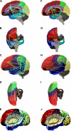
Figure 1
Figure 1
Functional connectivity (FC) for MDD patients compared with healthy controls for resting state (left) and task-related fMRI studies (right). Notes: A/F: sagittal, lateral view; b/g: sagittal, anterior view; C/H: lateral view of the surface; D/I: sagittal, dorsal view; E/J: lateral view, insula displayed by removing the opercular brain segments. The orange lines represent increased, the blue lines decreased FC. The circles indicate an altered self-referential connectivity (ie, within the region). Colors of the brain regions are coded according to Table 1. Abbreviations: fMRI, functional magnetic resonance imaging; MDD, major depressive disorder.
Figure 2
Figure 2
Synopsis of findings on FC in MDD compared to healthy controls using fMRI/resting state. Notes: The matrix shows the findings on connectivity alterations between the 17 brain regions (see Table 1), separated for decreased connectivity (lower triangle, blue) and increased connectivity (upper triangle, orange). The numbers in the cells specify the number of studies where this kind of altered connectivity was found. Abbreviations: FC, functional connectivity; fMRI, functional magnetic resonance imaging; MDD, major depressive disorder.
Figure 3
Figure 3
Histogram of the 17 regions found in fMRI/resting state studies showing the number of studies that found a region to have an altered FC with another region. Notes: Blue: decreased connectivities in MDD compared to healthy controls; orange: increased connectivities in MDD compared to healthy controls. Abbreviations: FC, functional connectivity; fMRI, functional magnetic resonance imaging; MDD, major depressive disorder.
Figure 4
Figure 4
Synopsis of findings on functional connectivity in MDD compared to healthy controls using task-related fMRI. Notes: The matrix shows the findings on FC alterations between the 17 brain regions (see Table 1), separated for decreased connectivity (lower triangle, blue) and increased connectivity (upper triangle, orange). The numbers in the cells specify the number of studies where this kind of altered FC was found. Abbreviations: FC, functional connectivity; fMRI, functional magnetic resonance imaging; MDD, major depressive disorder.
Figure 5
Figure 5
Histogram of the 17 regions found in fMRI/task-related studies showing the number of studies that found a region to have an altered FC with another region. Notes: Blue: decreased connectivities in MDD compared to healthy controls; orange: increased FC in MDD compared to healthy controls. Abbreviations: FC, functional connectivity; fMRI, functional magnetic resonance imaging; MDD, major depressive disorder.

References

    1. Arnold JF, Zwiers MP, Fitzgerald DA, et al. Fronto-limbic microstructure and structural connectivity in remission from major depression. Psychiatry Res. 2012;204(1):40–48. - PubMed
    1. Guo W, Liu F, Xue Z, et al. Decreased interhemispheric coordination in treatment-resistant depression: a resting-state FMRI study. PLoS One. 2013;8(8):e71368. - PMC - PubMed
    1. Horn DI, Yu C, Steiner J, et al. Glutamatergic and resting-state functional connectivity correlates of severity in major depression – the role of pregenual anterior cingulate cortex and anterior insula. Front Syst Neurosci. 2010;4:33. - PMC - PubMed
    1. Salomons TV, Dunlop K, Kennedy SH, et al. Resting-State Cortico-Thalamic-Striatal Connectivity Predicts Response to Dorsomedial Pre-frontal rTMS in Major Depressive Disorder. Neuropsychopharmacology. 2014;39(2):488–498. - PMC - PubMed
    1. Bear MF, Connors BW, Paradiso MA. Neuroscience: exploring the brain. 2. Baltimore: Lippincott, Williams & Wilkins; 2001.

LinkOut - more resources