. 2018 Oct 23:11:7333-7343.
doi: 10.2147/OTT.S167872. eCollection 2018.

Down-regulation of CXCL11 inhibits colorectal cancer cell growth and epithelial-mesenchymal transition

Affiliations

Down-regulation of CXCL11 inhibits colorectal cancer cell growth and epithelial-mesenchymal transition

Yu Jie Gao et al. Onco Targets Ther. .

Abstract

Background: The poor prognosis of colorectal cancer (CRC) largely results from local invasion and tumor metastases. Epithelial-mesenchymal transition (EMT) is a key step in the progression of solid tumors and plays a vital role in tumor metastasis. Recent studies demonstrate that C-X-C motif chemokine 11 (CXCL11) is involved in various cancers' progression. However, its biological activity in CRC needs deeper exploration.

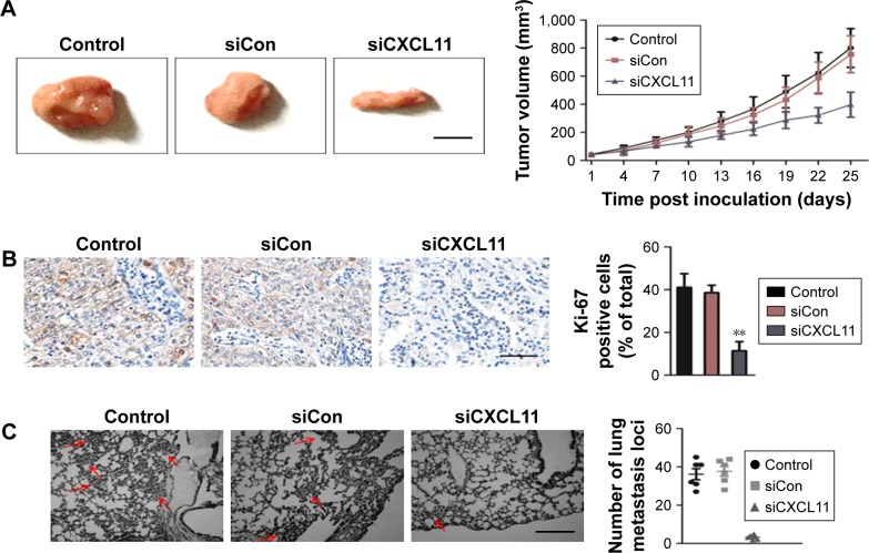
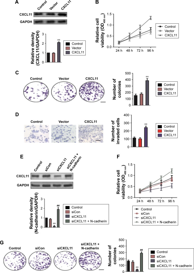
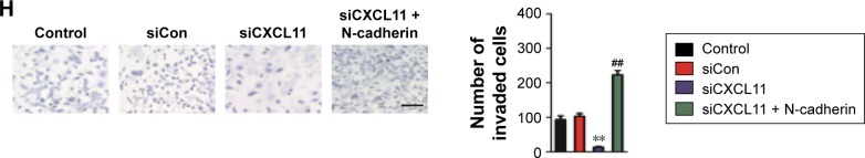
Methods: The level of CXCL11 in CRC tissues and cell lines was determined using the quantitative real-time PCR (qRT-PCR) assay. The MTT, colony formation, wound healing and Transwell invasion assays were applied to assess the role of CXCL11 in CRC cell growth, migration and invasion, in vitro, respectively. A xenograft model was constructed to analyze the function of CXCL11 in CRC cell growth in vivo.

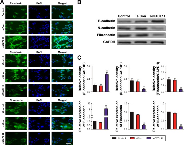
Results: CXCL11 was over-expressed in CRC tissues and cell lines. Repression of CXCL11 significantly inhibited CRC cell migration, invasion and EMT in vitro. In addition, down-regulation of CXCL11 reduced CRC cell growth and metastasis in vivo. Finally, we revealed that repression of CXCL11 inhibited the metastatic ability of CRC cell in a N-cadherin dependent manner.

Conclusion: In summary, this study explicates the oncogenic activities of CXCL11 in CRC cell growth and metastasis.

Keywords: CXCL11; EMT; colon cancer; invasion; migration.


Conflict of interest statement

Disclosure The authors report no conflicts of interest in this work.

Figures

Figure 1
Figure 1
The expression level of CXCL11 is over-regulated in CRC. Notes: (A) The level of CXCL11 in CRC tissues (T) and matched normal tissues (N) was detected by qRT-PCR assay. **P<0.01 compare to normal (N). (B) Immunohistochemical staining of CXCL11 in CRC tissues and paired surrounding normal tissues. Scale bars set at 100 µm. (C) The expression of CXCL11 in human colonic epithelial cells HCoEpiC and the CRC cell lines (SW480, SW116, HT-29 and COLO-205) was detected by Western blot assay. (D) CXCL11 gene examination in CRC (Oncomine database). Box plots originated from gene expression data in Oncomine compared with the expression of CXCL11 gene in the normal (left plot) and CRC tissue (right plot). (E) CXCL11 expression in the normal tissue and CRC tissue. Images were photographed from the Human Protein Atlas (http://www.proteinatlas.org) online database. Abbreviations: CRC, colorectal cancer; GADPH, Glyceraldehyde 3-phosphate dehydrogenase; N, normal tissues; T, tumor tissues; qRT-PCR, quantitative real-time PCR.
Figure 2
Figure 2
CXCL11 regulates SW480 cells growth and colony formation. Notes: (A) SW480 cells were transfected with the control siRNA or siCXCL11 and the expression of CXCL11 was determined by the Western blot assay. (B) SW480 cells were incubated with primary antibody against CXCL11 and stained with anti-rabbit FITC-conjugated secondary antibody. Blue depicts the nucleus and green depicts CXCL11. Magnification ×100, scale bar represents 200 µm. (C) Effects of CXCL11 siRNA on cell viability of SW480. Cells were transfected with CXCL11 siRNA and absorbance was detected at 24 hours, 48 hours, 72 hours and 96 hours, post transfection (left panel). The percentage of cell viability was normalized to control cells (right panel). (D) Down-expression of CXCL11 inhibited colony formation of SW480 cell. **P<0.01 compared to control cell. Scale bar represents 1 cm. Abbreviations: FITC, fluorescein isothiocyanate; GADPH, Glyceraldehyde 3-phosphate dehydrogenase; siRNA, small interfering RNA; siCXCL, small interfering RNA targeting C-X-C motif chemokine 11.
Figure 3
Figure 3
CXCL11 knocked-down inhibits SW480 cells migration and invasion. Notes: (A) SW480 cells were transfected with siCon or siCXCL11 and cell migration and invasion measured using a wound healing assay. Down-regulation of CXCL11 inhibited the migration of SW480 cells in vitro. The percentage of migration closure rate was calculated. Magnification ×100, scale bar represents 100 µm. (B) The transwell invasion assay was utilized to detect the invasion of CXCL11 down-regulation SW480 cells. Magnification ×100, scale bar represents 100 µm. **P<0.01 compared with control cell. Abbreviations: siCon, siRNA Control; siCXCL, small interfering RNA targeting C-X-C motif chemokine 11.
Figure 4
Figure 4
Down-expression of CXCL11 inhibits EMT of SW480 cell. Notes: (A) SW480 cells were transfected with siCon or siCXCL11 and the expression of epithelial marker E-cadherin and mesenchymal markers (N-cadherin and fibronectin) were examined by indirect immunofluorescence assay. Magnification ×200, scale bar represents 100 µm. (B) The expression of the EMT markers in SW480 cells was determined by western blotting analysis. (C) SW480 cell was transiently transfected with siCon or siCXCL11, and the mRNA level of EMT-associated markers was detected by qRT-PCR. **P<0.01 compare to control cell. Abbreviations: EMT, epithelial-mesenchymal transition; GADPH, Glyceraldehyde 3-phosphate dehydrogenase; qRT-PCR, quantitative real-time PCR; siCXCL11, siRNA CXCl11; EMT, epithelial-mesenchymal transition; siCon, siRNA Control; siCXCL, small interfering RNA targeting C-X-C motif chemokine 11.
Figure 5
Figure 5
Down-expression of CXCL11 inhibits SW480 cell growth and metastasis in vivo. Notes: (A) Representative pictures of tumor from parental and siCXCL11 SW480 cells transplanted into nude mice. The tumor volume was calculated at indicated days. Scale bar represents 1 cm. (B) Cell proliferation was detected by Ki-67 immunohistochemistry in xenografts. Percentage of Ki-67 positive cells was shown. Magnification ×100, scale bar represents 200 µm. **P<0.01 compared to the control. (C) Representative picture of H&E staining of metastasis foci on lung tissue (red arrowheads represent metastasis focus). The number of metastatic nodules in the lung sections was calculated. Magnification ×100, scale bar represents 100 µm. Abbreviations: H&E, hematoxylin-eosin staining; siCon, siRNA Control; siCXCL, small interfering RNA targeting C-X-C motif chemokine 11.
Figure 6
Figure 6
N-cadherin re-expression attenuates siCXCL11 mediated inhibition of proliferation, colony formation and mobility in SW480 cell. Notes: (A) The expression of CXCL11 in SW480 cells was detected by Western blotting. (B) Growth curves of SW480 cells that transfected with vectors containing CXCL11. (C) Colony formation assay of the CXCL11 over-expressing SW480 cells. Scale bar represents 1 cm. (D) Transwell invasion assay of SW480 cells in vitro. Magnification ×100, scale bar represents 100 µm. (E) SW480 cells were co-transfected with siCXCL11 and N-cadherin. The expression of N-cadherin was determined by the Western blotting assay. (F) Growth curves of SW480 cells that co-transfected with N-cadherin and siCXCL11. (G) Colony formation assays of the CXCL11 over-expressing SW480 cells. Scale bar represents 1 cm. (H) Transwell invasion assays of the SW480 cells in vitro. Magnification ×100, scale bar represents 100 µm. **P<0.01 compared to the control, ##P<0.01 compared to the cells transfected with siCXCL11 alone. Abbreviations: siCon, siRNA Control; siCXCL, small interfering RNA targeting C-X-C motif chemokine 11.
Figure 6
Figure 6
N-cadherin re-expression attenuates siCXCL11 mediated inhibition of proliferation, colony formation and mobility in SW480 cell. Notes: (A) The expression of CXCL11 in SW480 cells was detected by Western blotting. (B) Growth curves of SW480 cells that transfected with vectors containing CXCL11. (C) Colony formation assay of the CXCL11 over-expressing SW480 cells. Scale bar represents 1 cm. (D) Transwell invasion assay of SW480 cells in vitro. Magnification ×100, scale bar represents 100 µm. (E) SW480 cells were co-transfected with siCXCL11 and N-cadherin. The expression of N-cadherin was determined by the Western blotting assay. (F) Growth curves of SW480 cells that co-transfected with N-cadherin and siCXCL11. (G) Colony formation assays of the CXCL11 over-expressing SW480 cells. Scale bar represents 1 cm. (H) Transwell invasion assays of the SW480 cells in vitro. Magnification ×100, scale bar represents 100 µm. **P<0.01 compared to the control, ##P<0.01 compared to the cells transfected with siCXCL11 alone. Abbreviations: siCon, siRNA Control; siCXCL, small interfering RNA targeting C-X-C motif chemokine 11.

Similar articles

Cited by

References

    1. Zhu J, Liu M, Liu Y, Zhang Y, Yang B, Zhang W. Zoledronic acid regulates autophagy and induces apoptosis in colon cancer cell line CT26. Biomed Res Int. 2017;2017:7203584–7203586. - PMC - PubMed
    1. Arivazhagan R, Lee J, Bayarsaikhan D, et al. MicroRNA-340 inhibits the proliferation and promotes the apoptosis of colon cancer cells by modulating REV3L. Oncotarget. 2018;9(4):5155–5168. - PMC - PubMed
    1. Peng Q, Lin K, Chang T, et al. Identification of genomic expression differences between right-sided and left-sided colon cancer based on bioinformatics analysis. Onco Targets Ther. 2018;11:609–618. - PMC - PubMed
    1. Qin Y, Huo Z, Song X, Chen X, Tian X, Wang X. mir-106a regulates cell proliferation and apoptosis of colon cancer cells through targeting the PTEN/PI3K/AKT signaling pathway. Oncol Lett. 2018;15(3):3197–3201. - PMC - PubMed
    1. Spalinger MR, Manzini R, Hering L, et al. PTPN2 regulates inflammasome activation and controls onset of intestinal inflammation and colon cancer. Cell Rep. 2018;22(7):1835–1848. - PMC - PubMed