Review
. 2018 Oct 30:9:1533.
doi: 10.3389/fphys.2018.01533. eCollection 2018.

The Pathogenesis and Therapies of Striated Muscle Laminopathies

Affiliations
Review

The Pathogenesis and Therapies of Striated Muscle Laminopathies

Astrid Brull et al. Front Physiol. .

Abstract

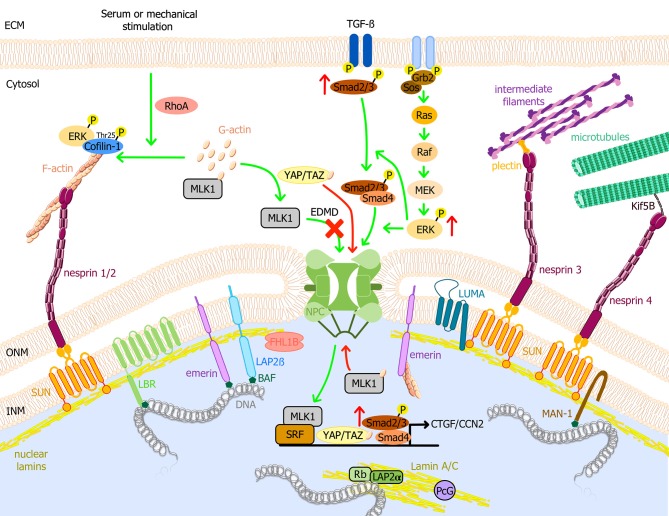
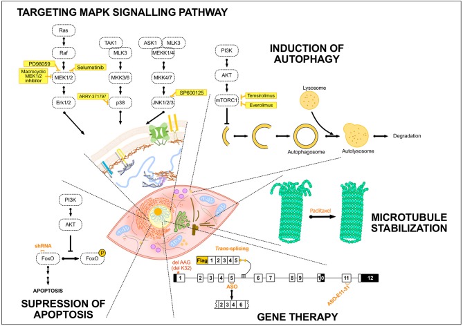
Emery-Dreifuss muscular dystrophy (EDMD) is a genetic condition characterized by early contractures, skeletal muscle weakness, and cardiomyopathy. During the last 20 years, various genetic approaches led to the identification of causal genes of EDMD and related disorders, all encoding nuclear envelope proteins. By their respective localization either at the inner nuclear membrane or the outer nuclear membrane, these proteins interact with each other and establish a connection between the nucleus and the cytoskeleton. Beside this physical link, these proteins are also involved in mechanotransduction, responding to environmental cues, such as increased tension of the cytoskeleton, by the activation or repression of specific sets of genes. This ability of cells to adapt to environmental conditions is altered in EDMD. Increased knowledge on the pathophysiology of EDMD has led to the development of drug or gene therapies that have been tested on mouse models. This review proposed an overview of the functions played by the different proteins involved in EDMD and related disorders and the current therapeutic approaches tested so far.

Keywords: A-type lamins; Emery-Dreifuss muscular dystrophy (EDMD); cardiomyopathy; emerin; luma; muscular dystrophy (MD); nesprin protein; nuclear envelope (NE).


Figures

Figure 1
Figure 1
Schematic model of the nuclear envelope proteins and their potential roles in EDMD physiopathology. Nuclear lamins form a meshwork underneath the INM connected with the cytoplasm. It interacts with TM proteins of the nuclear envelope, i.e., emerin, LBR, LAP2, SUN1/2, and MAN1, and with several transcription factors such as Rb. Through the LINC complex, A-type lamins interact with actin microfilaments, microtubules, and cytoplasmic intermediate filaments, connecting the nuclear lamina to the extracellular matrix. MAPK pathways are important transduction cascades initiated by extracellular mitogens, growth factors and cytokines at the cell surface and finalized to the nucleus to control gene expression, regulating cell proliferation and differentiation, survival and apoptosis. LMNA mutations causing EDMD have been related to the activation of ERK, JNK, and p38α cascades, leading to the posterior activation of CTGF/CCN2 via TGF-ß/Smad signaling pathway and cofilin-1, the latter leading to actin depolymerization. After serum or mechanical stimulation, RhoA promotes cytoplasmic actin polymerization, causing the release of MKL1 from cytoplasmic G-actin. MKL1 translocates into the nucleus and together with SRF induces gene expression. In addition, emerin facilitates polymerization of nuclear actin, reducing the nuclear export of MKL1 to the cytoplasm. In EDMD cells, emerin mislocalizes and is unable to modulate nuclear actin polymerization. G-actin binds to MKL1 and it is exported from the nucleus, impairing gene expression. YAP and TAZ are key transcription factors for cell proliferation. YAP/TAZ activation causes their nuclear accumulation, promoting cell proliferation, and inhibiting differentiation. Nuclear localization of YAP/TAZ is increased in patient myoblasts with LMNA mutations. Green arrows indicate reported pathways and red arrows indicate altered EDMD reported pathways. BAF, barrier-to-autointegration factor; ECM, Extracellular matrix; EDMD, Emery-Dreifuss muscular dystrophy; ERK, extracellular signal-regulated kinase; F-actin, actin filament; G-actin, globular actin; Grb2, growth factor receptor-bound protein 2; INM, inner nuclear membrane; Kif5B, kinesin family member 5B; LAP, lamin associated protein; LBR, lamin B receptor; MAPK, mitogen-activated protein kinase; MKL1, megakaryoblastic leukemia 1; NPC, nuclear pore complex; ONM, outer nuclear membrane; Raf, proto-oncogen serine/threonine-protein kinase; Rb, retinoblastoma protein; RhoA, ras homolog family member A; Sos, son of sevenless homolog 1; SRF, serum response factor; TGF-ß, transforming growth factor ß; YAP, yes-associated protein 1; TAZ, transcriptional coactivator with PDZ-binding motif.
Figure 2
Figure 2
Summary of the potential treatments tested so far in EDMD. Schematic drawing representing several treatment strategies that have been developed for EDMD. The portrayed treatments target diverse mechanisms occurring either in the cytoplasm, e.g., autophagy in the lysosomes, or in the nucleus, including exon-skipping or trans-splicing strategies. The treatment strategies addressed to EDMD have generally been developed to target MAPK signaling pathway or to induce autophagy and these include different inhibitors of components of these pathways such as selumetinib or temsirolimus. To target apoptosis, the utilization of shRNA specific to Fox O1 and 3 has been developed. Others treatments and strategies such as the use of paclitaxel to stabilize microtubules or gene therapy to convert the mutant transcript into a normal transcript or remove an in-frame exon containing a mutation have been studied. AKT, protein kinase B; ASK1, apoptosis signal-regulating kinase 1; ASO, antisense oligonucleotide; ERK, extracellular signal-regulated kinase; FoxO, forkhead box O; MEK1/2, MAPK/ERK kinase 1/2; MEKK1/4, mitogen-activated protein kinase kinase kinase 1/4; MKK3/6, mitogen-activated protein kinase kinase 3/6; MLK3, mitogen-activated protein kinase kinase kinase 11; mTORC1, mammalian target of rapamycin complex 1; PI3K, Phosphoinositide 3-kinase; Raf, proto-oncogen serine/threonine-protein kinase; TAK1, mitogen-activated protein kinase kinase kinase 7.

References

    1. Aaronson R. P., Blobel G. (1975). Isolation of nuclear pore complexes in association with a lamina. Proc. Natl. Acad. Sci. U.S.A. 72, 1007–1011. 10.1073/pnas.72.3.1007 - DOI - PMC - PubMed
    1. Aebi U., Cohn J., Buhle L., Gerace L. (1986). The nuclear lamina is a meshwork of intermediate-type filaments. Nature 323, 560–564. 10.1038/323560a0 - DOI - PubMed
    1. Alsheimer M., von Glasenapp E., Schnolzer M., Heid H., Benavente R. (2000). Meiotic lamin C2: the unique amino-terminal hexapeptide GNAEGR is essential for nuclear envelope association. Proc. Natl. Acad. Sci. U.S.A. 97, 13120–13125. 10.1073/pnas.240466597 - DOI - PMC - PubMed
    1. Arimura T., Helbling-Leclerc A., Massart C., Varnous S., Niel F., Lacene E., et al. . (2005). Mouse model carrying H222P-Lmna mutation develops muscular dystrophy and dilated cardiomyopathy similar to human striated muscle laminopathies. Hum. Mol. Genet. 14, 155–169. 10.1093/hmg/ddi017 - DOI - PubMed
    1. Arimura T., Sato R., Machida N., Bando H., Zhan D. Y., Morimoto S., et al. . (2010). Improvement of left ventricular dysfunction and of survival prognosis of dilated cardiomyopathy by administration of calcium sensitizer SCH00013 in a mouse model. J. Am. Coll. Cardiol. 55, 1503–1505. 10.1016/j.jacc.2009.10.065 - DOI - PubMed

LinkOut - more resources