Review
. 2018 Oct 30:9:2463.
doi: 10.3389/fimmu.2018.02463. eCollection 2018.

How ERAP1 and ERAP2 Shape the Peptidomes of Disease-Associated MHC-I Proteins

Affiliations
Review

How ERAP1 and ERAP2 Shape the Peptidomes of Disease-Associated MHC-I Proteins

José A López de Castro. Front Immunol. .

Abstract

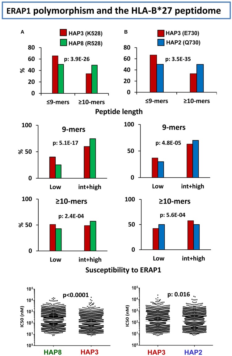
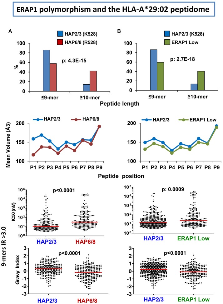
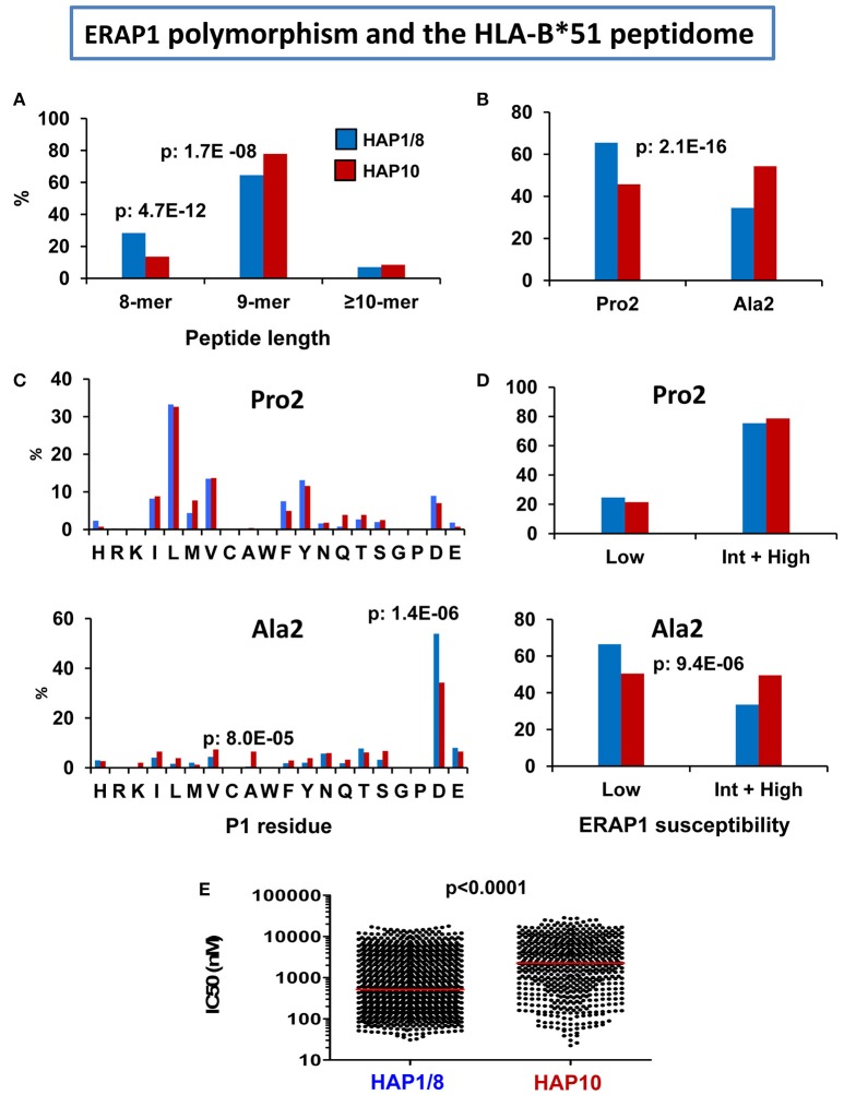
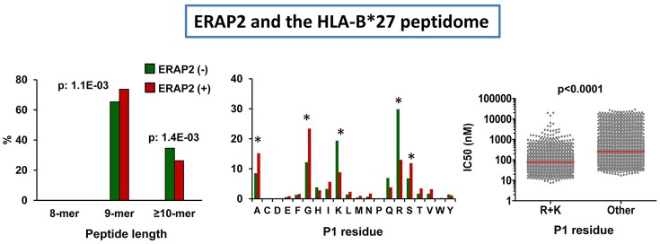
Four inflammatory diseases are strongly associated with Major Histocompatibility Complex class I (MHC-I) molecules: birdshot chorioretinopathy (HLA-A*29:02), ankylosing spondylitis (HLA-B*27), Behçet's disease (HLA-B*51), and psoriasis (HLA-C*06:02). The endoplasmic reticulum aminopeptidases (ERAP) 1 and 2 are also risk factors for these diseases. Since both enzymes are involved in the final processing steps of MHC-I ligands it is reasonable to assume that MHC-I-bound peptides play a significant pathogenetic role. This review will mainly focus on recent studies concerning the effects of ERAP1 and ERAP2 polymorphism and expression on shaping the peptidome of disease-associated MHC-I molecules in live cells. These studies will be discussed in the context of the distinct mechanisms and substrate preferences of both enzymes, their different patterns of genetic association with various diseases, the role of polymorphisms determining changes in enzymatic activity or expression levels, and the distinct peptidomes of disease-associated MHC-I allotypes. ERAP1 and ERAP2 polymorphism and expression induce significant changes in multiple MHC-I-bound peptidomes. These changes are MHC allotype-specific and, without excluding a degree of functional inter-dependence between both enzymes, reflect largely separate roles in their processing of MHC-I ligands. The studies reviewed here provide a molecular basis for the distinct patterns of genetic association of ERAP1 and ERAP2 with disease and for the pathogenetic role of peptides. The allotype-dependent alterations induced on distinct peptidomes may explain that the joint association of both enzymes and unrelated MHC-I alleles influence different pathological outcomes.

Keywords: Behçet's disease; ERAP; MHC; ankylosing spondylitis; antigen processing; inflammatory diseases; psoriasis; uveitis.


Figures

Figure 1
Figure 1
Major features of the peptidomes of disease-associated MHC-I molecules. Length distribution and % residue frequencies at P1, P2, and PC of B*27:05, A*29:02, B*51:01, and C*06:02 ligands (N: 1,117, 4,931, 1,620, and 1,287 peptides, respectively). The data are from the following references: B*27:05 (79), A*29:02 (66), B*51:01 (80), and C*06:02 (81).
Figure 2
Figure 2
Influence of ERAP1 polymorphism on the HLA-B*27 peptidome. (A) Effects of K528R. Comparison of the B*27:05 ligands over-represented (>1-fold) in a cell line (LG2, N: 2215 peptides) expressing the Hap3 variant of ERAP1 (red), with those over-represented in a cell line (C1R05, N: 2801) expressing the Hap8 variant (green). Hap3 and Hap8 differ by R127P, I276M, and K528R, but the two former changes have much less influence on peptide trimming. The comparisons involve peptide length (upper panel), joint frequencies of P1 residues with low or intermediate+high susceptibility to ERAP1 among 9-mers (second panel) or longer peptides (third panel), and theoretical affinity of the total ligands, with the medians indicated by bars. (B) Effects of E730Q. Comparison of the B*27:05 ligands over-represented (>1-fold) in the LG2 cell line (Hap3: red), with those over-represented in LCL 6370 (blue) expressing the Hap2 variant (N: 2,444 and 2,869 peptides, respectively). Hap3 and Hap2 differ only by the E730Q change. Conventions are as in (A). The statistical significance of the changes is indicated by the p-values, as estimated by the χ2 (three upper panels) or Mann–Whitney tests (lower panels). These data were originally published in reference (102).
Figure 3
Figure 3
Influence of ERAP1 on the A*29:02 peptidome. (A) Comparison of the A*29:02 ligands over-represented (>3-fold) in a cell line (PF97387, N: 292 peptides) expressing the Hap2/Hap3 variants of ERAP1 (blue), with those equally over-represented in a cell line (MOU, N: 383) expressing the Hap6/Hap8 variants (red). Both haplotype combinations differ by the R127P and K528R changes. The comparisons involve peptide length (upper panel), mean side chain volume at each peptide position among the 9-mers in these peptide sets (second panel), theoretical affinity of these 9-mers, with the medians indicated by bars (third panel), and hydropathy of the same peptides, estimated by the Grand Average of Hydropathy (GRAVY) index (lower panel). (B) Comparison of the A*29:02 ligands over-represented (>3-fold) in PF97387 (N: 446 peptides), with those equally over-represented in the cell line SWEIG (N: 390 peptides) expressing very low levels of ERAP1 (green). All conventions are as in (A). The statistical significance of the changes is indicated by the p-values, as estimated by the χ2 (upper panels) or Mann–Whitney tests (lower panels). These data were originally published in reference (66).
Figure 4
Figure 4
Influence of ERAP1 polymorphism on the HLA-B*51 peptidome. Comparison of the HLA-B*51:01 ligands from the transfectant cell line 721.221-B*51 (N: 1,271 peptides) expressing the Hap1/Hap8 variants of ERAP1 (blue), with the B*51:08 ligands from LCL BCH-30 (N: 624 peptides) expressing the Hap10 variant (red). The comparisons involve (A) peptide length distribution, (B) percent of peptides with Pro2 or Ala2, (C) Percent residue frequencies at P1 among peptides with Pro2 (upper panel) or Ala2 (lower panel), (D) percent of peptides with P1 residues showing low or intermediate+high susceptibility to ERAP1 trimming among peptides with Pro2 (upper panel) or Ala2 (lower panel), (E) theoretical affinity of B*51:01 and B*51:08 ligands for their respective B*51 subtypes, with the medians indicated by bars. The statistical significance of the changes is indicated by the p-values, as estimated by the χ2 (A–D) or Mann–Whitney tests (E). These data were originally published in reference (103).
Figure 5
Figure 5
Influence of ERAP2 on the HLA-B*27 peptidome. Comparison of the B*27:05 ligands over-represented (>1.5-fold) in the ERAP2-negative LCL 10151 (N: 764 peptides) expressing the Hap2 variant of ERAP1 (green), with those over-represented in the ERAP2 positive LCL 6370 (N: 817) expressing the same ERAP1 variant (red). The comparisons involve peptide length distribution (left panel), percent frequencies of P1 residues (middle panel), and theoretical affinity of HLA-B*27 ligands (N: 4,945), with N-terminal basic or other residues, with the medians indicated by bars. The statistical significance of the changes is indicated by the p-values (left and right panels) or asterisks (middle panel), as estimated by the χ2 (two left panels) or Mann–Whitney tests (right panel). The data in the two left panels and those in the right panel were originally published in Martin-Esteban et al. (101, 106), respectively.
Figure 6
Figure 6
Influence of ERAP2 on the A*29:02 peptidome. Comparison of the A*29:02 ligands over-represented (>1.5-fold) in the mock-transfected ERAP2-negative LCL PF97387 (PG-GFP, N: 839 peptides) expressing the Hap2/Hap3 variants of ERAP1 (green), with those (red) over-represented in the same ERAP2-transfected LCL (PF-ERAP2, N: 990 peptides). The comparisons involve peptide length distribution (left panel), joint frequencies of P1 residues showing low or intermediate+high susceptibility to ERAP2 trimming (middle panel) or showing low+intermediate or high hydropathy (right panel). The statistical significance of the changes is indicated by the p-values, as estimated by the χ2 test. These data were originally published in reference (93).

References

    1. Deitiker P, Atassi MZ. MHC genes linked to autoimmune disease. Crit Rev Immunol. (2015) 35:203–51. 10.1615/CritRevImmunol.2015014510 - DOI - PubMed
    1. Kuiper JJ, Van Setten J, Ripke S, Van 't SR, Mulder F, Missotten T, et al. A genome-wide association study identifies a functional ERAP2 haplotype associated with birdshot chorioretinopathy. Hum Mol Genet. (2014) 23:6081–7. 10.1093/hmg/ddu307 - DOI - PMC - PubMed
    1. Herbort CP, Jr, Pavesio C, LeHoang P, Bodaghi B, Fardeau C, Kestelyn P, et al. Why birdshot retinochoroiditis should rather be called ‘HLA-A29 uveitis’? Br J Ophthalmol. (2017) 101:851–5. 10.1136/bjophthalmol-2016-309764 - DOI - PMC - PubMed
    1. Brewerton DA, Hart FD, Nicholls A, Caffrey M, James DC, Sturrock RD. Ankylosing spondylitis and HL-A 27. Lancet (1973) 1:904–7. 10.1016/S0140-6736(73)91360-3 - DOI - PubMed
    1. Schlosstein L, Terasaki PI, Bluestone R, Pearson CM. High association of an HL-A antigen, W27, with ankylosing spondylitis. N Engl J Med. (1973) 288:704–6. 10.1056/NEJM197304052881403 - DOI - PubMed

Publication types

MeSH terms

Substances