

Synovial sarcoma of bone: Sarcoma typically of soft tissues presenting as a primary bone tumor
- PMID: 30425775
- PMCID: PMC6231113
- DOI: 10.1016/j.radcr.2018.10.026
Synovial sarcoma of bone: Sarcoma typically of soft tissues presenting as a primary bone tumor
Abstract
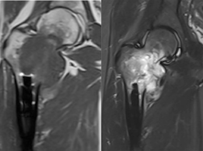
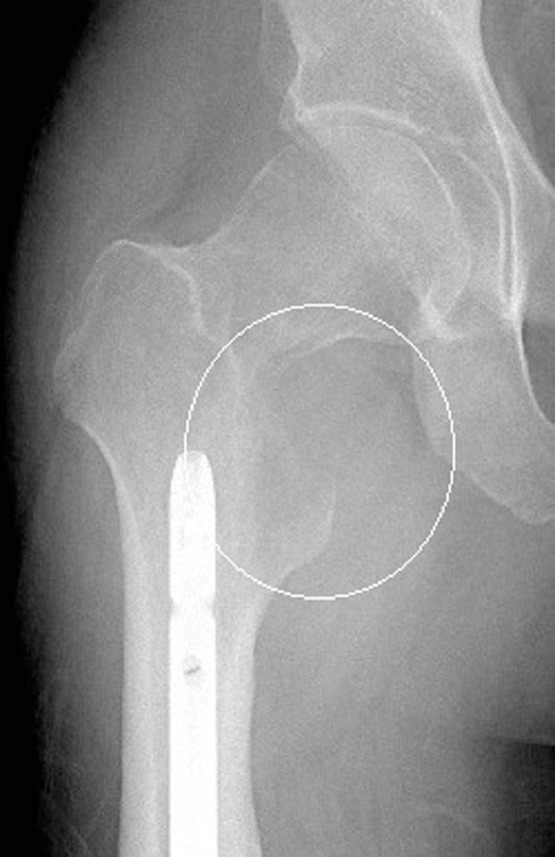
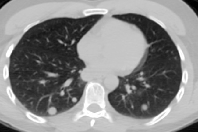
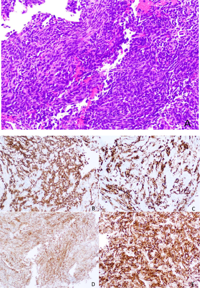
Synovial sarcoma typically presents as periarticular soft tissue mass in adolescent and young adult patients. Very rarely, soft tissue sarcomas may arise primarily within bone posing a significant diagnostic challenge as primary osseous malignancies such as osteosarcoma and metastatic disease are much more common. While tissue sampling with immunohistochemical and genetic testing are required for definitive diagnosis, radiologists and orthopedic oncologists should consider alternate etiologies when typical imaging features of more common bone tumors are not identified. As an example, we present a 33-year-old male referred with a pathologic hip fracture proven to represent primary synovial sarcoma of bone.
Keywords: Primary bone tumor; Synovial sarcoma.
Figures
Similar articles
-
Lymphatic dissemination of bone and soft tissue sarcomas: a lymphographic investigation.Acta Radiol Suppl. 1976;349:1-84. Acta Radiol Suppl. 1976. PMID: 206099 Review.
-
Primary Intraosseous Synovial Sarcoma with Molecular Confirmation: Expanding and Clarifying the Spectrum of This Rare Neoplasm.Case Rep Pathol. 2020 Jan 28;2020:5492754. doi: 10.1155/2020/5492754. eCollection 2020. Case Rep Pathol. 2020. PMID: 32082672 Free PMC article.
-
Fluorescence in Situ Hybridization (FISH) for Differential Diagnosis of Soft Tissue Sarcomas.Asian Pac J Cancer Prev. 2018 Mar 27;19(3):655-660. doi: 10.22034/APJCP.2018.19.3.655. Asian Pac J Cancer Prev. 2018. PMID: 29580035 Free PMC article.
-
Primary pulmonary synovial sarcoma: Diagnosis on squash smears.J Cytol. 2015 Jan-Mar;32(1):56-8. doi: 10.4103/0970-9371.155240. J Cytol. 2015. PMID: 25948950 Free PMC article.
-
Soft Tissue Tumors Rarely Presenting Primary in Bone; Diagnostic Pitfalls.Surg Pathol Clin. 2017 Sep;10(3):705-730. doi: 10.1016/j.path.2017.04.013. Epub 2017 Jun 29. Surg Pathol Clin. 2017. PMID: 28797510 Review.
Cited by
-
Case Report: Primary Intraosseous Poorly Differentiated Synovial Sarcoma of the Femur.Front Oncol. 2022 Mar 16;12:754131. doi: 10.3389/fonc.2022.754131. eCollection 2022. Front Oncol. 2022. PMID: 35372059 Free PMC article.
-
Knowledge of cytogenetic analysis for synovial sarcoma in sarcomatoid variant renal cell carcinoma.Clin Case Rep. 2021 Nov 7;9(11):e05052. doi: 10.1002/ccr3.5052. eCollection 2021 Nov. Clin Case Rep. 2021. PMID: 34765219 Free PMC article.
-
Synovial sarcoma: the misdiagnosed sarcoma.EFORT Open Rev. 2024 Mar 5;9(3):190-201. doi: 10.1530/EOR-23-0193. EFORT Open Rev. 2024. PMID: 38457918 Free PMC article. Review.
-
Synovial Sarcoma: A Clinical Review.Curr Oncol. 2021 May 19;28(3):1909-1920. doi: 10.3390/curroncol28030177. Curr Oncol. 2021. PMID: 34069748 Free PMC article. Review.
-
Insidious Synovial Sarcoma of Bone in a Patient with Rheumatoid Arthritis.Rev Bras Ortop (Sao Paulo). 2021 Nov 11;59(Suppl 1):e31-e33. doi: 10.1055/s-0041-1739172. eCollection 2024 Jul. Rev Bras Ortop (Sao Paulo). 2021. PMID: 39027174 Free PMC article.
References
-
- American Cancer Society Website Cancers that develop in young adults. 2018. https://www.cancer.org/cancer/cancer-in-young-adults/cancers-in-young-ad... [cited 2018 Sept 18]; available at.
-
- Kransdorf M.J., Murphey M.D. Imaging of soft tissue tumors. 2nd ed. Lippincott Williams & Wilkins; Philadelphia, PA: 2006. Malignant soft tissue tumors; pp. 7–37.
-
- Kransdorf M.J. Malignant soft-tissue tumors in a large referral population: distribution of diagnoses by age, sex, and location. Am J Roentgenol. 1995;164:129–134. - PubMed
Publication types
LinkOut - more resources
Full Text Sources
Research Materials
