

Histological analysis of post-eruption tooth wear adaptations, and ontogenetic changes in tooth implantation in the acrodontan squamate Pogona vitticeps
- PMID: 30425900
- PMCID: PMC6230436
- DOI: 10.7717/peerj.5923
Histological analysis of post-eruption tooth wear adaptations, and ontogenetic changes in tooth implantation in the acrodontan squamate Pogona vitticeps
Abstract
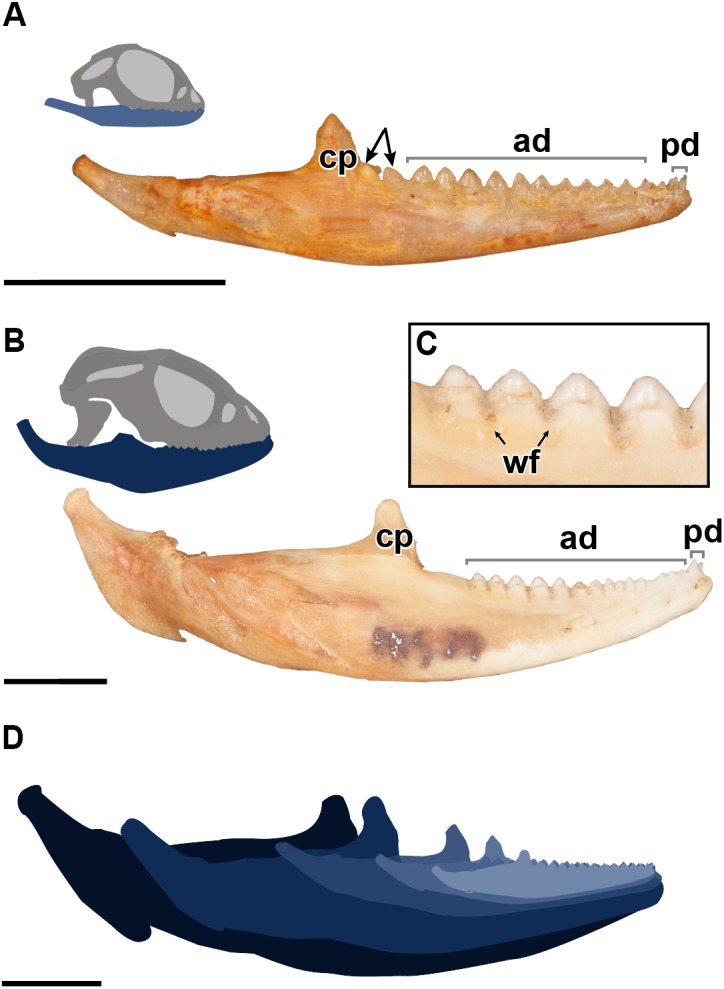
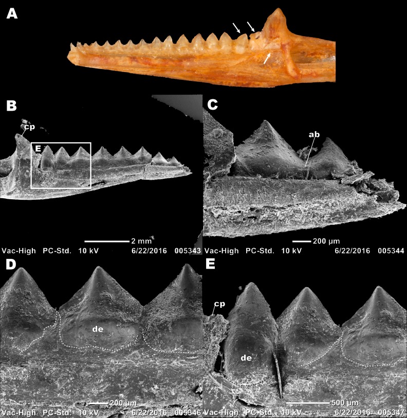
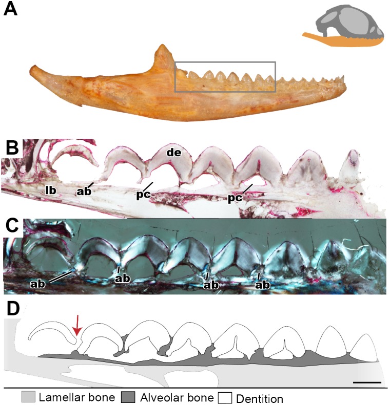
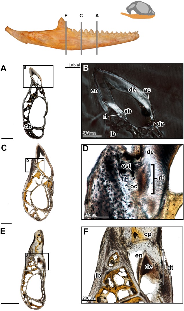
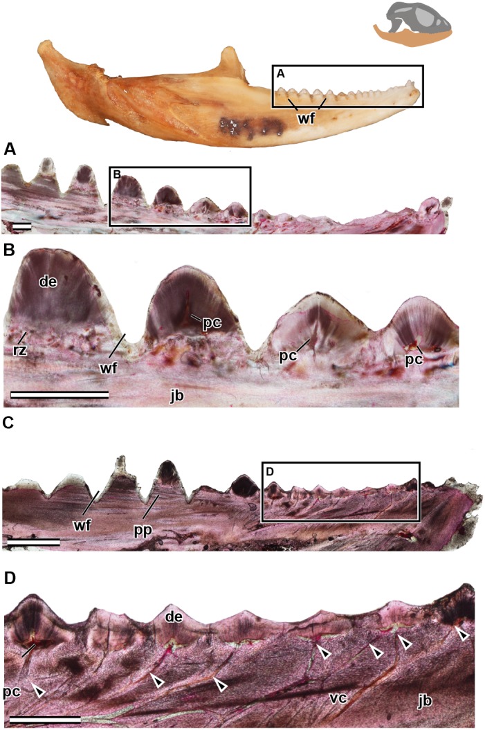
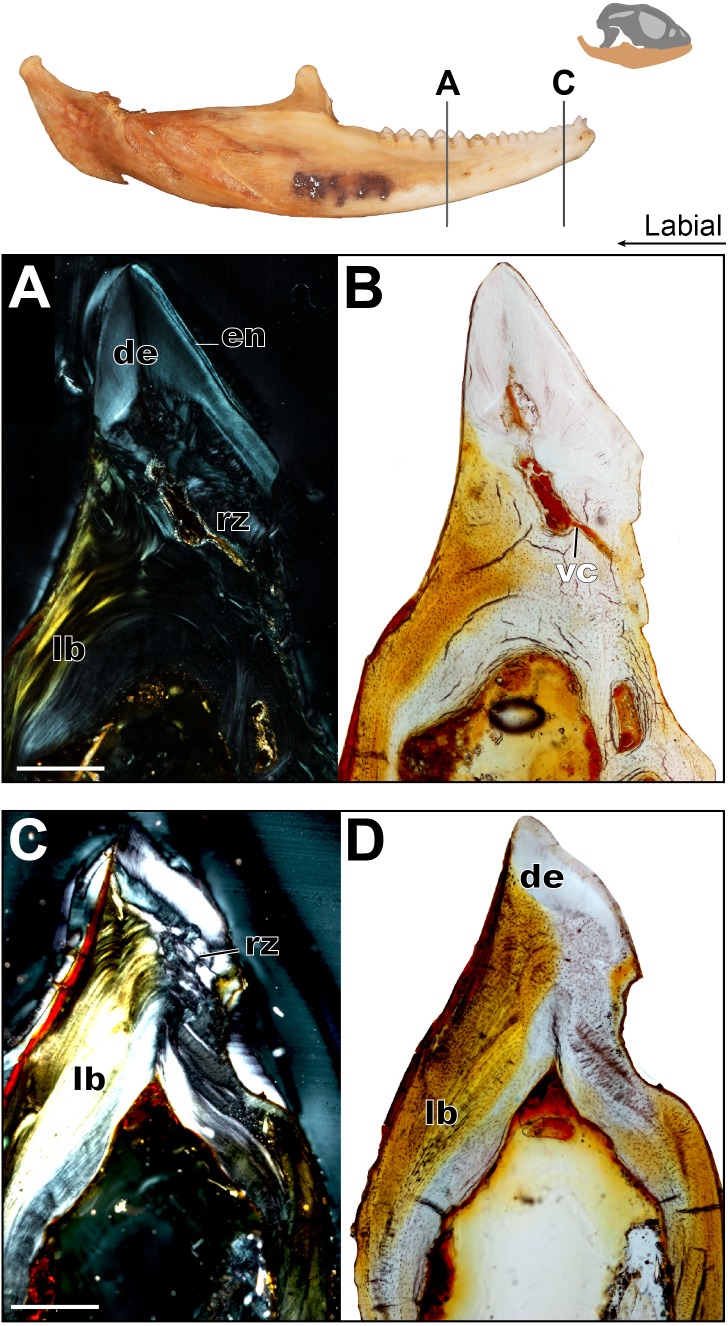
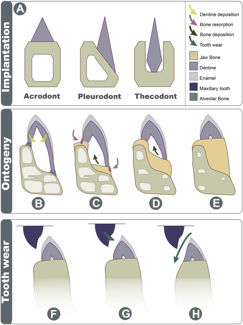
Teeth have been a focus of research in both extinct and extant taxa alike; a significant portion of dental literature is concerned with dental patterning and replacement. Most non-mammalian vertebrates continuously replace their dentition but an anomalous group of squamates has forgone this process in only having one tooth generation; these squamates all have apically implanted teeth, a condition known as acrodonty. Acrodont dentition and various characteristics attributed to it, including a lack of replacement, have often been defined ambiguously. This study explores this type of implantation through histology in the ontogeny of the acrodont agamid Pogona vitticeps. The non-replacing teeth of this squamate provides an opportunity to study wear adaptations, maintenance of occlusion in a non-mammalian system, and most importantly post-eruption changes in the tooth bone interface. In this study the post-eruption changes combined with dental wear likely gives the appearance of acrodont implantation.
Keywords: Acrodont; Histology; Ontogeny; Squamate; Tooth wear.
Conflict of interest statement
The authors declare there are no competing interests.
Figures
Similar articles
-
The Permian reptile Opisthodontosaurus carrolli: a model for acrodont tooth replacement and dental ontogeny.J Anat. 2018 Mar;232(3):371-382. doi: 10.1111/joa.12754. Epub 2017 Dec 6. J Anat. 2018. PMID: 29210080 Free PMC article.
-
Squamates as a model to understand key dental features of vertebrates.Dev Biol. 2024 Dec;516:1-19. doi: 10.1016/j.ydbio.2024.07.011. Epub 2024 Jul 26. Dev Biol. 2024. PMID: 39069116 Review.
-
Skull Development, Ossification Pattern, and Adult Shape in the Emerging Lizard Model Organism Pogona vitticeps: A Comparative Analysis With Other Squamates.Front Physiol. 2018 Mar 28;9:278. doi: 10.3389/fphys.2018.00278. eCollection 2018. Front Physiol. 2018. PMID: 29643813 Free PMC article.
-
Bite force data suggests relationship between acrodont tooth implantation and strong bite force.PeerJ. 2020 Jun 30;8:e9468. doi: 10.7717/peerj.9468. eCollection 2020. PeerJ. 2020. PMID: 32656000 Free PMC article.
-
Current Perspectives on Tooth Implantation, Attachment, and Replacement in Amniota.Front Physiol. 2018 Nov 21;9:1630. doi: 10.3389/fphys.2018.01630. eCollection 2018. Front Physiol. 2018. PMID: 30519190 Free PMC article. Review.
Cited by
-
Tooth attachment and pleurodont implantation in lizards: Histology, development, and evolution.J Anat. 2021 May;238(5):1156-1178. doi: 10.1111/joa.13371. Epub 2020 Dec 29. J Anat. 2021. PMID: 33372719 Free PMC article.
-
Squamate egg tooth development revisited using three-dimensional reconstructions of brown anole (Anolis sagrei, Squamata, Dactyloidae) dentition.J Anat. 2020 Jun;236(6):1004-1020. doi: 10.1111/joa.13166. Epub 2020 Feb 13. J Anat. 2020. PMID: 32056203 Free PMC article.
-
A new sphenodontian (Diapsida: Lepidosauria) from the Upper Triassic (Norian) of Germany and its implications for the mode of sphenodontian evolution.BMC Ecol Evol. 2024 Mar 16;24(1):35. doi: 10.1186/s12862-024-02218-1. BMC Ecol Evol. 2024. PMID: 38493125 Free PMC article.
-
Multiple evolutionary origins and losses of tooth complexity in squamates.Nat Commun. 2021 Oct 14;12(1):6001. doi: 10.1038/s41467-021-26285-w. Nat Commun. 2021. PMID: 34650041 Free PMC article.
-
The developmental origins of heterodonty and acrodonty as revealed by reptile dentitions.Sci Adv. 2021 Dec 17;7(51):eabj7912. doi: 10.1126/sciadv.abj7912. Epub 2021 Dec 17. Sci Adv. 2021. PMID: 34919438 Free PMC article.
References
-
- Ananjeva N, Smirina E. Growth layers in bones and acrodont teeth of the agamid lizard Laudakia stoliczkana (Blanford, 1875) (Agamidae, Sauria) Amphibia-Reptilia. 2007;28:193–204. doi: 10.1163/156853807780202512. - DOI
-
- Benton MJ. Tooth form, growth, and function in Triassic rhynchosaurs (Reptilia, Diapsida) Palaeontology. 1984;27(Part 4):737–776.
-
- Berkovitz BK, Shellis RP. The teeth of non-mammalian vertebrates. Academic Press; Cambridge: 2016.
LinkOut - more resources
Full Text Sources
