. 2018 Nov 13;4(1):132.
doi: 10.1186/s40792-018-0539-8.

Two cases of radiation-associated angiosarcoma of the breast

Affiliations

Two cases of radiation-associated angiosarcoma of the breast

Yuki Nomoto et al. Surg Case Rep. .

Abstract

Background: The incidence of radiation-associated angiosarcoma (RAA) of the breast has been increasing, and its prognosis is reportedly poor. It is important to remove tumor tissues completely to prevent recurrence.

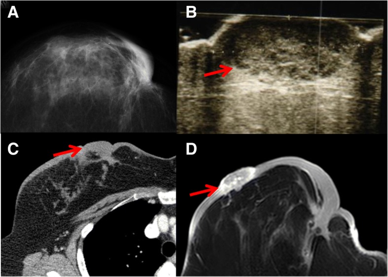
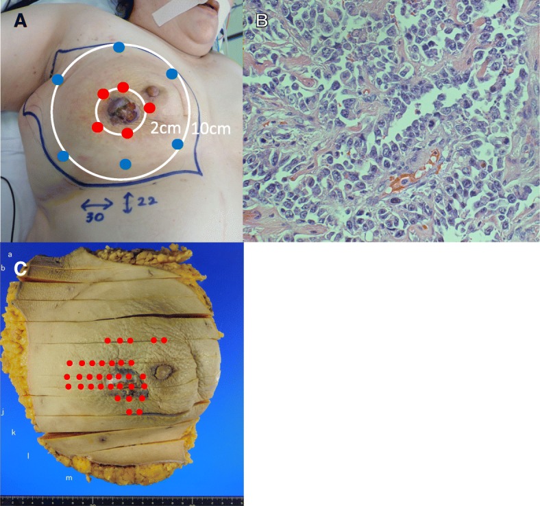
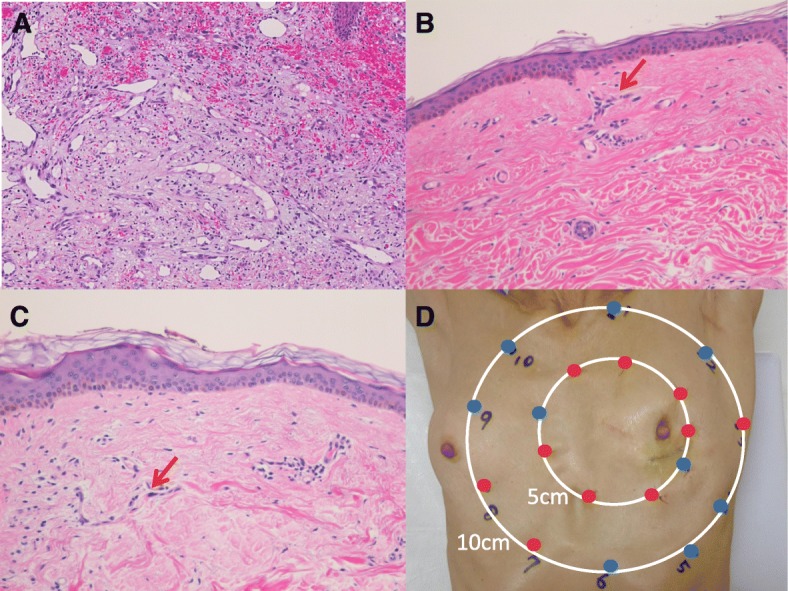
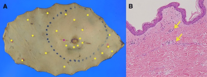
Case presentation: We report two cases of patients with RAA of the breast. Both patients had a nodule in their remaining breast a few years after undergoing breast-conserving surgery and radiation therapy for breast cancer. The nodules were diagnosed as angiosarcoma by skin biopsy and open biopsy, respectively. To determine the extent of lesion spread, mapping biopsy was performed before surgery. Both patients underwent mastectomy, extensive skin resection, and split skin grafting. Pathological findings showed that their tumors could be completely resected. After surgery, chemotherapy was performed.

Conclusion: In our cases, no local or distant recurrence has been detected in either patient for over 4 years. We identified the range of tumor invasion by preoperative mapping biopsy and completely resected all tumor tissue.

Keywords: Angiosarcoma; Breast cancer; Breast-conserving treatment; Radiation therapy; Secondary angiosarcoma.


Conflict of interest statement

Ethics approval and consent to participate

Not applicable.

Consent for publication

Not applicable.

Competing interests

The authors declare that they have no competing interests.

Publisher’s Note

Springer Nature remains neutral with regard to jurisdictional claims in published maps and institutional affiliations.

Figures

Fig. 1
Fig. 1
Imaging findings (case 1). a Mammography. b Breast ultrasound. c Computed tomography. d Magnetic resonance imaging. Arrow: tumor
Fig. 2
Fig. 2
Macroscopic and pathological findings (case 1). a Mapping biopsy was performed at 2 cm and 10 cm from the nodule. Red dots indicate positivity for tumor cells, and blue dots indicate negativity. b Pathological image of the resected specimen. It was diagnosed as angiosarcoma (hematoxylin-eosin [H.E.] staining: × 400). c Red dots indicate where tumor cells were observed. Tumor invasion was observed in a wider area outside the nodule
Fig. 3
Fig. 3
Macroscopic and microscopic pathological findings (case 2). a The nodule was diagnosed as angiosarcoma by open biopsy (H.E. staining: × 20). b Mapping biopsy 5 cm from the surgical trace (H.E. staining: × 20). Arrow: atypical endothelial cells. c Mapping biopsy 10 cm from the surgical trace (H.E. staining: × 20). Arrow: atypical endothelial cells. d Mapping biopsy was performed 5 cm and 10 cm away from the surgical trace. The red dots indicate areas where atypical endothelial cells were observed, and blue dots indicate areas where no atypical endothelial cells were observed
Fig. 4
Fig. 4
Macroscopic findings of mapping biopsy and pathological examination (case 2). a A red dot indicates the surgical trace. Yellow dots indicate locations at which atypical endothelial cells were observed. b Atypical endothelial cells, in which the degree of atypia was less than that of the primary tumor (H.E. staining: × 20). Arrow: atypical endothelial cells

References

    1. Veronesi U, Cascinelli N, Mariani L, Greco M, Saccozzi R, Luini A, et al. Twenty-year follow-up of a randomized study comparing breast-conserving surgery with radical mastectomy for early breast cancer. N Engl J Med. 2002;347:1227–1232. doi: 10.1056/NEJMoa020989. - DOI - PubMed
    1. Yap J, Chuba PJ, Thomas R, Aref A, Lucas D, Severson RK, et al. Sarcoma as a second malignancy after treatment for breast cancer. Int J Radiat Oncol Biol Phys. 2002;52:1231–1237. doi: 10.1016/S0360-3016(01)02799-7. - DOI - PubMed
    1. Chesebro AL, Chikarmane SA, Gombos EC, Giardino AA. Radiation-associated angiosarcoma of the breast: what the radiologist needs to know. AJR Am J Roentgenol. 2016;207(1):217–225. doi: 10.2214/AJR.15.15888. - DOI - PubMed
    1. Chikarmane SA, Gombos EC, Jagadeesan J, Raut C, Jagannathan JP. MRI findings of radiation-associated angiosarcoma of the breast (RAS) J Magn Reson Imaging. 2015;42:763–770. doi: 10.1002/jmri.24822. - DOI - PMC - PubMed
    1. Brenn T, Fletcher CD. Postradiation vascular proliferations: an increasing problem. Histopathology. 2006;48:106–114. doi: 10.1111/j.1365-2559.2005.02293.x. - DOI - PubMed

LinkOut - more resources