. 2019 Mar;8(3):301-312.
doi: 10.1002/sctm.18-0056. Epub 2018 Nov 13.

Serum from Asthmatic Mice Potentiates the Therapeutic Effects of Mesenchymal Stromal Cells in Experimental Allergic Asthma

Affiliations

Serum from Asthmatic Mice Potentiates the Therapeutic Effects of Mesenchymal Stromal Cells in Experimental Allergic Asthma

Soraia C Abreu et al. Stem Cells Transl Med. 2019 Mar.

Abstract

Asthma is a chronic inflammatory disease characterized by airway inflammation and remodeling, which can lead to progressive decline of lung function. Although mesenchymal stromal cells (MSCs) have shown beneficial immunomodulatory properties in preclinical models of allergic asthma, effects on airway remodeling have been limited. Mounting evidence suggests that prior exposure of MSCs to specific inflammatory stimuli or environments can enhance their immunomodulatory properties. Therefore, we investigated whether stimulating MSCs with bronchoalveolar lavage fluid (BALF) or serum from asthmatic mice could potentiate their therapeutic properties in experimental asthma. In a house dust mite (HDM) extract asthma model in mice, unstimulated, asthmatic BALF-stimulated, or asthmatic serum-stimulated MSCs were administered intratracheally 24 hours after the final HDM challenge. Lung mechanics and histology; BALF protein, cellularity, and biomarker levels; and lymph-node and bone marrow cellularity were assessed. Compared with unstimulated or BALF-stimulated MSCs, serum-stimulated MSCs further reduced BALF levels of interleukin (IL)-4, IL-13, and eotaxin, total and differential cellularity in BALF, bone marrow and lymph nodes, and collagen fiber content, while increasing BALF IL-10 levels and improving lung function. Serum stimulation led to higher MSC apoptosis, expression of various mediators (transforming growth factor-β, interferon-γ, IL-10, tumor necrosis factor-α-stimulated gene 6 protein, indoleamine 2,3-dioxygenase-1, and IL-1 receptor antagonist), and polarization of macrophages to M2 phenotype. In conclusion, asthmatic serum may be a novel strategy to potentiate therapeutic effects of MSCs in experimental asthma, leading to further reductions in both inflammation and remodeling than can be achieved with unstimulated MSCs. Stem Cells Translational Medicine 2019;8:301&312.

Keywords: Animal models; Bone marrow stromal cells; Cell therapy; Eosinophils; Lung; Macrophages.


Conflict of interest statement

D.J.W. has received research funding from Athersys Inc., Biotek Inc., and United Therapeutics Inc. The other authors indicated no potential conflicts of interest.

Figures
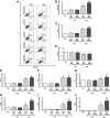
Figure 1
Figure 1
In vitro stimulation of mesenchymal stromal cells (MSCs) with serum yielded further cellular death and modified the MSC secretome. (A): Apoptosis was measured in MSCs by assessing the percentage of annexin‐V+/propidium iodide cells by flow cytometry. Representative plots for MSCs: unstimulated or stimulated with bronchoalveolar lavage fluid (BALF) or serum from control group (CTRL) and house dust mite (HDM)‐challenged mice for 24 hours. Gene expression of caspase‐3 (B), Bax (a proapoptotic Bcl‐2 family protein; C), Bcl‐2 (B‐cell lymphoma 2; D), transforming growth factor (TGF)‐β1 (E), interferon (IFN)‐γ (F), IL‐10 (G), tumor necrosis factor‐α‐stimulated gene 6 protein (TSG‐6); (H), indoleamine 2,3‐dioxygenase‐1 (IDO‐1); (I), and IL‐1 receptor antagonist (IL‐1RN); (J) was assessed by RT‐PCR in cells after 24 hours of each specific stimulation. Data represent relative gene expression calculated as a ratio of average expression of the target gene compared with the reference gene (GAPDH) and expressed as fold change relative to unstimulated MSCs. Bars represent mean ± SD. *, Significantly different from MSC (p < .05). **, Significantly different from BALF‐CTRL MSCs (p < .05). #, Significantly different from SERUM‐CTRL MSCs (p < .05). , Significantly different from BALF‐HDM MSCs (p < .05).
Figure 2
Figure 2
In vitro MSC stimulation with SERUM‐HDM induced further macrophage polarization to the M2 profile. Alveolar macrophages (105 cells per well) from HDM mice were cultured with conditioned media obtained from MSCs (105 cells per well) either unstimulated or stimulated with BALF or SERUM from CTRL and HDM‐challenged mice for 24 hours. Relative gene expression of (A) iNOS, (B) TNF‐α, (C) IL‐1β, (D) arginase‐2, (E) IL‐10, and (F) TGF‐β was calculated as a ratio of the average gene expression levels compared with the reference gene (GAPDH) and expressed as fold changes relative to MΦ group (alveolar macrophages cultured with conditioned media from unstimulated MSCs). Bars are means + SD of four wells per condition. *, Significantly different from MΦ (p < .05). **, Significantly different from MSC‐BALF‐CTRL (p < .05). #, Significantly different from MSC‐SERUM‐CTRL (p < .05). , Significantly different from MSC‐BALF‐HDM (p < .05). Abbreviations: MSC, mesenchymal stromal cell; iNOS, inducible nitric oxide synthase; IFN‐α, interferon‐α; IL, interleukin; TNF‐α, tumor necrosis factor‐α; TGF‐β, transforming growth factor‐β; HDM, house dust mite; BALF, bronchoalveolar lavage fluid.
Figure 3
Figure 3
Stimulation of MSCs with serum yielded further modulation of mediators in the lung in vivo. Protein levels of IL‐4, IL‐13, eotaxin, and IL‐10 in BALF. CTRL, phosphate‐buffered saline‐challenged mice; HDM, HDM‐challenged mice; SAL, HDM mice treated with saline; MSC, HDM mice treated with unstimulated MSCs; HDM‐MSC‐BALF, HDM mice treated with MSCs stimulated with BALF from asthmatic mice; HDM‐MSC‐SERUM, HDM mice treated with MSCs stimulated with serum from asthmatic mice. Boxes show the interquartile range (P25–P75) range, whiskers denote the range (minimum–maximum), and horizontal lines represent the median of eight animals per group. *, Significantly different from CTRL (p < .05). **, Significantly different from HDM‐SAL (p < .05). Abbreviations: MSCs, mesenchymal stromal cells; HDM, house dust mite; IL, interleukin; BALF, bronchoalveolar lavage fluid.
Figure 4
Figure 4
Ex vivo stimulation of MSCs with serum yielded further reduction of inflammation in the lungs in vivo. (A): Tissue cellularity: fractional area of mononuclear (MN, white bar) and polymorphonuclear (PMN, black bar) cells in lung tissue, (B) total leukocytes, (C) eosinophils, (D) lymphocytes, (E) macrophages, (F) neutrophils, (G) B220+ cells, (H) CD4+ cells, and (I) CD4+CD25+FoxP3+ cells in BALF. CTRL, phosphate‐buffered saline‐challenged mice; HDM, HDM‐challenged mice; SAL, HDM mice treated with saline; MSC, HDM mice treated with unstimulated MSCs; HDM‐MSC‐BALF, HDM mice treated with MSCs stimulated with BALF from asthmatic mice; HDM‐MSC‐SERUM, HDM mice treated with MSCs stimulated with serum from asthmatic mice. Data are presented as mean ± SD (A). Boxes show the interquartile (P25–P75) range, whiskers denote the range (minimum–maximum), and horizontal lines represent the median of eight animals per group (B–I). *, Significantly different from CTRL (p < .05). **, Significantly different from HDM‐SAL (p < .05). #, Significantly different from HDM‐MSC (p < .05). Abbreviations: BALF, bronchoalveolar lavage fluid; MSC, mesenchymal stromal cells; HDM, house dust mite.
Figure 5
Figure 5
Ex vivo stimulation of MSCs with serum yielded further reduction of lung remodeling in vivo. Collagen fiber content in (A) lung parenchyma and (B) airway. Representative photomicrographs of lung parenchyma (original magnification: ×200) and airways (original magnification: ×400) stained by the Picrosirius‐polarization method (C). CTRL, phosphate‐buffered saline‐challenged mice; HDM, HDM‐challenged mice; SAL, HDM mice treated with saline; MSC, HDM mice treated with unstimulated MSCs; HDM‐MSC‐BALF, HDM mice treated with MSCs stimulated with BALF from asthmatic mice; HDM‐MSC‐SERUM, HDM mice treated with MSCs stimulated with serum from asthmatic mice. Boxes show the interquartile (P25–P75) range, whiskers denote the range (minimum–maximum), and horizontal lines represent the median of eight animals per group. Scale bars: 100 μm. *, Significantly different from CTRL (p < .05). **, Significantly different from HDM‐SAL (p < .05). Abbreviations: BALF, bronchoalveolar lavage fluid; MSC, mesenchymal stromal cells; HDM, house dust mite.
Figure 6
Figure 6
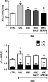
Ex vivo stimulation of MSCs with serum led to further improvement in lung mechanics in vivo. Static lung elastance (Est,L; upper panel), resistive (ΔP1,L), and viscoelastic (ΔP2,L) pressures (lower panel). CTRL, phosphate‐buffered saline‐challenged mice; HDM, HDM‐challenged mice; SAL, HDM mice treated with saline; MSC, HDM mice treated with unstimulated MSCs; HDM‐MSC‐BALF, HDM mice treated with MSCs stimulated with BALF from asthmatic mice; HDM‐MSC‐SERUM, HDM mice treated with MSCs stimulated with serum from asthmatic mice. Bars represent mean ± SD of eight animals per group. *, Significantly different from CTRL (p < .05). **, Significantly different from HDM‐SAL (p < .05). #, Significantly different from HDM‐MSC (p < .05). Abbreviations: BALF, bronchoalveolar lavage fluid; MSC, mesenchymal stromal cells; HDM, house dust mite.
Figure 7
Figure 7
Ex vivo stimulation of MSCs with serum led to further reductions of cellularity in bone marrow and mLN in vivo. (A): Total leukocytes, (B) eosinophils, (C) lymphocytes, (D) macrophages, (E) neutrophils in bone marrow, (F) total leukocytes, (G) CD4+ cells, and (H) B cells in mediastinal lymph nodes (mLN). CTRL, phosphate‐buffered saline‐challenged mice; HDM, HDM‐challenged mice; SAL, HDM mice treated with saline; MSC, HDM mice treated with unstimulated MSCs; HDM‐MSC‐BALF, HDM mice treated with MSCs stimulated with BALF from asthmatic mice; HDM‐MSC‐SERUM, HDM mice treated with MSCs stimulated with serum from asthmatic mice. Boxes show the interquartile (P25–P75) range, whiskers denote the range (minimum–maximum), and horizontal lines represent the median of eight animals per group. *, Significantly different from CTRL (p < .05). **, Significantly different from HDM‐SAL (p < .05). Abbreviations: BALF, bronchoalveolar lavage fluid; MSC, mesenchymal stromal cells; HDM, house dust mite.

References

    1. Global Strategy for Asthma Management and Prevention . Global Iniciative for Asthma; 2018. Available at https://ginasthma.org/2018‐gina‐report‐global‐strategy‐for‐asthma‐manage.... Accessed September 27, 2018.
    1. Royce SG, Tang ML. The effects of current therapies on airway remodeling in asthma and new possibilities for treatment and prevention. Curr Mol Pharmacol 2009;2:169–181. - PubMed
    1. Barnes PJ. New therapies for asthma: Is there any progress? Trends Pharmacol Sci 2010;31:335–343. - PubMed
    1. Papierniak ES, Lowenthal DT, Harman E. Novel therapies in asthma: Leukotriene antagonists, biologic agents, and beyond. Am J Ther 2013;20:79–103. - PubMed
    1. Cruz FF, Borg ZD, Goodwin M et al. Systemic administration of human bone marrow‐derived mesenchymal stromal cell extracellular vesicles ameliorates aspergillus hyphal extract‐induced allergic airway inflammation in immunocompetent mice. Stem Cells Translational Medicine 2015;4:1302–1316. - PMC - PubMed

Publication types