. 2018;18(74):255-264.
doi: 10.15557/JoU.2018.0037.

TRUS-guided drainage of the ectopic ureter entering the prostatic urethra and TRUS-guided transurethral neo-orifice formation using holmium laser

Affiliations

TRUS-guided drainage of the ectopic ureter entering the prostatic urethra and TRUS-guided transurethral neo-orifice formation using holmium laser

Waldemar Białek et al. J Ultrason. 2018.

Abstract

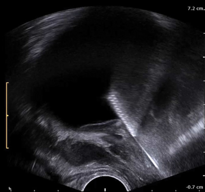
A fifty-nine year-old male was hospitalized for exacerbation of chronic pancreatitis. As a gigantic cyst of the pancreatic tail was identified, it was fused with the jejunal loop. Due to persistent fever and severe symptoms in the storage and voiding phases, the patient was referred to a urologist. Because transrectal ultrasound examination revealed a fluid collection resembling the left seminal vesicle filled with purulent material, a transrectal puncture procedure was performed. The analysis of computed tomography scans led to the diagnosis of duplicated collecting system of the left kidney with the enormous ureter of the upper moiety that entered the prostate gland. In order to permanently decompress the hydronephrosed upper moiety of the left kidney, the patient was deemed eligible for endoscopic treatment. A transurethral incision through the bladder wall and the adjacent segment of the ectopic ureter was made with holmium laser under transrectal ultrasonography guidance, thus creating a neo-orifice of this ureter.

Keywords: TRUS; ectopic ureter; holmium laser; neo-orifice; pancreatic cyst.


Conflict of interest statement

Conflict of interest

Authors do not report any financial or personal connections with other persons or organizations, which might negatively affect the contents of this publication and/or claim authorship rights to this publication.

Figures

Fig. 1.
Fig. 1.
Axial contrast-enhanced computed tomography. On the left, note a huge fluid collection with smooth outlines, impinging on the tail of the pancreas (interpreted as a cyst of the tail of the pancreas)
Fig. 2.
Fig. 2.
Axial computed tomography, arterial phase. The cystic lesion that compensates the upper left renal pole contains both dense material and gas
Fig. 3.
Fig. 3.
Axial computed tomography. Above the left kidney, there is a thick-walled fluid space with air-fluid level interpreted as a cyst of the tail of the pancreas
Fig. 4.
Fig. 4.
Transabdominal ultrasound of the urinary bladder in the transverse section. A collection with dense fluid material, measuring 28 × 49 mm, elevates the urinary bladder in the region of the trigone and left ureteral orifice. The lesion was interpreted as a seminal vesicle abscess. The examination was performed with a convex 6 MHz probe (MyLab C, Esaote, Italy)
Fig. 5.
Fig. 5.
A transrectal scan of the lesion that elevates the trigone of the urinary bladder on the left side. Owing to the dense contents, the location and the presence of incomplete septa, the collection was interpreted as a congested seminal vesicle filled with purulent material. The examination was performed with an endorectal 9 MHz probe (MyLab C, Esaote, Italy)
Fig. 6.
Fig. 6.
An image recorded during ultrasound-guided transrectal puncture of the lesion interpreted as the left seminal vesicle (endorectal 9 MHz probe, MyLab C, Esaote, Italy)
Fig. 7.
Fig. 7.
An ultrasound image of the left kidney with a collection of dense fluid in the cephalad position. The lesion displacing the tail of the pancreas, spleen and the left kidney was interpreted as a cyst of the tail of the pancreas. The examination was performed with a convex 6 MHz probe (MyLab C, Esaote, Italy)
Fig. 8.
Fig. 8.
Magnetic resonance imaging conducted after the puncture procedure, aspiration of the dense purulent material from the left ectopic ureter and lavage with an amikacin solution. An axial scan performed at the level of the base of the prostate gland presents a narrowing segment of the ectopic ureter that goes beyond the midline to enter the prostatic urethra between the seminal colliculus and the neck of the urinary bladder on the right side
Fig. 9.
Fig. 9.
A transrectal prostate image in the sagittal section. A cystic lesion in the neighborhood of the prostatic urethra corresponds with the cross-section of the distal segment of the ectopic ureter. It was considered to make an incision and dilatation of the ureter end, narrowed along the fragment of 3 mm, from the side of the prostatic urethra. Another option was to make an incision on the neck of the urinary bladder at hour 6 to the depth of at least 5 mm so that the distal segment of the ectopic ureter, when healed, could open on the border of the neck and trigone of the urinary bladder. Due to the risk of re-stricure and the impossibility of leaving a DJ ureteral stent inserted in this location for the time of the healing period, it was proposed to join the distal segment of the ectopic ureter and urinary bladder between the bladder neck and the opening of the normal lower moiety ureter. The examination was performed with a transrectal end-fire probe with the frequency of 6 MHz (ProFocus Ultraview, BK Medical, Denmark)
Fig. 10.
Fig. 10.
A sagittal image of the prostate and urinary bladder. The laser resectoscope tip can be noticed in the urinary bladder. The typical stripes on the resectoscope tip in ultrasonography represent the perforated coat of the tool. The examination was performed with a transrectal end-fire probe with the frequency of 12 MHz (ProFocus Ultraview, BK Medical, Denmark)
Fig. 11.
Fig. 11.
Thanks to transrectal imaging, one may trace the resectoscope tip on the ultrasound monitor. The image from the resectoscope camera enables assessment of the urinary bladder mucosa, while ultrasound shows sections through the entire wall of the bladder and its neighborhood, and presents the relationship of the resectoscope with the structures adjacent to the bladder. In this case, ultrasound guidance enables the application of the laser fiber to the bladder wall in order to make an incision precisely above the perivesical segment of the ectopic ureter. Due to the differences in ultrasound wave scattering in different tissues, the resectoscope tip is seen as bent, which should be considered an artifact. The examination was performed with a transrectal end-fire probe with the frequency of 12 MHz (ProFocus Ultraview, BK Medical, Denmark)
Fig. 12.
Fig. 12.
An endoscopic image presenting the site where the bladder wall incision is made using holmium laser, above the perivesical segment of the ectopic ureter. In the upper part of the image, one can notice the orifice of the non-dilated ureter of the lower moiety of the left kidney
Fig. 13.
Fig. 13.
An endoscopic image recorded during dilation of the canal that joins the perivesical segment of the ectopic ureter and the bladder, which is supposed to be a neoorifice of the upper moiety ureter. The fiber diameter was 400 µm, and the holmium laser light had power of 10 W
Fig. 14.
Fig. 14.
Ultrasound imaging enables continuous monitoring of the laser fiber tip location during energy emission. In this case, the moment of the penetration of the laser fiber tip into the ectopic ureter lumen was captured. It is accompanied by the appearance of a hyperechoic gas trail, forming at the tip of the fiber on the background of transsonic urine. The examination was performed with a transrectal end-fire probe with the frequency of 12 MHz (ProFocus Ultraview, BK Medical, Denmark)
Fig. 15.
Fig. 15.
The transverse section through the neo-orifice of the ureter to the urinary bladder and the perivesical part of the upper moiety ureter. The examination was performed with a transrectal end-fire probe with the frequency of 12 MHz (ProFocus Ultraview, BK Medical, Denmark)
Fig. 16.
Fig. 16.
A color Doppler image shows a urine stream flowing into the urinary bladder through the neo-orifice. The examination was performed with a transrectal end-fire probe with the frequency of 12 MHz (ProFocus Ultraview, BK Medical, Denmark)
Fig. 17.
Fig. 17.
The image encompasses the perivesical segments of the upper and lower moiety ureters as the peristaltic wave is passing through the lower moiety ureter (located further from the transducer head). The emptied upper moiety ureter is twice as thick as the lower moiety ureter. The examination was performed with a transrectal end-fire probe with the frequency of 12 Mhz (ProFocus Ultraview, BK Medical, Denmark) after implantation of the balloon Foley catheter into the urinary bladder
Fig. 18.
Fig. 18.
The image encompasses the lower part of the upper moiety ureter. In the ureter, approximately 15 mm from the neo-orifice, there is a fold of the ureter wall that counteracts retrograde urine flow from the bladder. The examination was performed with a transrectal end-fire probe with the frequency of 12 Mhz (ProFocus Ultraview, BK Medical, Denmark) 11 months after neo-orifice formation

Similar articles

References

    1. Amatulle P, Kane RA, Bradley F: Ureteral duplication anomaly with ectopic intraprostatic insertion. J Ultrasound Med 1997; 16: 231–233. - PubMed
    1. Fannin O, Cammack JT, Crotty KL, Neal DE Jr: Bilateral single ectopic ureters: Diagnosis using transrectal ultrasound. J Urol 1993; 150: 1229–1231. - PubMed
    1. Hubosky SG, Davis JW, Given RW, Fabrizio MD: Robotically assisted radical laparoscopic prostatectomy in a patient with ectopic ureteral insertion into the prostate. J Robot Surg 2007; 1: 85–88. - PMC - PubMed
    1. Kim B, Kawashima A, Ryu JA, Takahashi N, Hartman RP, King BF Jr: Imaging of the seminal vesicle and vas deferens. Radiographics 2009; 29: 1105–1121. - PubMed
    1. Kuligowska E, Keller E, Ferrucci JT: Treatment of pelvic abscesses: Value of one-step sonographically guided transrectal needle aspiration and lavage. AJR Am J Roentgenol 1995; 164: 201–206. - PubMed

LinkOut - more resources