Review
. 2019 Jan 1;99(1):513-554.
doi: 10.1152/physrev.00001.2018.

Building and Regenerating the Lung Cell by Cell

Affiliations
Review

Building and Regenerating the Lung Cell by Cell

Jeffrey A Whitsett et al. Physiol Rev. .

Abstract

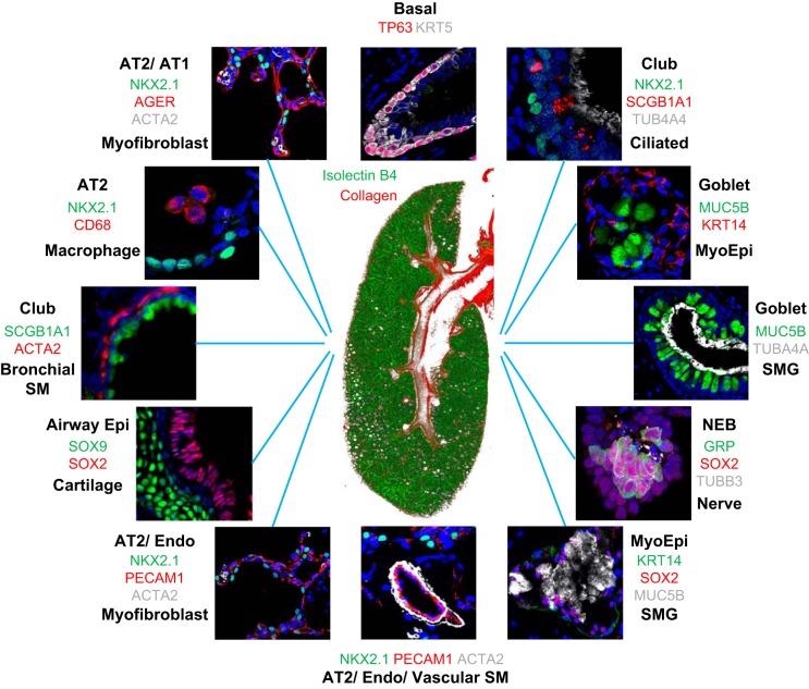
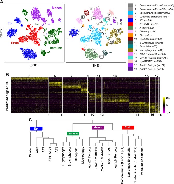
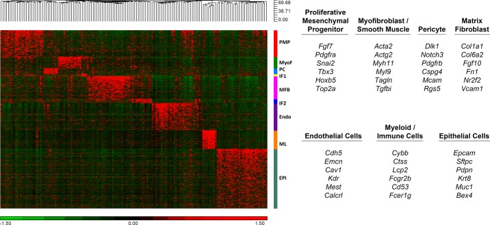
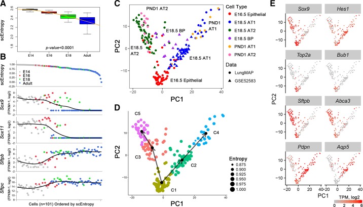
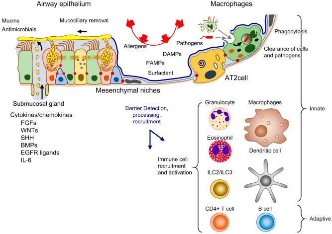
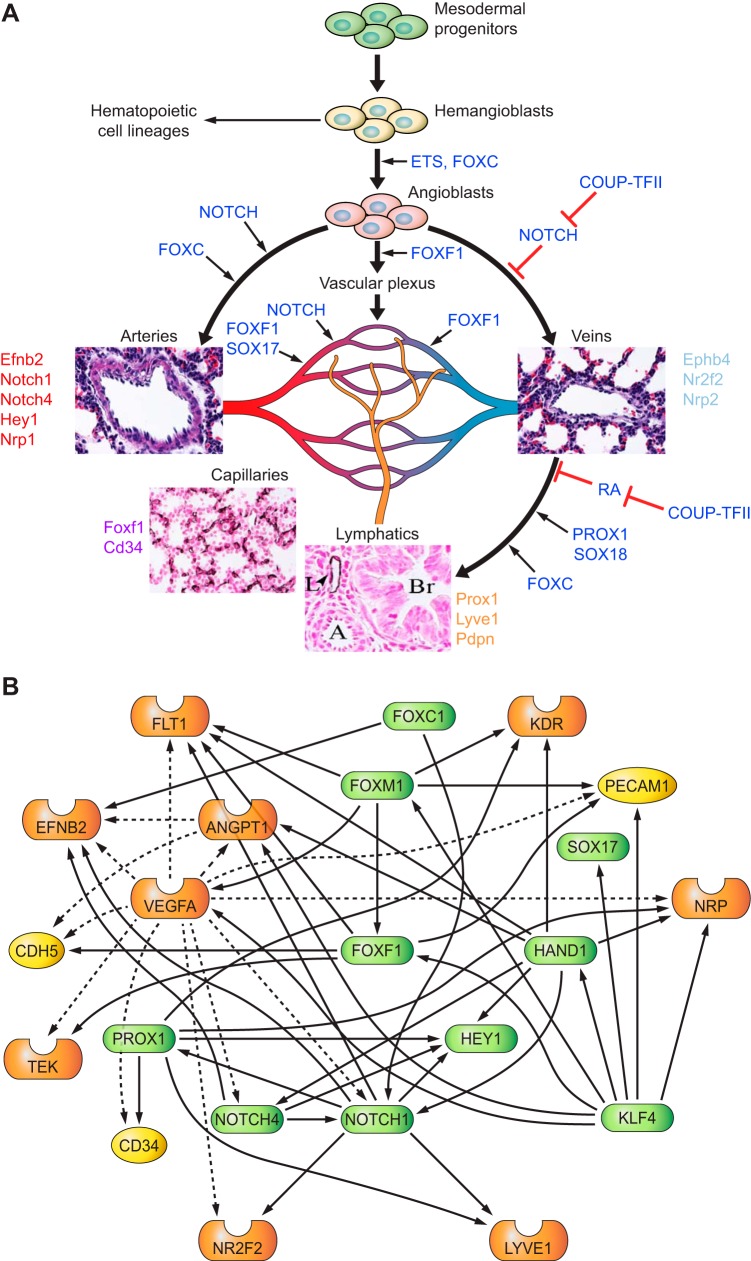
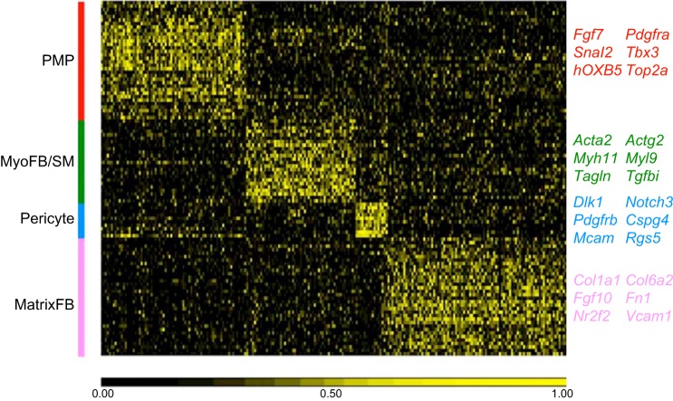
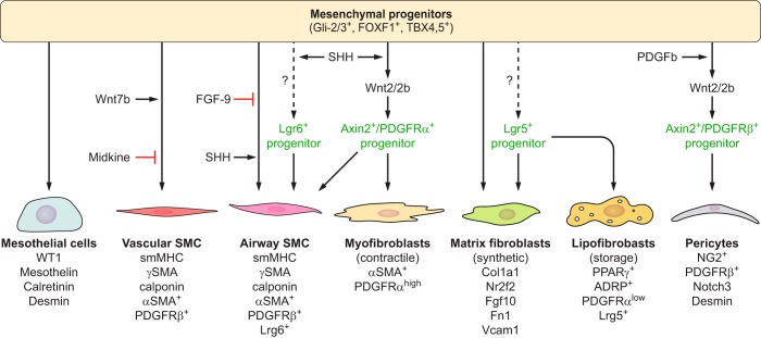
The unique architecture of the mammalian lung is required for adaptation to air breathing at birth and thereafter. Understanding the cellular and molecular mechanisms controlling its morphogenesis provides the framework for understanding the pathogenesis of acute and chronic lung diseases. Recent single-cell RNA sequencing data and high-resolution imaging identify the remarkable heterogeneity of pulmonary cell types and provides cell selective gene expression underlying lung development. We will address fundamental issues related to the diversity of pulmonary cells, to the formation and function of the mammalian lung, and will review recent advances regarding the cellular and molecular pathways involved in lung organogenesis. What cells form the lung in the early embryo? How are cell proliferation, migration, and differentiation regulated during lung morphogenesis? How do cells interact during lung formation and repair? How do signaling and transcriptional programs determine cell-cell interactions necessary for lung morphogenesis and function?


Conflict of interest statement

No conflicts of interest, financial or otherwise, are declared by the authors.

Figures

FIGURE 1.
FIGURE 1.
Diverse cells and structures of the mammalian lung. At the center is an image of the right lobe of the mouse lung on PN3, in the early alveolar period of morphogenesis. Green indicates endothelial cells of the pulmonary vasculature, and red marks the second harmonic image of collagen in the main bronchus, subsegmental bronchi, and pulmonary artery (red) at the center of the figure. Diverse pulmonary cell types and their niches are shown by fluorescence antibody staining as indicated by the colors that correspond to the antibodies used to stain each cell type. Images are available on the LungImage website (https://research.cchmc.org/lungimage/?page_id=21726) and include examples of cells and structures shared by mouse and human pulmonary tissues.
FIGURE 2.
FIGURE 2.
Single cell RNA analysis identifies multiple pulmonary cell types. A: four major cell types (left panel) and 18 subtypes (right panel) were identified by RNA analysis using “Drop-seq” of single cells (n = 8,090) from mouse lung at postnatal day 3 (PND3). B: heatmap shows the expression of the predicted cell type signature genes corresponding to cell type. Numerical values 3–18 represent cell clusters defined in A. C: hierarchical clustering reconstructs major lung cell types predicted from the RNA data. Endo, endothelial cells; Mesen, mesenchymal cells; Epi, epithelial cells; Immune, immune cells; FB, fibroblast cells; AT1, alveolar type 1 cells; AT2, alveolar type 2 cells; MyoFB, myofibroblast cells; SMC, smooth muscle cells; MatrixFB, matrix fibroblast cells.
FIGURE 3.
FIGURE 3.
Heatmap of gene expression patterns from single cell RNA-sequencing. Signature genes identifying major lung cell types were predicted from single cell RNA-seq analysis from E16.5 fetal mouse lungs and represented in 2D heatmaps (data are available at https://research.cchmc.org/pbge/lunggens/celltype_E16_p3.html). Nine major cell clusters are labeled with the color bar at the right side of the heatmap. PMP, proliferative mesenchymal progenitor; MyoF, myofibroblast; IF1, intermediate fibroblast 1; MFB, matrix fibroblast; Endo, endothelial cell; ML, myeloid cell; Epi, epithelial cell. Representative signature genes identifying seven major cell types are listed in the right panel.
FIGURE 4.
FIGURE 4.
SLICE reconstructs cell differentiation lineages using single-cell RNA-seq data. A: single cell entropy (scEntropy) of mouse alveolar type 2 (AT2, n = 101) cells decreased during the perinatal period. RNA data from epithelial cells from E14.5, E16.5, E18.5, and adult mouse lung (327) were used to compute entropy. B: the decrease in scEntropies of AT2 cells correlates with increased AT2 cell differentiation. Expression of early progenitor cell markers (Sox9 and Sox11) and mature AT2 markers (Sftpb and Sftpc) were used to validate the order predicted by scEntropies. C: predicted differentiation path of AT1 and AT2 cells from E16.5, E18.5, and postnatal day 1 (PND1) is shown. D: a branched differentiation model of AT1 and AT2 cell differentiation from bipotent progenitors was inferred using SLICE (109). E: an inferred differentiation model was produced using known cell selective marker genes.
FIGURE 5.
FIGURE 5.
Transcriptional network initiating pulmonary morphogenesis and differentiation. Lung buds and the tracheal stalk form between E9 and E10 from the ventral region of the foregut endoderm of the mouse embryo. The transcription factor TTF-1 (NKX2–1), shown at E10 (green), marks cells that form the initial lung buds. Transcription factors SOX17 and FOXA2 mark the differentiation of the early endoderm before lung bud specification. During branching morphogenesis (E12), epithelial cells migrate and proliferate to form the major conducting airways, indicated by expression of SOX2. SOX9 and high levels of TTF-1 mark peripheral acinar bud epithelial cells that will form the alveoli after birth. Alveolarization (shown at PN7) occurs from birth to ~28 days in the mouse, creating the extensive gas-exchange region typical of the mammalian lung. During the canalicular-saccular period of development, airway epithelial cell differentiation, influenced by the transcription factors in red, produce ciliated, basal, goblet, and club cells. Alveolar AT2 (TTF-1) and AT1 cells (HOPX) are derived from SOX9 expressing progenitors. [A part of this figure was created by Dr. John Shannon and used with permission. Another part of this figure, used with permission, was from Whitsett et al. (354).]
FIGURE 6.
FIGURE 6.
Differentiation of the embryonic foregut endoderm. A: foregut endodermal cells respond to bone morphogenetic protein 4 (BMP4), sonic hedgehog (SHH), and fibroblast growth factor (FGF) signaling along the dorsal-ventral axis of the common esophageal (SOX2 in blue) and lung (NKX2–1 in red) tubules. The tubules migrate into the splanchnic mesenchyme (FOXF1 in green). B: Noggin, from the notochord, inhibits BMP4, maintaining SOX2 expression in esophageal cells. Retinoic acid mediates SHH signaling that activates Gli2/3 in the splanchnic mesenchyme, activating Wnt2/2b and BMP4 that maintains NKX2–1 expression in the epithelium required for lung specification. Maintenance of the lung bud requires FGF10 produced by the mesenchyme and β-catenin signaling in the epithelium that regulate the patterning of the mesenchyme (green) and the epithelium (red). C: complex paracrine signaling regulates branching morphogenesis. During the embryonic to canalicular periods of lung development, respiratory epithelial cells migrate and proliferate as airways and peripheral acini are formed. Epithelial cells from the peripheral lung buds proliferate and migrate in response to FGF10 gradients produced by the mesenchyme that are counterregulated by Spry1,2 to limit proliferation. FGF, WNT, SHH, and BMP signaling regulates growth and patterning of the lung buds in a transcriptional network by which ETV5 regulates SHH in the epithelium, activating Gli2/3, FoxF1, and TBX proteins in the mesenchyme, to control expression of Wnt2/2b and FGF-10. Retinoic acid influences SHH and renders the endoderm responsive to NKX2–1. After separation of trachea and esophagus, SOX2 is re-expressed in conducting airways. SOX9 marks peripheral acinar cells that ultimately differentiate into AT1 and AT2 cells to form the alveoli. (A courtesy of Dr. Aaron Zorn, used with permision.)
FIGURE 7.
FIGURE 7.
Stages of branching morphogenesis. Left panel shows a posterior view of the mouse lung (red) and the esophagus (blue) at E10. Images of embryonic and postnatal mouse lung from E14 to the alveolar period on postnatal day 14 are shown in A–D. Major stages of lung morphogenesis are shared in the human and mouse. In A–D, lung epithelial cells (thyroid transcription factor-1, TTF-1) are shown in green, endothelial cells in red (endomucin), and smooth muscle myofibroblasts in purple (α-smooth muscle actin, SMA). Lung morphogenesis proceeds from a solid branched organ to the open alveolar structures after birth. Major conducting airways are formed by branching morphogenesis from the embryonic to canalicular period of fetal development. Sacculation and alveolarization are completed in the perinatal and postnatal period, creating the gas exchange region. Insets show higher magnifications. (Left image courtesy of Dr. Aaron Zorn, used with permission.)
FIGURE 8.
FIGURE 8.
Genetic networks regulating ciliated cell differentiation in conducting airways and submucosal glands. Basal and club progenitor cells differentiate into ciliated cells that line the majority of the conducting airways and the ducts of submucosal glands (A). In the absence of NOTCH, Gmnc is induced, activating a transcriptional network that directs centriole replication and the synthesis of the structural proteins forming motile cilia. Expression and assembly of ciliary proteins is regulated by Foxj1, and associated transcription factors, Rfx and Myb. B and C: confocal images of human lung. Ciliated cells are shown in the ducts of submucosal glands and along the conducting airways identified by TubA4A (in white). Club cells, marked by SCGB1A1 (red), are less abundant in human than mouse airways. Basal cells expressing TP63 and KRT5 or KRT14 (B and D) are progenitors of ciliated cells in the human airways. [A from Whitsett and Alenghat (353).]
FIGURE 9.
FIGURE 9.
Genetic networks regulating goblet cell differentiation. Goblet cells produce mucins, e.g., MUC5B and MUC5AC (green), in submucosal glands and airways (A–D). Basal and club cells expressing Sox2, TP63, and Grhl2 are the primary progenitors from which goblet cells differentiate in response to environmental, infectious, and inflammatory signals. Active NOTCH signaling and JAK/STAT, in part via STAT6, activates SPDEF (or Sam pointed domain Ets-like factor) that regulates gene expression and differentiation of goblet cells and production of mucins. Factors activating or inhibiting SPDEF and goblet cell differentiation are shown. Goblet cells express cytokines and chemokines regulating Th2 innate immunity in the lung.
FIGURE 10.
FIGURE 10.
Transcriptional networks regulating differentiation of alveolar epithelial cells during the saccular stage of mouse lung development. A: NKX2–1 (aka TTF-1, green) identifies epithelial cells in acinar tubules at the canalicular stage of mouse lung development (E16.5). Endothelial cells of the pulmonary microvasculature are shown by EMCN (red) (A and B). NKX2–1 (blue-green) identifies AT2 cells in mouse alveoli at PN28 (B) at which time alveolar capillary networks are stained by EMCN (red) (C). AT1 (AGER in red) and AT2 cells (NKX2–1, green) are shown in human lung tissues at 4 mo of age. Acta2 (white) stains myofibroblasts seen at alveolar tips (C). Transcription factors influencing AT2 and AT1 differentiation are shown in the schematic.
FIGURE 11.
FIGURE 11.
Structure of the alveolar gas-exchange region. A: gas exchange is facilitated by creation of a vast surface area lined primarily by AT1 cells (HOPX, in white) and AT2 cells (ABCA3 in red) or NKX2–1 in green (C). AT2 cells synthesize and secrete pulmonary surfactant lipids and proteins that reduce surface tension in the alveoli after birth. SMA (green) marks vascular smooth muscle. B: surfactant components are secreted into the alveoli and recycled by AT2 epithelial cells. Surfactant is catabolized by alveolar macrophages by processes regulated by GM-CSF. Proteins selectively expressed by AT1 or AT2 cells are listed on the right of the panel. D: an electron micrograph demonstrates lamellar bodies containing surfactant lipids and proteins in AT2 epithelial cells. AT1 cells line the majority of the alveolar gas exchange surface, coming into close contact with endothelial cells in the pulmonary microvasculature to facilitate transport of O2 and CO2 to red blood cells (RBC). [Modified from Whitsett and Alenghat (353).]
FIGURE 12.
FIGURE 12.
Diverse progenitor cells repair the conducting airway epithelium. The conducting airway surface and submucosal glands are shown. Basal cells (SOX2, TP63) are the primary progenitor-stem cells in the pseudostratified regions of the conducting airways. Basal cells in the ducts of submucosal glands and myoepithelial cells in the submucosal glands also serve as progenitors from which ciliated, basal, and secretory cells are produced. Secretory (club) cells also serve as proliferative progenitors following injury. Signaling and transcriptional networks regulating cell migration, proliferation, and redifferentiation following lung injury are shown. Bronchoalveolar stem cells (BASCs) and lineage negative epithelial progenitors (LNEPs) represent additional progenitor cells types (not shown).
FIGURE 13.
FIGURE 13.
Intracellular signaling during repair of the respiratory epithelium. The respiratory tract is constantly exposed to microbial pathogens, particles, and toxicants and is capable of remarkable regeneration to maintain and restore the lung structure and function following injury. Mucociliary clearance, barrier function, production of antimicrobial proteins, and robust innate and acquired immune systems protect the lung from injury. Epithelial cells, innate immune lymphocytes, macrophages, monocytes, lymphocytes, and neutrophils respond to pathogens with robust and diverse cytokine, chemokine, and growth factor expression to recruit inflammatory cells, in turn, enhancing lung repair. Paracrine signaling and direct cell-cell interactions among multiple cell types mediate responses to injury. A diversity of signaling molecules are generated during lung injury and repair which activate proliferation, migration, and differentiation to maintain epithelial barriers and restore lung function and homeostasis.
FIGURE 14.
FIGURE 14.
Signaling and transcriptional networks regulating endothelial cell lineages. A: heterogeneity of pulmonary endothelial cells. Mesoderm-derived bipotential hemangioblasts differentiate into hematopoietic and endothelial progenitor cells (angioblasts). Angioblasts give a rise to arterial, venal, lymphatic, and capillary endothelial cell lineages. Arterial endothelial cell fate is promoted by NOTCH activation. Chicken ovalbumin in upstream promoter transcription factor 2 (COUP-TFII) inhibits NOTCH, enhances venous endothelial differentiation, and cooperates with PROX1, FOXC1/2, and SOX18 to stimulate lymphatic endothelial differentiation. Development of pulmonary capillary (microvascular) endothelial cells is promoted by FOXF1, SOX17, and NOTCH. B: transcriptional network regulating pulmonary endothelial development. Predicted endothelial cell regulatory gene network consists of representative transcription factors (green), signaling molecules (orange), and target genes (yellow). The regulatory relationships between key regulators and their predicted targets are identified through literature mining using the Ingenuity knowledge base. Solid lines indicate direct regulations. Dashed lines indicate indirect regulation.
FIGURE 15.
FIGURE 15.
Identification of diverse pulmonary mesenchymal cells. Signature genes of 4 major lung mesenchymal cell types from E16.5 mouse lung identified via single cell RNA-seq analysis were represented in a 2-dimensional heatmap. Each row represents a single cell. Each column represents a signature gene. Cell clusters were labeled in the color bar at the left side of the heatmap. PMP, proliferative mesenchyme progenitors; MyoFB/SM, myofibroblast/smooth muscle; MatrixFB, matrix fibroblast. Representative signature genes were listed in the right panel colored as the corresponding cell types. Data are available online: https://research.cchmc.org/pbge/lunggens/celltype_E16_p3.html.
FIGURE 16.
FIGURE 16.
Signaling networks regulating mesenchymal cell differentiation. Mesenchymal progenitors expressing glioma-associated oncogene family zinc finger 2/3 (Gli2/3), forkhead box F1 (FOXF1), and T-box 4/5 (TBX4/5) differentiate into vascular smooth muscle cells (SMC), airway SMC, myofibroblasts, lipofibroblasts, matrix fibroblasts, pericytes, and mesothelial cells. Cell-selective markers for each of the mesenchymal cell types are provided. Differentiation of airway SMC and myofibroblasts occurs, in part, via intermediate progenitors expressing Lgr6 and Axin2/PDGFRα. Lgr5+ mesenchymal progenitors differentiate into lipofibroblasts and matrix fibroblasts. Axin2+/PDGFRβ+ progenitors give rise to pericytes. Differentiation of mesenchymal cell types is regulated by the wingless (WNT), sonic hedgehog (SHH), fibroblast growth factor (FGF), and platelet-derived growth factor (PDGF) signaling pathways as indicated in diagram.
FIGURE 17.
FIGURE 17.
Diverse fibroblasts build the alveolar scaffold. A: the electron micrograph shows the ultrastructure of an alveolar septae from the adult mouse lung. Most of the alveolar surface is covered by AT1 epithelial cell(s). Gas exchange occurs across the AT1-endothelial interface with alveolar capillaries. Matrix-, lipo-, and myofibroblasts are seen in the septal wall. A collagen-elastin bundle (matrix) is seen at the septal tip. B and C: confocal imaging of the mouse lung (PND3) is shown after injection with isolectin B4 visualizing endothelial cells of the alveolar capillary network. Surfaces of the alveoli were rendered from confocal images after staining the endothelium with isolectin B4 lectin after acquisition at 585–635 nm (green). Second harmonic imaging shows collagen bundles in red. (Figure courtesy of Dr. Matt Kofron, used with permission).
FIGURE 18.
FIGURE 18.
A predicted transcriptional network regulating differentiation of the pulmonary mesenchyme. A predicted mesenchymal cell gene regulatory network consists of representative transcription factors (green), signaling molecules (orange), and target genes (yellow). Regulatory relationships between key transcriptional regulators and signaling predicted targets were identified through literature mining using the Ingenuity knowledge database. Solid lines indicate direct regulatory relationships. Dashed lines indicate indirect regulatory relationships.

References

    1. Akeson AL, Cameron JE, Le Cras TD, Whitsett JA, Greenberg JM. Vascular endothelial growth factor-A induces prenatal neovascularization and alters bronchial development in mice. Pediatr Res 57: 82–88, 2005. doi:10.1203/01.PDR.0000148070.89006.3F. - DOI - PubMed
    1. Alejandre-Alcázar MA, Kwapiszewska G, Reiss I, Amarie OV, Marsh LM, Sevilla-Pérez J, Wygrecka M, Eul B, Köbrich S, Hesse M, Schermuly RT, Seeger W, Eickelberg O, Morty RE. Hyperoxia modulates TGF-beta/BMP signaling in a mouse model of bronchopulmonary dysplasia. Am J Physiol Lung Cell Mol Physiol 292: L537–L549, 2007. doi:10.1152/ajplung.00050.2006. - DOI - PubMed
    1. Alphonse RS, Vadivel A, Zhong S, McConaghy S, Ohls R, Yoder MC, Thébaud B. The isolation and culture of endothelial colony-forming cells from human and rat lungs [Correction in Nat Protoc 11: 192, 2016.]. Nat Protoc 10: 1697–1708, 2015. doi:10.1038/nprot.2015.107. - DOI - PubMed
    1. Ameis D, Khoshgoo N, Iwasiow BM, Snarr P, Keijzer R. MicroRNAs in Lung Development and Disease. Paediatr Respir Rev 22: 38–43, 2017. doi:10.1016/j.prrv.2016.12.002. - DOI - PubMed
    1. Anderson PJ, Lynch TJ, Engelhardt JF. Multipotent Myoepithelial Progenitor Cells Are Born Early during Airway Submucosal Gland Development. Am J Respir Cell Mol Biol 56: 716–726, 2017. doi:10.1165/rcmb.2016-0304OC. - DOI - PMC - PubMed

Publication types

MeSH terms

LinkOut - more resources