. 2019 Feb 1;26(1):45-53.
doi: 10.1093/dnares/dsy037.

3' Branch ligation: a novel method to ligate non-complementary DNA to recessed or internal 3'OH ends in DNA or RNA

Affiliations

3' Branch ligation: a novel method to ligate non-complementary DNA to recessed or internal 3'OH ends in DNA or RNA

Lin Wang et al. DNA Res. .

Abstract

Nucleic acid ligases are crucial enzymes that repair breaks in DNA or RNA during synthesis, repair and recombination. Various genomic tools have been developed using the diverse activities of DNA/RNA ligases. Herein, we demonstrate a non-conventional ability of T4 DNA ligase to insert 5' phosphorylated blunt-end double-stranded DNA to DNA breaks at 3'-recessive ends, gaps, or nicks to form a Y-shaped 3'-branch structure. Therefore, this base pairing-independent ligation is termed 3'-branch ligation (3'BL). In an extensive study of optimal ligation conditions, the presence of 10% PEG-8000 in the ligation buffer significantly increased ligation efficiency to more than 80%. Ligation efficiency was slightly varied between different donor and acceptor sequences. More interestingly, we discovered that T4 DNA ligase efficiently ligated DNA to the 3'-recessed end of RNA, not to that of DNA, in a DNA/RNA hybrid, suggesting a ternary complex formation preference of T4 DNA ligase. These novel properties of T4 DNA ligase can be utilized as a broad molecular technique in many important genomic applications, such as 3'-end labelling by adding a universal sequence; directional tagmentation for NGS library construction that achieve theoretical 100% template usage; and targeted RNA NGS libraries with mitigated structure-based bias and adapter dimer problems.

Keywords: NGS; T4 DNA ligase; molecular tool; novel ligation.


Figures

Figure 1
Figure 1
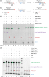
3′ Branch ligation by T4 DNA ligase at non-conventional DNA ends formed by nicks, gaps, and overhangs. (a) Schematic representation of ligation assay with different DNA accepter types. The blunt-end DNA donor (blue) is a synthetic, partially dsDNA molecule with dideoxy 3-termini (filled circles) to prevent DNA donor self-ligation. The long arm of the donor is 5′-phosporylated. The DNA acceptors were assembled using 2 or 3 oligos (black, red, and orange lines) to form a nick (without phosphates), a gap (1 or 8 nt), or a 36-nt 3-recessive end. All strands of the substrates are unphosphorylated, and the scaffold strand is 3 dideoxy protected. (b) Analysis of the size shift of ligated products of substrates 1, 2, 3, and 4, respectively, using a 6% denaturing polyacrylamide gel. Reactions were performed according to the optimized condition. The negative no-ligase controls (lanes 1, 3, 4, 6, 7, 9, 10, 12, and 13) were loaded at 1 or 0.5× volume of corresponding experimental assays. If ligation occurs, the substrate size is shifted up by 22 nt. Red arrowheads correspond to the substrate, and purple arrowheads correspond to donor-ligated substrates. Donor and substrate sequences in Supplementary Table S1. (c) Expected sizes of substrate and ligation product and approximate ligation efficiency in each experimental group. The intensity of each band was estimated using ImageJ and normalized by its expected size. Ligation efficiency was estimated by dividing the normalized intensity of ligated products by the normalized total intensity of ligated and unligated products.
Figure 2
Figure 2
Gel analysis of size shift of ligated products using 6% TBE polyacrylamide gel. Red arrowheads correspond to the substrate, and purple arrowheads correspond to donor-ligated substrates: substrate 5 (nick) (a), substrate 6 (1-nt gap) (b), substrate 7 (2-nt gap) (c), substrate 8 (3-nt gap), (d) and substrate 9 (3-recessive end) (e). Three DNA donors with different bases at the 5-end of the ligation junction (T, A, or GA) were examined. Donor and substrate sequences are summarized in Supplementary Table S1. (f) Table of ligation efficiency calculated based on normalized band intensity using ImageJ.
Figure 3
Figure 3
3′ Branch ligation at the 3′ end of RNA in DNA/RNA hybrid. Schematic representation of 3′-branch ligation on a DNA/RNA hybrid with a 20‐bp complimentary region. We tested whether blunt-end DNA donors would ligate to the 3-recessive end of DNA and/or to the 3-recessive end of RNA. DNA(ON-21) hybridizes with the RNA strand (a), whereas DNA(ON-23) cannot hybridize with the RNA strand (b). (c, d) Gel analysis of size shift of ligated products using 6% denaturing polyacrylamide gel. The red arrowheads correspond to the RNA substrate (29 nt), and the green arrowhead corresponds to DNA substrate (80 nt). The purple arrowhead corresponds to donor-ligated RNA substrates. If ligation occurs, the substrate size would shift up by 20 nt. (c) Lanes 1 and 2, experimental duplicates; lanes 7–10, no-ligase controls; 10% PEG was added with T4 DNA ligase. (d) Lane 1, no-ligase control; lanes 2, 3, and 8, T4 DNA ligase with 10% PEG; lanes 4, 5, and 9, T4 RNA ligase 1 with 20% DMSO; lanes 6, 7, and 10, T4 RNA ligase 2 with 20% DMSO.
Figure 4
Figure 4
(a–c) Schematic representation of three transposon tagmentation methods followed by PCR amplification using Pr-A (blue arrow) and Pr-B (green arrow). Two-transposon method (a); one Y transposon tagmentation with 3-gap filling (b); one-transposon method with adapter ligation at 3-gap (c). (d) Graph of amplification signal after purification using pr-A or pr-A with pr-B after the various tagmentation and gap ligation conditions.
Figure 5
Figure 5
Base distribution bias of Tn5-gap ligation (a), two transposons (b), and regular TA ligation (c). Only the first 20 bases from each end of the ligation are presented; adenine, blue; cytosine, orange; guanine, grey; thymine, yellow; the average and standard deviation of five-independent libraries are presented.

References

    1. Lehnman I.R. 1974, DNA ligase: structure, mechanism, and function, Science (80-.), 186, 790–7. - PubMed
    1. Tomkinson A.E., Mackey Z.B.. 1998, Structure and function of mammalian DNA ligases, Mutat . Res., 407, 1–9. - PubMed
    1. Timson D.J., Singleton M.R., Wigley D.B.. 2000, DNA ligases in the repair and replication of DNA, Mutat . Res., 460, 301–18. - PubMed
    1. Ho C.K., Wang L.K., Lima C.D., Shuman S.. 2004, Structure and mechanism of RNA ligase, Structure, 12, 327–39. - PubMed
    1. Tomkinson A.E., Vijayakumar S., Pascal J.M., Ellenberger T.. 2006, DNA ligases: structure, reaction mechanism, and function, Chem . Rev., 106, 687–99. - PubMed