. 2018 May 25:3:192-207.
doi: 10.1016/j.isci.2018.04.013. Epub 2018 Apr 22.

Mitochondria-Targeted Honokiol Confers a Striking Inhibitory Effect on Lung Cancer via Inhibiting Complex I Activity

Affiliations

Mitochondria-Targeted Honokiol Confers a Striking Inhibitory Effect on Lung Cancer via Inhibiting Complex I Activity

Jing Pan et al. iScience. .

Abstract

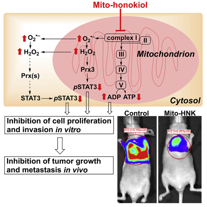
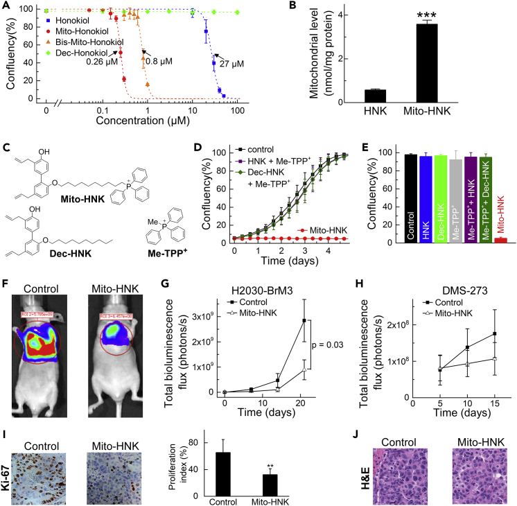
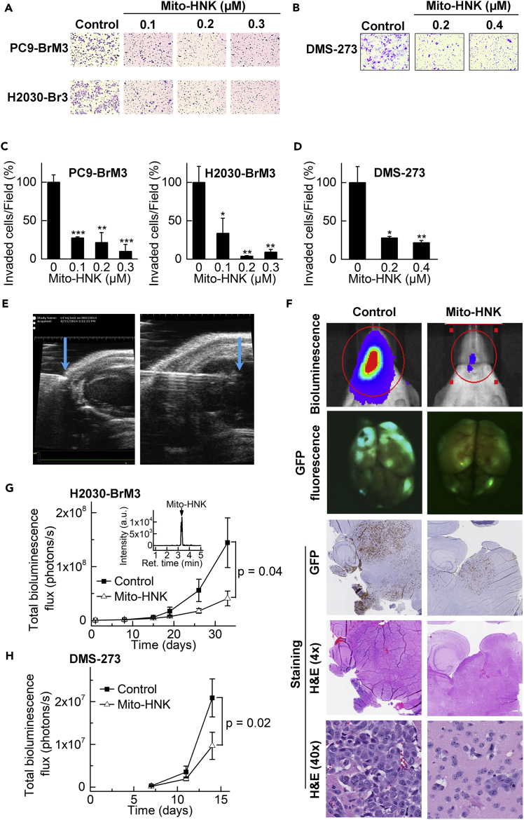
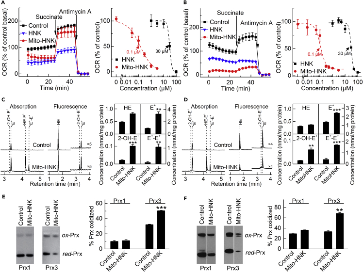
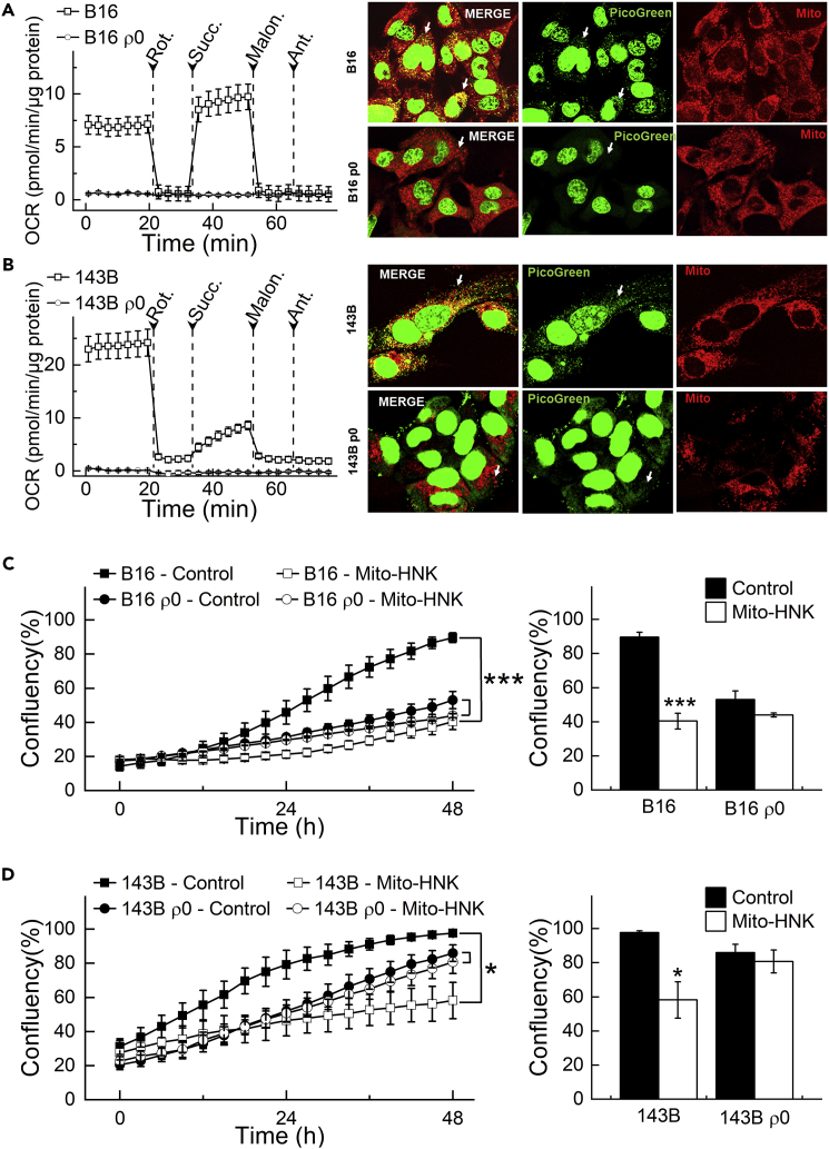
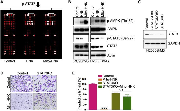
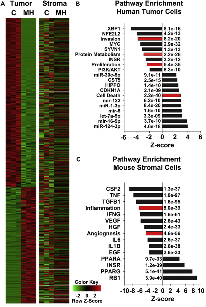
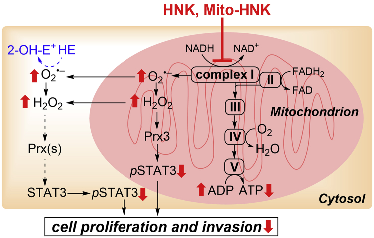
We synthesized a mitochondria-targeted honokiol (Mito-HNK) that facilitates its mitochondrial accumulation; this dramatically increases its potency and efficacy against highly metastatic lung cancer lines in vitro, and in orthotopic lung tumor xenografts and brain metastases in vivo. Mito-HNK is >100-fold more potent than HNK in inhibiting cell proliferation, inhibiting mitochondrial complex ?, stimulating reactive oxygen species generation, oxidizing mitochondrial peroxiredoxin-3, and suppressing the phosphorylation of mitoSTAT3. Within lung cancer brain metastases in mice, Mito-HNK induced the mediators of cell death and decreased the pathways that support invasion and proliferation. In contrast, in the non-malignant stroma, Mito-HNK suppressed pathways that support metastatic lesions, including those involved in inflammation and angiogenesis. Mito-HNK showed no toxicity and targets the metabolic vulnerabilities of primary and metastatic lung cancers. Its pronounced anti-invasive and anti-metastatic effects in the brain are particularly intriguing given the paucity of treatment options for such patients either alone or in combination with standard chemotherapeutics.

Keywords: Immunology; Medicinal and Aromatic Plants; Natural Product Chemistry.


Figures

None
Graphical abstract
Figure 1
Figure 1
Design, Synthesis, and Subchronic Toxicity Screen of Mito-HNK (A) Synthesis of Mito-HNK. Both mono-substituted (Mito-HNK) and double-substituted (Bis-Mito-HNK) mitochondria-targeted derivatives of honokiol were synthesized. The products were purified by HPLC and characterized by NMR and mass spectrometry. TPP+-linked compounds accumulate selectively in cancer cell mitochondria, according to the Nernst equation. (B and C) Examples of subchronic toxicity screen of Mito-HNK. (B) Measured body weight (squares, left axis) and rectal temperature (circles, right axis). (C) Latency to fall in the inverted screen test measured after 8 weeks of treatment. The 2×, 10×, and 20× doses relate to the effective dose (1× = 3.5 μmol/kg) and correspond to 7.5, 37.5, and 75 μmol/kg, respectively. Error bars represent standard deviation (SD), n = 5 mice per group. Error bars indicate SE.
Figure 2
Figure 2
Mito-HNK Inhibits the Proliferation and Tumor Growth of Lung Cancer Cells (A) Mito-HNK and bis-Mito-HNK inhibit the proliferation of H2030 cells at significantly lower concentrations than HNK. The IC50 values were determined at the point when untreated cells reached ∼95% confluence (∼4 days of incubation). (B) Treatment of H2030-BrM3 cells (0.1 μM, 24 hr) leads to significantly higher mitochondrial accumulation of Mito-HNK than HNK (***P < 0.001 vs. HNK). (C) The chemical structures of Mito-HNK and the control compounds (Dec-HNK and Me-TPP+) used for data in panels D and E. (D and E) The combinations of HNK or decyl-HNK with Me-TPP+ fail to reproduce the anti-proliferative effects of Mito-HNK. The traces recorded during real-time monitoring of cell confluence are shown in (D), and the cell confluence after 5 days of incubation with the compounds (1 μM) is shown in (E). (F) Representative bioluminescence live imaging of NSCLC orthotopic tumor growth in control or Mito-HNK-treated mice (3.75 μmol/kg each). (G) Quantitative data for bioluminescence imaging of the orthotropic growth of NSCLC (H2030-Br3M) cells (n = 6 per group, observation duration = 21 days). (H) Quantitative data for the bioluminescence imaging of the orthotropic growth of SCLC (DMS-273) cells (n = 6 per group, observation duration = 15 days). (I) Left panel, representative immunohistochemistry staining of H2030-BrM3 orthotopic lung tumors for Ki-67 in control and Mito-HNK-treated groups; right panel, quantitative estimation of cell proliferation showing percentage of Ki-67+ cells (**p < 0.01 versus control). (J) Representative H&E staining images of H2030-BrM3 orthotopic lung tumors taken from control and Mito-HNK groups. Error bars indicate SE.
Figure 3
Figure 3
Mito-HNK Inhibits Invasion and Brain Metastasis of Lung Cancer Cells (A and C) The anti-invasive effects of Mito-HNK on NSCLC lines H2030-BrM3 and PC9-BrM3 were assessed via the Boyden chamber invasion assay after 48-hr treatment. Representative images are shown in (A) and the quantitative data are shown in (C, *P < 0.05, **P < 0.01, ***P < 0.001 vs.control). (B and D) Representative images (B) and quantitative data (D) of the invasion assay for SCLC DMS-273 cells indicate that Mito-HNK is at least 100-fold more potent than HNK (*P < 0.05, **P < 0.01 vs.control). (E) High-resolution echocardiography to visualize the needle position within the left cardiac ventricle, for injecting lung cancer cells in the in vivo experiments to establish brain metastases. (F) Representative bioluminescence, GFP expression, and H&E staining images of brains taken from control, HNK, and Mito-HNK (3.75 μmol/kg each) mice. (G and H) Quantitative data for the bioluminescence imaging of brain metastases over time. Mito-HNK treatment was started 1 day after injection of H2030-BrM3 cells (G, n = 6 for control, and n = 5 for Mito-HNK, observation duration = 27 days) or DMS-273 cells (H, n = 7 each, observation duration = 14 days). (G, inset) shows the LC-MS trace from a brain extract indicating the presence of Mito-HNK (65 pmol/g of whole brain tissue). Error bars indicate SE.
Figure 4
Figure 4
Effects of Mito-HNK on Mitochondrial Complex I Activity, ROS, and the Redox State of Peroxiredoxins To measure complex I activity, cells were pretreated for 24 hr with Mito-HNK and HNK, the cell membrane was permeabilized, and OCR was measured upon the addition of mitochondrial substrates/inhibitors. (A and B) Both HNK (IC50 = 30 μM for both cell lines) and Mito-HNK (IC50 = 0.1 μM for both cell lines) inhibit complex I in H2030-BrM3 NSCLC cells (A) and DMS-273 SCLC cells (B). (C and D) The effect of Mito-HNK (1 μM, 24 hr treatment) on cellular ROS production, as measured by HPLC-based profiling of the oxidation products of the HE probe in H2030-BrM3 (C) and DMS-273 (D) cells. The compound 2-hydroxyethidium (2-OH-E+) is a specific product for superoxide and diethidium (E+-E+) is a marker product for one-electron oxidants. HPLC traces are shown in the left panels and the quantitative results in the right panels (**p < 0.01, ***p < 0.001). (E and F) The 24-hr treatment of H2030-BrM3 cells with 0.2 μM Mito-HNK (E) or DMS-273 cells with 0.3 μM Mito-HNK (F) leads to significant oxidation of mitochondrial Prx3, whereas the oxidation state of cytosolic Prx1 is not significantly affected (**P < 0.01, ***P < 0.001 vs.control). Error bars indicate SD.
Figure 5
Figure 5
Depletion of mtDNA Abrogates the Anti-Proliferative Effects of Mito-HNK in Lung Cancer Cells (A and B) Validation of the loss of activities of the mitochondrial complexes in ρ0 cells. (A and B, left panels) Permeabilized cells were assayed in medium containing 10 mM pyruvate and 1.5 mM malate (substrates for complex I) in mannitol and sucrose (MAS) buffer. The complex I-related oxygen consumption rate (OCR) was assayed immediately and verified by injecting rotenone (complex I inhibitor) as indicated. Then, complex II-related OCR was measured by supplying cells with succinate (substrate for complex II, 10 mM). Both malonate (complex II inhibitor, 10 mM) and antimycin A (complex III inhibitor, 20 μM) were injected where indicated. (A and B, right panels) Validation of the absence of mitochondria in ρ0 cells with PicoGreen staining for mtDNA. Arrows indicate mitochondria fibrillar network. (C and D) mtDNA depletion abrogates the anti-proliferative effects of Mito-HNK (0.4 μM, 48 hr) in both B16 and 143B ρ0 cells. (***P < 0.001 vs. B16 or 143b parental cells). Error bars indicate SD.
Figure 6
Figure 6
Role of STAT3 in the Anti-proliferative and Anti-invasive Effects of HNK and Mito-HNK in Lung Cancer Cells (A) Results of the receptor tyrosine kinase proteomic array of H2030-BrM3 cells treated with HNK (20 μM) or Mito-HNK (0.2 μM) for 24 hr. (B) Western blot analysis of the effect of HNK (20 μM) and Mito-HNK (0.2 μM) on AMPK and STAT3 phosphorylation status in PC9-BrM3 and H2030-BrM3 cells. (C) Western blot to verify knockout and knockdown of STAT3 in H2030-BrM3 cells. (D and E) STAT3 knockout abrogates the anti-invasive and anti-proliferation effects of Mito-HNK (0.2 μM) in H2030-BrM3 cells. (D) Representative images and (E) the quantitative results of the cell invasion assay, ***p < 0.001. Error bars indicate SE.
Figure 7
Figure 7
Analysis of Pathways Regulated by Mito-HNK Treatment in the Malignant Tumor Cells and Nonmalignant Stroma of H2030-BrM3 Brain Metastatic Lesions (A) RNAseq analysis to compare differentially expressed (DE) genes that regulate cell death in the malignant tumor and nonmalignant stroma of brain metastatic lesions of mice treated with the vehicle control (C) or Mito-HNK (MH). (B and C) Analysis of molecular pathways and cellular functions that are positively and negatively enriched (indicated by Z score) following Mito-HNK treatment in the malignant tumor cells (B) and nonmalignant stroma (C). The raw Z scores were normalized within tumor and stromal groups. Note that the significant enrichment of DE genes that induce cell death are highly upregulated only in the Mito-HNK-treated malignant tumor cells (Z score: +2.5; p < 10−57). p Values are indicated on the graphs. Black bars indicate molecular pathways, and red bars indicate cellular function pathways.
Figure 8
Figure 8
Proposed Mechanisms of the Antiproliferative and Anti-invasive Effects of HNK and Mito-HNK NADH dehydrogenase (complex I) plays a key role in regulating and maintaining mitochondrial function and energy production (ATP). Suppressing complex I activity can increase ROS production and signaling through ROS-associated pathways. We propose that one potential mechanism of action of Mito-HNK is that it inhibits complex I in lung cancer cells; stimulates ROS generation, which promotes oxidation of mitochondrial Prx3; activates AMPK; and inhibits STAT3ser727 phosphorylation and cell proliferation. The results indicate that Mito-HNK mediates these events more robustly and at much lower concentrations than does HNK.

References

    1. Arora S., Singh S., Piazza G.A., Contreras C.M., Panyam J., Singh A.P. Honokiol: a novel natural agent for cancer prevention and therapy. Curr. Mol. Med. 2012;12:1244–1252. - PMC - PubMed
    1. Asin-Cayuela J., Manas A.R.B., James A.M., Smith R.A.J., Murphy M.P. Fine-tuning the hydrophobicity of a mitochondria-targeted antioxidant. FEBS Lett. 2004;571:9–16. - PubMed
    1. Bai X., Cerimele F., Ushio-Fukai M., Waqas M., Campbell P.M., Govindarajan B., Der C.J., Battle T., Frank D.A., Ye K. Honokiol, a small molecular weight natural product, inhibits angiogenesis in vitro and tumor growth in vivo. J. Biol. Chem. 2003;278:35501–35507. - PubMed
    1. Berridge M.V., Dong L., Neuzil J. Mitochondrial DNA in tumor initiation, progression, and metastasis: role of horizontal mtDNA transfer. Cancer Res. 2015;75:3203–3208. - PubMed
    1. Bindoli A., Fukuto J.M., Forman H.J. Thiol chemistry in peroxidase catalysis and redox signaling. Antioxid. Redox Signal. 2008;10:1549–1564. - PMC - PubMed

LinkOut - more resources