. 2018 Nov 13;25(7):1953-1965.e4.
doi: 10.1016/j.celrep.2018.10.034.

DNA-PK Is Targeted by Multiple Vaccinia Virus Proteins to Inhibit DNA Sensing

Affiliations

DNA-PK Is Targeted by Multiple Vaccinia Virus Proteins to Inhibit DNA Sensing

Simon R Scutts et al. Cell Rep. .

Abstract

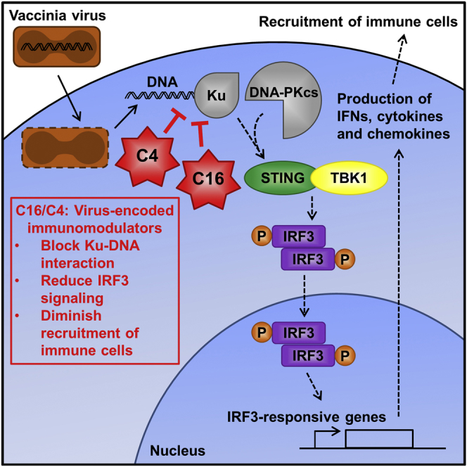
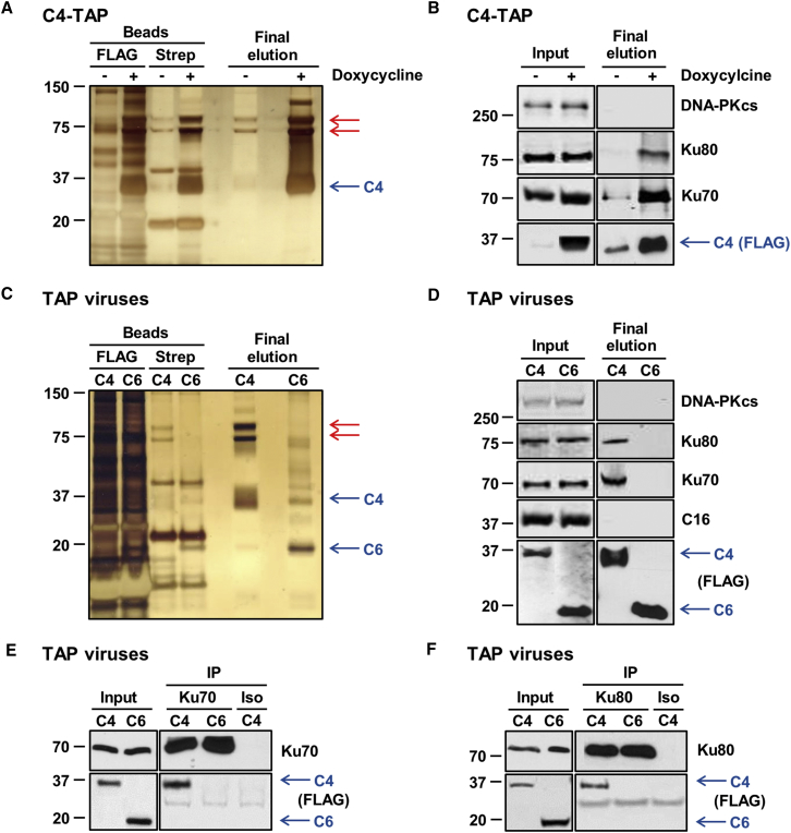
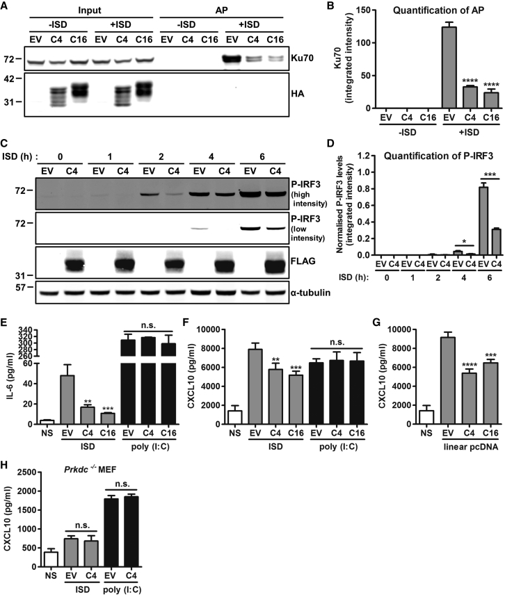
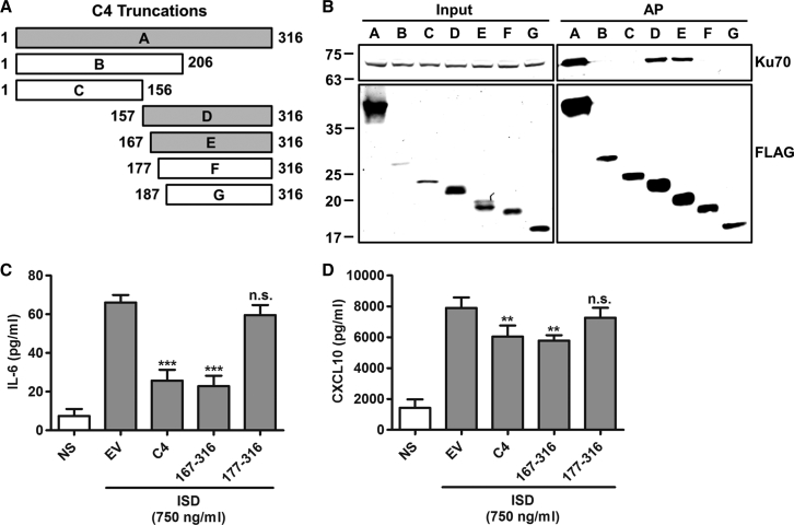
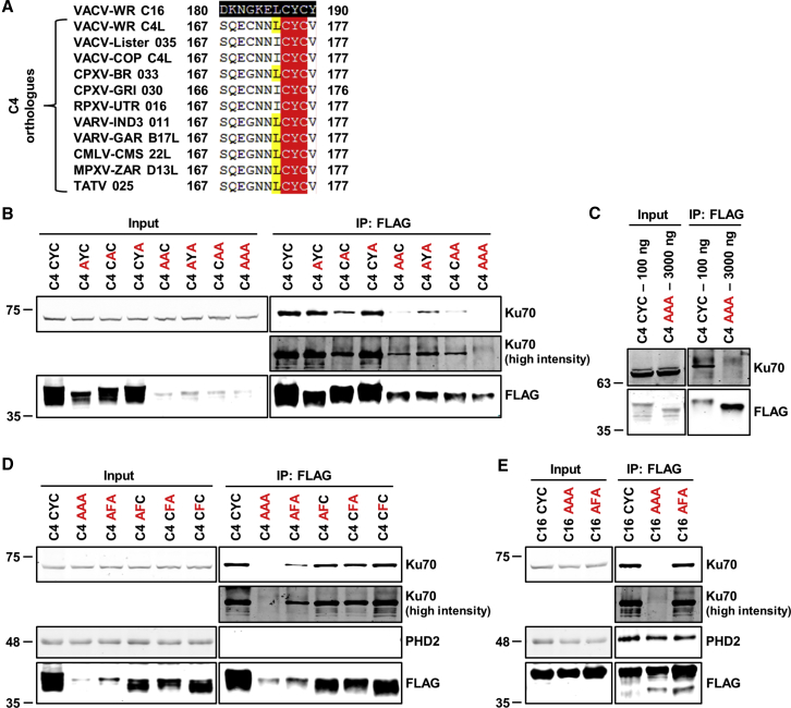
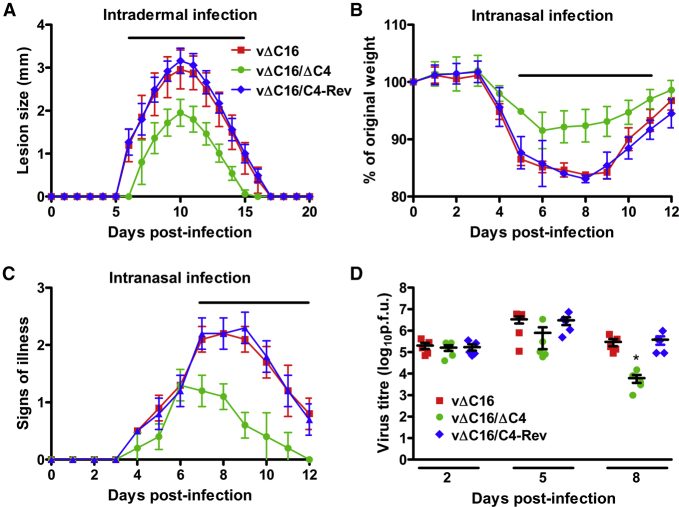
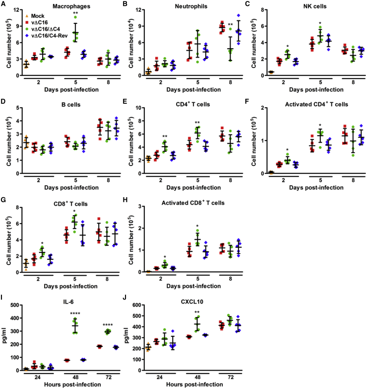
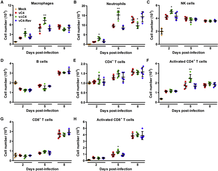
Virus infection is sensed by pattern recognition receptors (PRRs) detecting virus nucleic acids and initiating an innate immune response. DNA-dependent protein kinase (DNA-PK) is a PRR that binds cytosolic DNA and is antagonized by vaccinia virus (VACV) protein C16. Here, VACV protein C4 is also shown to antagonize DNA-PK by binding to Ku and blocking Ku binding to DNA, leading to a reduced production of cytokines and chemokines in vivo and a diminished recruitment of inflammatory cells. C4 and C16 share redundancy in that a double deletion virus has reduced virulence not seen with single deletion viruses following intradermal infection. However, non-redundant functions exist because both single deletion viruses display attenuated virulence compared to wild-type VACV after intranasal infection. It is notable that VACV expresses two proteins to antagonize DNA-PK, but it is not known to target other DNA sensors, emphasizing the importance of this PRR in the response to infection in vivo.

Keywords: DNA protein kinase; DNA sensing; IRF3 signaling; immune evasion; pattern recognition receptor; protein C4; vaccinia virus; virulence factor.


Figures

None
Graphical abstract
Figure 1
Figure 1
C4 Co-immunoprecipitates with the Ku Heterodimer (A) Tandem affinity purification of TAP-tagged C4. The C4-C′TAP HEK293 TRex cell line was induced with 2 μg/ml doxycycline for 24 hr. Protein lysates from induced and uninduced cells were then subjected to tandem affinity purification. After elution, proteins from beads and supernatant (final elution) were separated by SDS-PAGE and visualized by silver staining. Red arrows point to bands of interest observed in the final elution. (B) Whole cell lysates (inputs) and final elution proteins from (A) were analyzed by immunoblotting with antibodies against the indicated proteins. (C) Tandem affinity purification of TAP-tagged C4 in the context of infection. HEK293T cells were infected with 2 plaque-forming units (PFU)/cell of vC4-TAP or vC6-TAP for 16 hr, and protein lysates were subjected to tandem affinity purification, separated by SDS-PAGE, and visualized by silver staining. Red arrows point to bands of interest observed in the final elution. (D) Protein samples from (C) were analyzed by immunoblotting with antibodies against the indicated proteins. (E and F) Confirmation of the C4 interaction with Ku70 and Ku80. HEK293T cells were infected with 2 PFU/cell of vC4-TAP or vC6-TAP for 16 hr. Pre-cleared lysates were then immunoprecipitated with (E) anti-Ku70, (F) anti-Ku80, or isotype control antibodies (Iso). Input and precipitated protein complex (IP) samples were analyzed by SDS-PAGE and immunoblotting. The positions of molecular mass markers are shown (kDa).
Figure 2
Figure 2
C4 Inhibits DNA Sensing (A) DNA affinity purification. HEK293T cells were transfected with 2 μg/ml of EV or plasmids encoding HA-tagged C4 or C16 for 24 hr, followed by 7.5 μg/ml of 45-base pair (bp) biotinylated immunostimulatory DNA (ISD) for 1 hr. Cells were lysed, and streptavidin beads were incubated with cytoplasmic fractions to purify the biotinylated DNA and associated proteins. Samples were analyzed by SDS-PAGE and immunoblotting with Ku70 and HA antibodies. (B) Integrated intensity of Ku70 AP from (A) from three experimental replicates was calculated by infrared imaging by using a Li-Cor Odyssey scanner. (C) P-IRF3 immunoblotting. HeLa cells were transfected for 24 hr with EV or C4-TAP plasmid and then stimulated by transfection with 7.5 μg/ml of 180-bp ISD at the time point indicated. Whole cell lysates were analyzed by immunoblotting with antibodies against the indicated proteins. (D) Integrated intensity of P-IRF3 from (C) was normalized to corresponding α-tubulin levels from three experimental replicates by using a Li-Cor Odyssey scanner. (E and F) MEFs were mock transfected (NS), transfected with 80 ng/ml EV or plasmids encoding TAP-tagged C4 or C16, and cotransfected with 750 ng/ml ISD or 100 ng/ml poly (I:C) for 24 hr. Levels of (E) IL-6 and (F) CXCL10 in supernatants were measured by ELISA, performed in quadruplicate. (G) MEFs were mock transfected (NS), transfected with 80 ng/ml EV or plasmids encoding TAP-tagged C4 or C16, and cotransfected with 750 ng/ml EV pcDNA4/TO linearized previously with BamHI and NotI. After 24 hr, levels of CXCL10 in supernatants were measured by ELISA in quadruplicate. (H) Prkdc−/− MEFs were mock transfected (NS), transfected with 80 ng/ml EV or TAP-tagged C4 plasmid, and cotransfected with 750 ng/ml ISD or 100 ng/ml poly (I:C) for 24 hr. Levels of CXCL10 in supernatants were measured by ELISA, performed in quadruplicate. Data are represented as ±SD. ∗∗p < 0.01, ∗∗∗p < 0.001, ∗∗∗∗p < 0.0001, n.s. = non-significant. Each experiment was performed three times and one representative experiment is shown.
Figure 3
Figure 3
The C-Terminal Domain of C4 Is Sufficient for the Inhibition of DNA Sensing (A) Schematic of C4 truncation mutants showing C4 fragments capable (gray bars) and not capable (white bars) of binding Ku70. (B) Affinity purification of C4 truncations. TAP-tagged full-length C4 (a) and amino acid residues 1–206 (b), 1–156 (c), 157–316 (d), 167–316 (e), 177–316 (f), and 187–316 (g) were expressed by transfection in HEK293T cells and affinity purified with Strep-Tactin beads. Whole cell lysates (input) and affinity purified proteins (AP) were separated by SDS-PAGE and immunoblotted with anti-FLAG and anti-Ku70 antibodies. (C and D) MEFs were mock transfected (NS), transfected with 80 ng/ml EV or plasmids encoding TAP-tagged full-length C4 or C4 amino acid residues 167–316 or 177–316, and cotransfected with 750 ng/ml ISD. After 24 hr, levels of (C) IL-6 and (D) CXCL10 in supernatants were measured by ELISA. Data are represented as ±SD. ∗∗p < 0.01, ∗∗∗p < 0.001, n.s. = non-significant. Each experiment was performed three times and one representative experiment is shown.
Figure 4
Figure 4
Site-Directed Mutagenesis of Three Conserved Residues in C16 and C4 Abrogates Binding to Ku (A) Amino acid residues CYC are conserved between C4 (C174, Y175, and C176) and C16 (C187, Y188, and C189). Protein sequence alignment of C4 residues 167–177 from VACV strain WR and corresponding residues of C16 and other C4 orthologs of various orthopoxviruses. Black shading represents the sequence others are aligned to. Red and yellow shading indicates identical and highly similar residues, respectively. GenBank accession numbers: VACV-WR C16, YP_232892.1; VACV-WR C4L, YP_232906.1; VACV-Lister 021, ABD52470.1; VACV-COP C4L, AAA47996.1; CPXV-BR 033, NP_619822.1; CPXV-GRI 030, CAA64101.1; RBPX-UTR 016, AAS49729.1; VARV-IND3 011, NP_042055.1; VARV-GAR B17L, CAB54611.1; CMLV-CMS 22L, AAG37478.1; MPXV-ZAR D13L, NP_536443.1; and TATV 025, YP_717332.1. (B) Mutagenesis of C4 residues CYC alters binding to Ku. TAP-tagged wild-type C4 (CYC) and C4 mutants as indicated were expressed in HEK293T cells and immunoprecipitated with anti-FLAG agarose beads. Proteins were separated by SDS-PAGE and analyzed by immunoblotting with anti-Ku70 and imaged with high and low intensity, anti-FLAG, and anti-C4 antibodies. (C) Mutagenesis of CYC to AAA abrogates binding to Ku. Different quantities of wild-type C4 plasmid were transfected into HEK293T cells to obtain comparable expression with the C4 AAA mutant, and then lysates were subjected to immunoprecipitation with anti-FLAG agarose beads and analyzed by immunoblotting. (D and E) The aromatic ring of tyrosine in CYC of (D) C4 and (E) C16 is a critical component of this interaction. Plasmids encoding indicated TAP-tagged proteins were transfected into HEK293T cells and protein complexes were immunoprecipitated with anti-FLAG agarose beads. Immunoblotting was performed with the indicated antibodies. Immunoblots are representative of three independent experiments.
Figure 5
Figure 5
Absence of Both C16 and C4 Leads to Attenuation of Virus Virulence In Vivo (A) Intradermal infection. C57BL/6 mice (n = 5) were infected i.d. with 104 PFU in both ears of the indicated viruses. Lesion sizes were measured daily with a micrometer. (B–D) Intranasal infection. BALB/c mice (n = 5) were infected intranasally with 1 × 105 PFU per mouse of the indicated viruses and their weights and signs of illness were measured daily. (B) Weights. Data are expressed as a percentage of the mean weight of the same group of animals on day 0 ± SD. (C) Signs of illness. The mean ± SD score of each group of animals is shown. (D) The viral titers in lungs were determined by plaque assay. The horizontal bars indicate days where the lesion size, weight loss, or signs of illness induced by vΔC16/ΔC4 were statistically different (p < 0.05) from both vΔC16 and vΔC16/C4-Rev. The figure legend shown in (A) applies to (B) and (C). Each experiment was performed twice and one representative experiment is shown.
Figure 6
Figure 6
Deletion of Both C16 and C4 Inhibits Recruitment of Immune Cells and Reduces Production of IL-6 and CXCL10 In Vivo Groups of five BALB/c mice were infected intranasally with 1 × 105 PFU per mouse of the indicated viruses and at the indicated times p.i. (A–H) Mice were sacrificed and cells were extracted from (A and B) BAL fluid and (C–H) lungs, counted, and analyzed by flow cytometry. (A) Macrophages, (B) neutrophils, (C) NK cells, and (D) B cells were identified and quantified by fluorescence-activated cell sorting (FACS). (E–H) The number and activation status (CD69+) of CD4+ and CD8+ T cells were analyzed by FACS. (I and J) At the indicated times p.i., mice were killed and BAL fluid was extracted, and levels of (I) IL-6 and (J) CXCL10 were measured by ELISA (n = 5). Data are represented as means of cell counts ± SD. p < 0.05 and ∗∗p < 0.01 for vΔC16/ΔC4, compared with both vΔC16 and vΔC16/C4-Rev. Each experiment was performed twice and one representative experiment is shown.
Figure 7
Figure 7
Deletion of C4 from Wild-Type Western Reserve VACV Increases Recruitment of Innate Immune Cells and Activation of T Cells BALB/c mice were infected intranasally with 5 × 103 PFU per mouse of the indicated viruses and at the indicated times p.i.; groups (n = 5) were sacrificed and cells were extracted from (A and B) BAL fluid and (C–H) lungs, counted, and analyzed by flow cytometry. (A) Macrophages, (B) neutrophils, (C) NK cells, and (D) B cells were identified and quantified by FACS. (E–H) The number and activation status (CD69+) of CD4+ and CD8+ T cells were analyzed by FACS. Data are represented as means of cell counts ± SD. p < 0.05 and ∗∗p < 0.01 for vΔC4, compared with both vC4 and vC4-Rev. Each experiment was performed twice and one representative experiment is shown.

Similar articles

Cited by

References

    1. Ablasser A., Bauernfeind F., Hartmann G., Latz E., Fitzgerald K.A., Hornung V. RIG-I-dependent sensing of poly(dA:dT) through the induction of an RNA polymerase III-transcribed RNA intermediate. Nat. Immunol. 2009;10:1065–1072. - PMC - PubMed
    1. Ahn J., Barber G.N. Self-DNA, STING-dependent signaling and the origins of autoinflammatory disease. Curr. Opin. Immunol. 2014;31:121–126. - PubMed
    1. Alcamí A., Smith G.L. A soluble receptor for interleukin-1 beta encoded by vaccinia virus: a novel mechanism of virus modulation of the host response to infection. Cell. 1992;71:153–167. - PubMed
    1. Almine J.F., O’Hare C.A.J., Dunphy G., Haga I.R., Naik R.J., Atrih A., Connolly D.J., Taylor J., Kelsall I.R., Bowie A.G. IFI16 and cGAS cooperate in the activation of STING during DNA sensing in human keratinocytes. Nat. Commun. 2017;8:14392. - PMC - PubMed
    1. Bartlett N., Symons J.A., Tscharke D.C., Smith G.L. The vaccinia virus N1L protein is an intracellular homodimer that promotes virulence. J. Gen. Virol. 2002;83:1965–1976. - PubMed

Publication types

MeSH terms

LinkOut - more resources