. 2018 Nov:37:281-293.
doi: 10.1016/j.ebiom.2018.10.054. Epub 2018 Nov 11.

A renal-cerebral-peripheral sympathetic reflex mediates insulin resistance in chronic kidney disease

Affiliations

A renal-cerebral-peripheral sympathetic reflex mediates insulin resistance in chronic kidney disease

Wei Cao et al. EBioMedicine. 2018 Nov.

Erratum in

Abstract

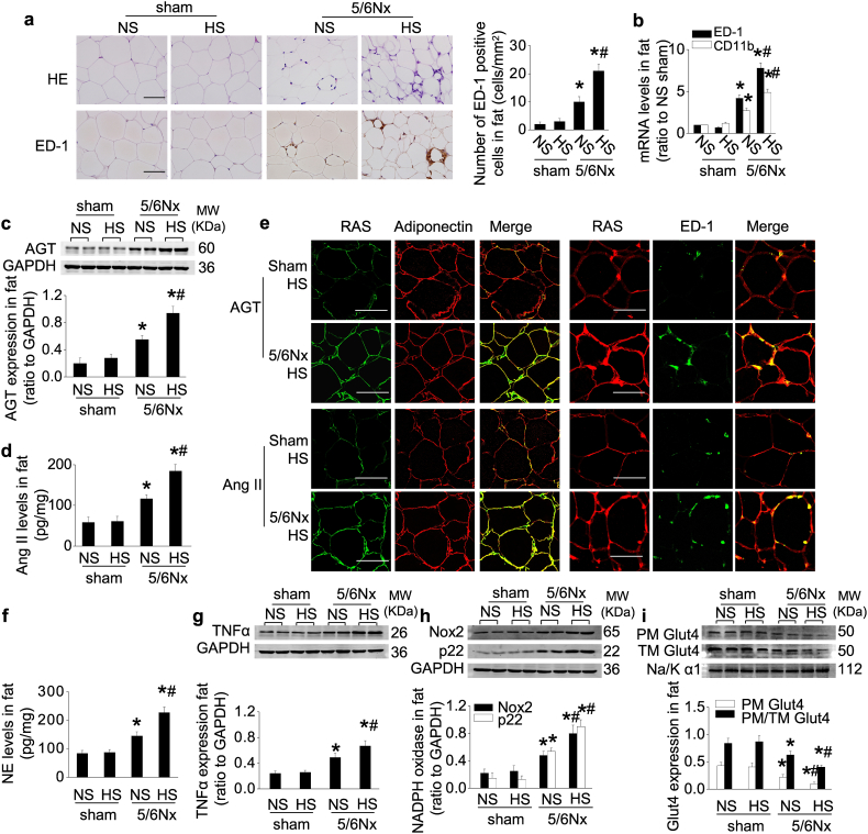
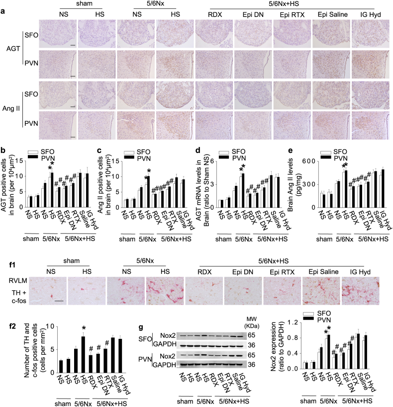
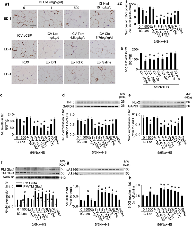
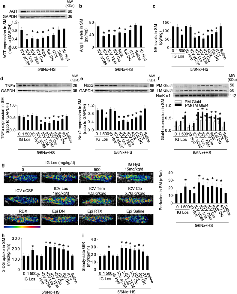
Background: Insulin resistance (IR) complicates chronic kidney disease (CKD). We tested the hypothesis that CKD activates a broad reflex response from the kidneys and the white adipose tissue to impair peripheral glucose uptake and investigated the role of salt intake in this process.

Methods: 5/6-nephrectomized rats were administered normal- or high-salt for 3 weeks. Conclusions were tested in 100 non-diabetic patients with stage 3-5 CKD.

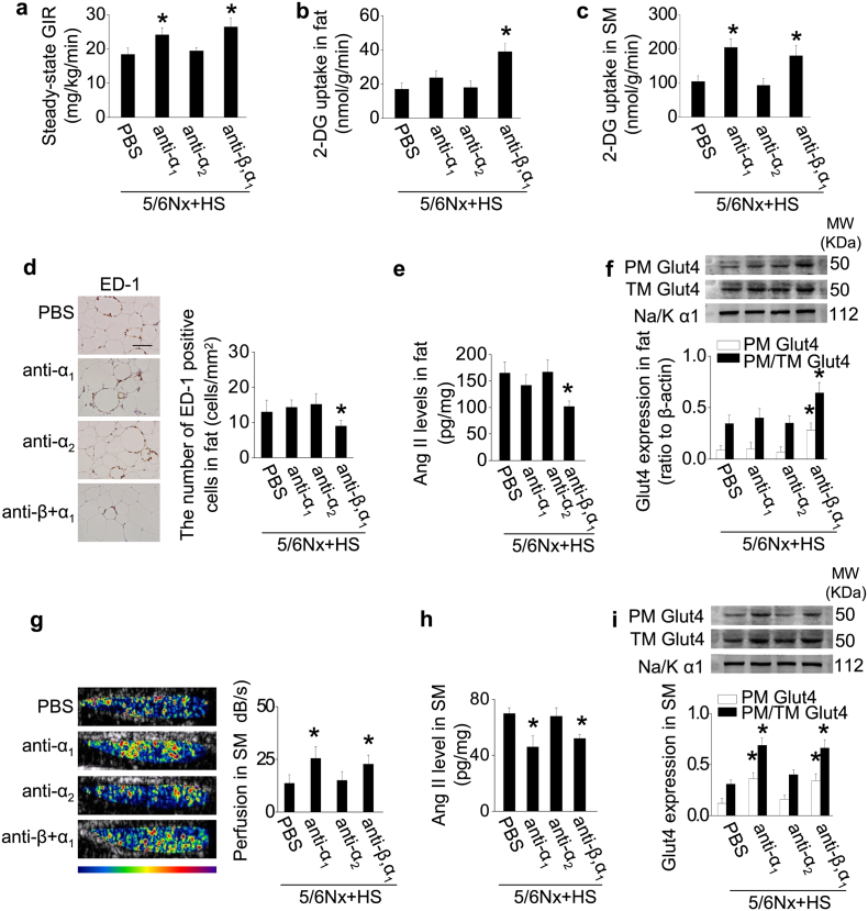
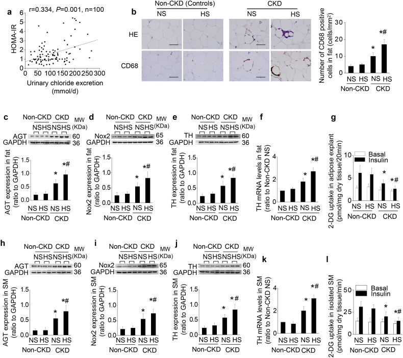
Findings: High-salt in 5/6-nephrectomized rats decreased insulin-stimulated 2-deoxyglucose uptake >25% via a sympathetic nervous system (SNS) reflex that linked the IR to reactive oxygen species (ROS) and the renin-angiotensin system (RAS) in brain and peripheral tissues. Salt-loading in CKD enhanced inflammation in adipose tissue and skeletal muscle, and enhanced the impairment of insulin signaling and Glut4 trafficking. Denervation of the kidneys or adipose tissue or deafferentation of adipose tissue improved IR >40%. In patients with non-diabetic CKD, IR was positively correlated with salt intake after controlling for cofounders (r = 0.334, P = 0.001) and was linked to activation of the RAS/SNS and to impaired glucose uptake in adipose tissue and skeletal muscle, all of which depended on salt intake.

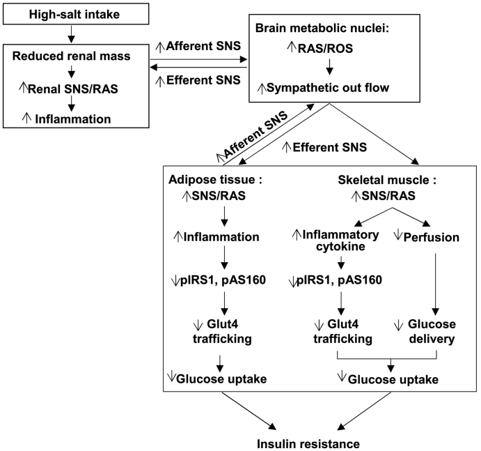
Interpretation: CKD engages a renal/adipose-cerebral-peripheral sympathetic reflex that activates the RAS/ROS axes to promote IR via local inflammation and impaired Glut4 trafficking that are enhanced by high-salt intake. The findings point to a role for blockade of RAS or α-and-β-adrenergic receptors to reduce IR in patients with CKD. FUND: National Natural Science Foundation of China.

Keywords: Adipose tissue; Chronic kidney disease; Insulin resistance; Renin-angiotensin system; Salt; Sympathetic reflex.


Figures

Unlabelled Image
Graphical abstract
Fig. 1
Fig. 1
CKD in rats impairs glucose uptake in adipose tissue and skeletal muscle and results in IR that are enhanced by high dietary salt. (a) Homeostasis model assessment of IR (HOMA-IR). (b) Steady-state glucose infusion rate (GIR) during hyperinsulinemic-euglycemic clamp. (c) Basal and insulin-stimulated whole-body glucose disposal. (d) Basal and insulin-stimulated hepatic glucose production. (e and f) Insulin-stimulated 2-deoxy-glucose (DG) uptake in adipose tissue (e) and skeletal muscle (SM) (f). (g–i) Serum levels of norepinephrine (NE) (g), 8-iso-prostane (h) and TNFα (i). Data are expressed as mean ± SD (n = 6 in each group). Effects of salt-loading in all panels except d are significantly higher in 5/6Nx rats than sham-operated rats (two-way ANOVA P < 0.05). *P < 0.05 vs sham rats with the same salt intake, #P < 0.05 vs 5/6Nx rats on normal-salt diet by unpaired t-test. NS, normal salt; HS, high salt.
Fig. 2
Fig. 2
CKD in rats activates the RAS and SNS in adipose tissue, resulting in inflammation and impaired Glut4 trafficking that are enhanced by high dietary salt. (a and b) Adipose macrophage infiltration: representative photos of HE staining and immunohistochemical staining of ED-1, quantitative analysis of ED-1-positive cells (a), and mRNA levels of ED-1 and CD11b (b) in adipose tissue. (c and d) Protein levels of AGT (c) and Ang II (d) in adipose tissue. (e) Localization of adipose AGT or Ang II determined by double-staining with an antibody against AGT or Ang II and an antibody recognizing adiponectin (marker of adipocyte) or ED-1 (marker of macrophage). (f–h) Norepinephrine (NE) level (f), and expression of TNFα (g) and NADPH oxidase subunit Nox2 and p22phox (h) in adipose tissue. (i) Glut4 expression in extracted plasma membrane (PM) and the ratio of PM to total membrane (TM) Glut4 under insulin stimulation. Scale bar = 100 μm. Data are expressed as mean ± SD (n = 6 in each group). Effects of salt-loading in all panels are significantly higher in 5/6Nx rats than sham-operated rats (two-way ANOVA P < 0.05). *P < 0.05 vs sham rats with the same salt intake, #P < 0.05 vs 5/6Nx rats on normal-salt diet by unpaired t-test.
Fig. 3
Fig. 3
CKD in rats activates RAS and SNS in skeletal muscle, reduces muscle perfusion, and impairs Glut4 trafficking that are enhanced by high dietary salt. (a and b) Protein levels of AGT (a) and Ang II (b) in skeletal muscle (SM). (c) Localization of muscle AGT or Ang II determined by double-staining with an antibody against AGT or Ang II and an antibody recognizing dystrophy (marker of myocyte) or Griffonia simplicifolia I lection (marker of microvessel). (d–f) Norepinephrine (NE) level (d), and expression of TNFα (e) and NADPH oxidase subunit Nox2 and p22phox (f) in SM. (g) Glut4 expression in extracted plasma membrane (PM) and the ratio of PM to total membrane (TM) Glut4 under insulin stimulation. (h) Hindlimb perfusion measured by contrast-enhanced ultrasound under insulin stimulation. Scale bar = 100 μm. Data are expressed as mean ± SD (n = 6 in each group). Effects of salt-loading in all panels are significantly higher in 5/6Nx rats than sham-operated rats (two-way ANOVA P < 0.05). *P < 0.05 vs sham rats with the same salt intake, #P < 0.05 vs 5/6Nx rats on normal-salt diet by unpaired t-test.
Fig. 4
Fig. 4
CKD in rats activates the cerebral RAS and TH expression via signals from the innervated kidney and adipose tissue that are enhanced by high dietary salt. (a–e) Central RAS expression in sham and 5/6Nx rats with normal- or high-salt intake. Renal denervation (RDX), denervation of epididymal adipose tissue (Epi DN), or deafferentation of epididymal adipose tissue with resiniferatoxin (Epi RTX) downregulates expression of central RAS in the subfornical organ (SFO) and paraventricular nucleus (PVN): representative photos of immunohistochemistry staining (a), quantitation of AGT or Ang II-positive cells (b&c), mRNA levels of AGT (d), and the protein levels of Ang II in brain (e). (f) Expression of tyrosine hydroxylase (TH) in c-fos positive neurons in rostral ventrolateral medulla (RVLM). (g) Expression of Nox2 in brain. Scale bar = 100 μm. Data are expressed as mean ± SD (n = 6 in each group). Effects of salt-loading in all panels are significantly higher in 5/6Nx rats than sham-operated rats (two-way ANOVA P < 0.05). *P < 0.05 vs 5/6Nx rats on normal-salt diet, #P < 0.05 vs 5/6Nx rats on high-salt diet by unpaired t-test. Epi Saline, injection of vehicle (saline) into bilateral epididymal fat pads.
Fig. 5
Fig. 5
Activation of adipose RAS and impaired Glut4 trafficking in 5/6Nx rats with high-salt intake are prevented by blockade of a renal-cerebral-adipose sympathetic reflex. (a) Increased adipose macrophage infiltration in 5/6Nx rats under high-salt condition was inhibited by intracerebroventricular administration (ICV) of losartan or clonidine, denervation of the kidneys (RDX) or epididymal fat pads (Epi DN), or deafferentation of epididymal fat pads with resiniferatoxin (Epi RTX). (b) Protein levels of Ang II in adipose tissue. (c–e) Norepinephrine (NE) level (c), and expression of TNFα (d) and Nox2 (e) in adipose tissue. (f) Glut4 expression in extracted plasma membrane (PM) and the ratio of PM to total membrane (TM) Glut4 under insulin stimulation. (g) Insulin-stimulated phosphorylation of AS160 in adipose tissue. (h) Insulin-stimulated 2-deoxy-glucose (DG) uptake in adipose tissue. Scale bar = 100 μm. Data are expressed as mean ± SD (n = 6 in each group). *P < 0.05 vs 5/6Nx rats under high-salt condition given vehicle (0 mg/kg/d inhibitor) by unpaired t-test.
Fig. 6
Fig. 6
Activation of the RAS/ROS and impaired Glut4 trafficking in the skeletal muscle of 5/6Nx rats with high-salt intake are prevented by blockade of a renal/adipose-cerebral-muscle sympathetic reflex. (a and b) Overexpression of skeletal muscle (SM) AGT (a) and Ang II (b) in 5/6Nx rats under high-salt condition was inhibited by intracerebroventricular administration (ICV) of losartan or clonidine, denervation of the kidneys (RDX) or epididymal fat pads (Epi DN), or deafferentation of epididymal fat pads with resiniferatoxin (Epi RTX). (c–e) Norepinephrine (NE) level(c), and expression of TNFα (d) and Nox2 (e) in SM. (f) Glut4 expression in extracted plasma membrane (PM) and the ratio of PM to total membrane (TM) Glut4 under insulin stimulation. (g) Hindlimb perfusion measured by contrast-enhanced ultrasound under insulin stimulation. (h) Insulin-stimulated 2-deoxy-glucose (DG) uptake in SM. (i) Steady-state glucose infusion rate (GIR) during hyperinsulinemic-euglycemic clamp. Data are expressed as mean ± SD (n = 6 in each group). *P < 0.05 vs 5/6Nx rats under high-salt condition given vehicle (0 mg/kg/d inhibitor) by unpaired t-test.
Fig. 7
Fig. 7
Adrenergic receptors mediate IR in rats with CKD under high-salt condition. To further confirm the role of SNS in IR in 5/6Nx rats under high-salt condition, CKD rats on high-salt diet were treated with α1-adrenergic receptor (AR) antagonist (anti-α1; prazosin), or α2-AR antagonist (anti-α2; atipamezole), or β- and α1-AR antagonist (anti-β,α1; carvedilol) for 3 weeks. (a) Steady-state glucose infusion rate (GIR) during hyperinsulinemic-euglycemic clamp. (b and c) Insulin-stimulated 2-deoxy-glucose (DG) uptake in adipose tissue (b) and skeletal muscle (SM) (c). (d) Macrophage infiltration in adipose tissue: representative photos of immunohistochemical staining of ED-1, and quantitative analysis of ED-1-positive cells. (e) Levels of Ang II in adipose tissue. (f) Adipose Glut4 expression in extracted plasma membrane (PM) and the ratio of PM to total membrane (TM) Glut4 under insulin stimulation. (g) Hindlimb perfusion measured by contrast-enhanced ultrasound under insulin stimulation. (h) Levels of Ang II in SM. (I) SM Glut4 expression in extracted PM and the ratio of PM to TM Glut4 under insulin stimulation. Scale bar = 100 μm. Data are expressed as mean ± SD (n = 6 in each group). *P < 0.05 vs 5/6Nx rats under high-salt condition given vehicle (PBS) by unpaired t-test.
Fig. 8
Fig. 8
The level of salt intake determines IR in non-diabetic patients with stage 3–5 CKD. (a) Correlation between daily urinary chloride excretion and the score of HOMA-IR after adjusting for age, body mass index, triglyceride, eGFR, systolic blood pressure, and caloric intake in 100 non-diabetic patients with CKD stage 3–5 using a partial correlation analysis. (b–l) To evaluate the alterations in adipose tissue and skeletal muscle (SM) in CKD under high-salt condition, tissue samples from predialysis patients with normal-salt (<5 g/day) or high-salt (>10 g/day) intake are compared with age- and salt intake-matched non-CKD patients (controls). Compared to CKD patients with normal salt consumption, CKD patients with high-salt consumption exhibit increased adipose inflammation (b), overexpression of AGT, Nox2, and TH in adipose tissue (c–f) and skeletal muscle (h–k), and impaired insulin-stimulated glucose uptake in adipose tissue explants (g) and isolated muscle strips (l). Scale bar =100 μm. Data are expressed as mean ± SD in b–l (n = 6 in each group). Effects of salt-loading in all panels are significantly higher in CKD patients than non-CKD patients (two-way ANOVA P < 0.05). *P < 0.05 vs non-CKD patients (controls), #P < 0.05 vs CKD patients with normal salt diet in b-l by unpaired t-test.
Fig. 9
Fig. 9
The schematic diagram summarizing the steps that link a CKD in rats to insulin resistance. CKD activates the afferent sympathetic drive from the kidney and adipose tissue to the peripheral tissues that reduces the insulin-stimulated glucose uptake. This acts in a positive feedback mode to interlink the RAS/ROS systems in the brain with the RAS/ROS system in the tissues responsible for glucose disposal (adipose tissue and skeletal muscle) and thereby to mediate insulin resistance in CKD, and its enhancement by a high-salt intake.

Similar articles

Cited by

References

    1. de Boer I.H., Zelnick L., Afkarian M. Impaired glucose and insulin homeostasis in moderate-severe CKD. J Am Soc Nephrol. 2016;27(9):2861–2871. - PMC - PubMed
    1. Xu H., Huang X., Arnlov J. Clinical correlates of insulin sensitivity and its association with mortality among men with CKD stages 3 and 4. Clin J Am Soc Nephrol. 2014;9(4):690–697. - PMC - PubMed
    1. Shinohara K., Shoji T., Emoto M. Insulin resistance as an independent predictor of cardiovascular mortality in patients with end-stage renal disease. J Am Soc Nephrol. 2002;13(7):1894–1900. - PubMed
    1. Kaartinen K., Syrjanen J., Porsti I. Insulin resistance and the progression of IgA glomerulonephritis. Nephrol Dial Transplant. 2007;22(3):778–783. - PubMed
    1. Spoto B., Pisano A., Zoccali C. Insulin resistance in chronic kidney disease: A systematic review. Am J Physiol Renal Physiol. 2016;311(6) [F1087-F108] - PubMed

MeSH terms