. 2018 Dec 1;26(22):5962-5972.
doi: 10.1016/j.bmc.2018.11.004. Epub 2018 Nov 5.

Modifying aroylhydrazone prochelators for hydrolytic stability and improved cytoprotection against oxidative stress

Affiliations

Modifying aroylhydrazone prochelators for hydrolytic stability and improved cytoprotection against oxidative stress

Qin Wang et al. Bioorg Med Chem. .

Abstract

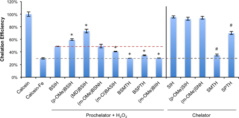
BSIH ((E)-N'-(2-(4,4,5,5-tetramethyl-1,3,2-dioxaborolan-2-yl)benzylidene)isonicotinohydrazide) is a prodrug version of the metal chelator SIH ((E)-N'-(2-hydroxybenzylidene)isonicotinohydrazide) in which a boronate group prevents metal chelation until reaction with hydrogen peroxide releases SIH, which is then available for sequestering iron(III) and inhibiting iron-catalyzed oxidative damage. While BSIH has shown promise for conditionally targeting iron sequestration in cells under oxidative stress, the yield of SIH is limited by the fact that BSIH exists in cell culture media as an equilibrium mixture with its hydrolysis products isoniazid and 2-formylphenyl boronic acid. In the current study, several BSIH analogs were evaluated for their hydrolytic stability, reaction outcomes with H2O2, and prochelator-to-chelator conversion efficiency. Notably, the para-methoxy derivative (p-OMe)BSIH ((E)-N'-(5-methoxy-2-(4,4,5,5-tetramethyl-1,3,2-dioxaborolan-2-yl)benzylidene)isonicotinohydrazide) and the meta-, para-double substituted (MD)BSIH ((E)-N'-((6-(4,4,5,5-tetramethyl-1,3,2-dioxaborolan-2-yl)benzo[d][1,3]dioxol-5-yl)methylene)isonicotinohydrazide) showed 1.3- and 1.9-fold improved hydrolytic stability compared to BSIH, respectively, leading to a 22 and 50% increase in chelator released. Moreover, both prochelators were found to protect retinal pigment epithelial cells stressed with either H2O2 or paraquat insult.

Keywords: Chelating agent; Cytoprotection; Fenton chemistry; Iron; Oxidative stress.


Figures

Figure 1.
Figure 1.
Calcein fluorescence assay with chelators and H2O2-activated prochelators. Buffered solutions of prochelators were pre-incubated overnight with excess H2O2 then diluted to 4 μM and equilibrated with 2 μM calcein-Fe3+ for 2 h prior to measurement of calcein fluorescence emission. Chelation efficiency was expressed as a percentage of the calcein emission of the free calcein control. The grey dashed line indicates the residual fluorescence response of 2 μM calcein quenched by 1 equiv of Fe3+. The red dashed line indicates the ~50% calcein emission restored by the reaction mixture of BSIH and H2O2. Data are presented as mean ± SD, n = 3; statistical significance (t-test), * p < 0.05, compared to the BSIH group. # p < 0.05, compared to the SIH group.
Figure 2.
Figure 2.
Hydrolysis of (p-OMe)BSIH observed by 1H NMR spectroscopy. The aromatic regions of 1H NMR spectra of (a) (p-OMe)BSIH (10 mM in DMSO-d6) (b) (p-OMe)BSIH (500 μM in D2O, with 5% DMSO-d6 after 1-h equilibration) and (c) INH (500 μM in D2O, with 5% DMSO-d6). The arrows indicate the peaks corresponding to the aromatic protons of isoniazid (red).
Figure 3.
Figure 3.
Hydrolytic stability of (m-OMe)BIIH analyzed by 1H NMR spectroscopy. The aromatic regions of 1H NMR spectra of (a) (m-OMe)BIIH (10 mM in DMSO-d6) and (b) (m-OMe)BIIH (500 μM in D2O, with 5% DMSO-d6).
Figure 4.
Figure 4.
The hydrolysis equilibria of 100 μM (a) (p-OMe)BSIH and (c) (MD)BSIH observed by UV-Vis spectrophotometry; spectra of reaction mixtures of 100 μM INH with100 μM of (b) (p-OMe)FBA or (d) (MD)FBA, pH 7.4 over the course of 30 min.
Figure 5.
Figure 5.
The reactions of prochelators (p-OMe)BSIH and (MD)BSIH with H2O2 observed by 1H NMR. The aromatic regions of 1H NMR spectra of (a) the reaction mixture of (p-OMe)BSIH (500 μM in D2O, with 5% DMSO-d6) with H2O2 (25 mM) for 12 h (b) (p-OMe)SIH (500 μM in D2O, with 5% DMSO-d6) (c) INH (500 μM in D2O, with 5% DMSO-d6) (d) the reaction mixture of (MD)BSIH (200 μM in D2O, with 2% DMSO-d6) with H2O2 (10 mM) for 12 h. See main text for description of arrows and peak labels.
Figure 6.
Figure 6.
Inherent cytotoxicity of selected aroylhydrazone prochelators after 72-h incubation in ARPE-19 retinal pigment epithelial cells. Error bars represent standard deviations from triplicate runs (n = 3).
Figure 7.
Figure 7.
Cytoprotective effects of aroylhydrazone prochelators from oxidative damage induced by H2O2 in ARPE-19 cells. ARPE-19 cells were pre-incubated with various concentrations of aroylhydrazone prochelators for 5 h prior to exposure to H2O2. Cell viability was measured 19 h after peroxide treatment and expressed as percentage of the untreated control group (100%). Error bars represent standard deviations from triplicate runs (n = 3).
Figure 8.
Figure 8.
Cytoprotective effects of aroylhydrazone prochelators against oxidative damage induced by 10 mM paraquat in ARPE-19 cells. Cell viability was measured after 48 h and expressed as percentage of the untreated control group (100%). Error bars represent standard deviations from triplicate runs (n = 3).
Chart 1.
Chart 1.
Synthetic scheme and structures of prochelators used in this study. Structures of the corresponding chelators replace the boronic ester or boronic acid with an OH functional group; names of chelators mirror those of the prochelators, just without the “B”.
Scheme 1.
Scheme 1.
Key steps involved in BSIH hydrolytic degradation, peroxide activation, and Fe3+ chelation.

Similar articles

Cited by

References

    1. Stockwell BR; Friedmann Angeli JP; Bayir H; Bush AI; Conrad M; Dixon SJ; Fulda S; Gascón S; Hatzios SK; Kagan VE; Noel K; Jiang X; Linkermann A; Murphy ME; Overholtzer M; Oyagi A; Pagnussat GC; Park J; Ran Q; Rosenfeld CS; Salnikow K; Tang D; Torti FM; Torti SV; Toyokuni S; Woerpel KA; Zhang DD. Ferroptosis: A Regulated Cell Death Nexus Linking Metabolism, Redox Biology, and Disease. Cell 2017; 171: 273–285. - PMC - PubMed
    1. Zecca L; Youdim MB; Riederer P; Connor JR; Crichton RR. Iron, brain ageing and neurodegenerative disorders. Nat. Rev. Neurosci 2004; 5: 863–873. - PubMed
    1. Kell D Towards a unifying, systems biology understanding of large-scale cellular death and destruction caused by poorly liganded iron: Parkinson’s, Huntington’s, Alzheimer’s, prions, bactericides, chemical toxicology and others as examples. Arch. Toxicol 2010; 84: 825–889. - PMC - PubMed
    1. Zhou T; Ma Y; Kong X; Hider RC. Design of iron chelators with therapeutic application. Dalton Trans 2012; 41: 6371–6389 - PubMed
    1. Dunaief JL. Iron induced oxidative damage as a potential factor in age-related macular degeneration: The Cogan Lecture. Invest. Ophthalmol. Vis. Sci 2006; 47: 4660–4664. - PubMed

Publication types