. 2018 Dec;30(12):3038-3057.
doi: 10.1105/tpc.18.00641. Epub 2018 Nov 14.

Analysis of Two New Arabinosyltransferases Belonging to the Carbohydrate-Active Enzyme (CAZY) Glycosyl Transferase Family1 Provides Insights into Disease Resistance and Sugar Donor Specificity

Affiliations

Analysis of Two New Arabinosyltransferases Belonging to the Carbohydrate-Active Enzyme (CAZY) Glycosyl Transferase Family1 Provides Insights into Disease Resistance and Sugar Donor Specificity

Thomas Louveau et al. Plant Cell. 2018 Dec.

Abstract

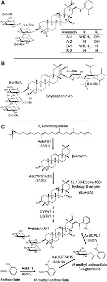
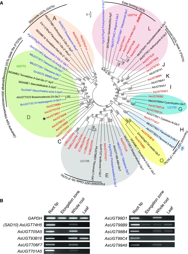
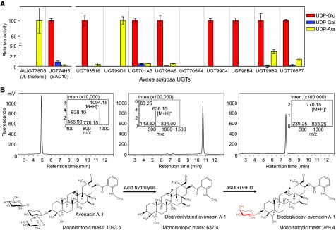
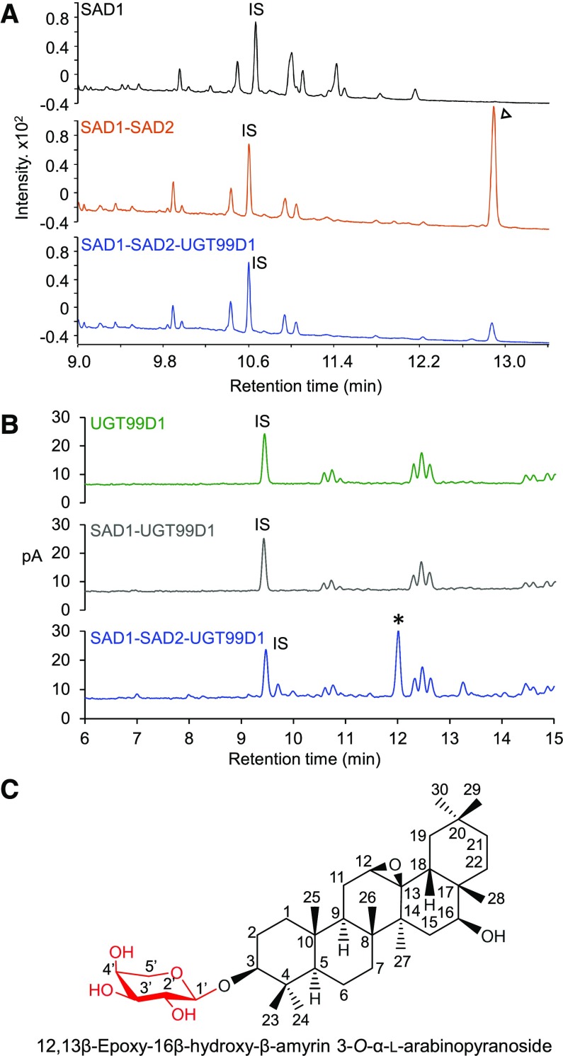
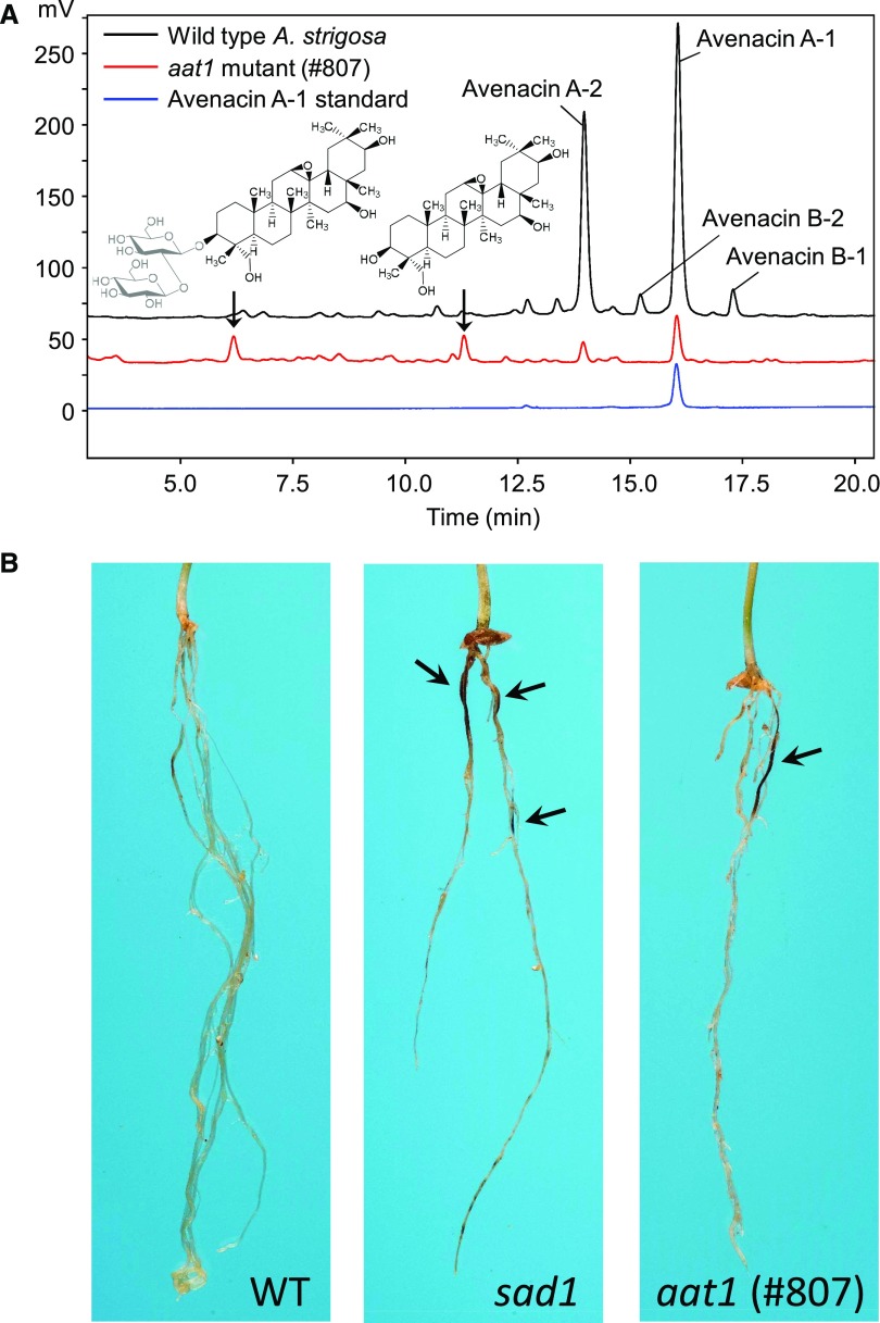
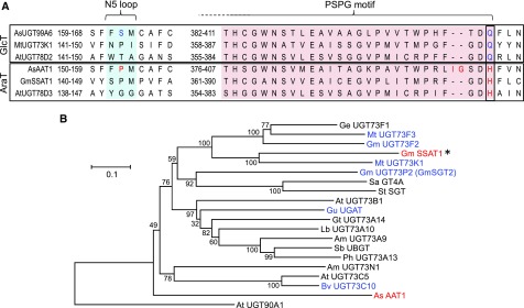
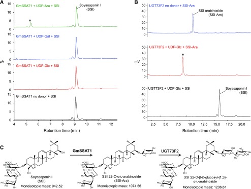
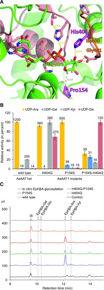
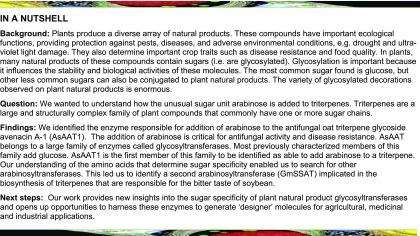
Glycosylation of small molecules is critical for numerous biological processes in plants, including hormone homeostasis, neutralization of xenobiotics, and synthesis and storage of specialized metabolites. Glycosylation of plant natural products is usually performed by uridine diphosphate-dependent glycosyltransferases (UGTs). Triterpene glycosides (saponins) are a large family of plant natural products that determine important agronomic traits such as disease resistance and flavor and have numerous pharmaceutical applications. Most characterized plant natural product UGTs are glucosyltransferases, and little is known about enzymes that add other sugars. Here we report the discovery and characterization of AsAAT1 (UGT99D1), which is required for biosynthesis of the antifungal saponin avenacin A-1 in oat (Avena strigosa). This enzyme adds l-Ara to the triterpene scaffold at the C-3 position, a modification critical for disease resistance. The only previously reported plant natural product arabinosyltransferase is a flavonoid arabinosyltransferase from Arabidopsis (Arabidopsis thaliana). We show that AsAAT1 has high specificity for UDP-β-l-arabinopyranose, identify two amino acids required for sugar donor specificity, and through targeted mutagenesis convert AsAAT1 into a glucosyltransferase. We further identify a second arabinosyltransferase potentially implicated in the biosynthesis of saponins that determine bitterness in soybean (Glycine max). Our investigations suggest independent evolution of UDP-Ara sugar donor specificity in arabinosyltransferases in monocots and eudicots.


Figures

Figure 1.
Figure 1.
Triterpene Glycoside Structures and Avenacin Biosynthesis. (A) Avenacins: antifungal defense compounds from oat. (B) Soyasaponin Ab, a triterpene glycoside associated with bitterness and anti-feedant activity in soybean. (C) Current understanding of the avenacin biosynthetic pathway. The major avenacin, A-1, is synthesized from the linear isoprenoid precursor 2,3-oxidosqualene. 2,3-Oxidosqualene is cyclized by the triterpene synthase AsbAS1 (SAD1) to the pentacyclic triterpene scaffold β-amyrin (Haralampidis et al., 2001). β-Amyrin is then oxidized to EpHβA by the cytochrome P450 enzyme AsCYP51H10 (SAD2; Geisler et al., 2013). Subsequent, as-yet uncharacterized modifications involve a series of further oxygenations and addition of a branched trisaccharide moiety at the C-3 position, initiated by introduction of an l-Ara. Acylation at the C-21 position is performed by the AsSCPL1 (SAD7). The acyl donor used by AsSCPL1 is N-methyl anthranilate glucoside, which is generated by the methyl transferase AsMT1 and the glucosyl transferase AsUGT74H5 (SAD10; Mugford et al., 2009, 2013; Owatworakit et al., 2013).
Figure 2.
Figure 2.
Mining for Candidate Avenacin Glycosyltransferase Genes (A) Phylogenetic tree of UGTs expressed in A. strigosa root tips (red;listed in Supplemental Table 3). Characterized triterpenoid glycosyltransferases from other plant species (blue) and other biochemically characterized plant UGTs (black) are also included (listed in Supplemental Table 4). The UGT groups are as defined by Ross, et al. (2001). Some of the most common groups of enzyme activities are indicated. The tree was constructed using the Neighbor Joining method with 1000 bootstrap replicates (% support for branch points shown). The scale bar indicates 0.1 substitutions per site at the amino acid level. The alignment file is available as Supplemental Data Set 1. (B) Expression profiles of selected oat UGT genes (RT-PCR). Tissues were collected from 3-d-old A. strigosa seedlings. The characterized avenacin biosynthetic gene AsUGT74H5 (Sad10) and the GAPDH housekeeping gene are included as controls.
Figure 3.
Figure 3.
Biochemical Characterization of Candidate Oat UGTs (A) Evaluation of sugar donor specificity of recombinant oat UGTs using the universal acceptor TCP. Relative activities with different sugar nucleotide donors are shown. Conversion of TCP to TCP glycoside was monitored by spectrophotometry at 405 nm. The previously characterized oat N-methyl anthranilate glucosyltransferase AsUGT74H5 (SAD10) and Arabidopsis flavonoid arabinosyltransferase UGT78D3 were included as controls. Values are means of three biological replicates; error bars represent sds. (B) LC-MS profiles for avenacin A-1 (left), deglycosylated avenacin A-1 (middle), and the product generated by incubation of deglycosylated avenacin A-1 with AsUGT99D1 (right) (detection by fluorescence; excitation and emission wavelengths 353 nm and 441 nm, respectively). The hydrolyzed avenacin product is shown with an intact 12,13-epoxide (*), but this epoxide may have rearranged to a ketone under the acidic hydrolysis conditions resulting in deglycosylated 12-oxo-avenacin A-1, as observed in Geisler et al. (2013).
Figure 4.
Figure 4.
Transient Expression of AsAAT1 in N. benthamiana (A) GC-MS analysis of extracts from agro-infiltrated N. benthamiana leaves. Comparison of the metabolite profiles of leaves expressing SAD1 and SAD2 (red) or SAD1, SAD2, and UGT99D1 (blue). EpHβA, identified previously as the coexpression product of SAD1 and SAD2, is indicated by an arrowhead (Geisler et al., 2013). The upper chromatogram consists of a control from leaves expressing GFP only (black). (B) HPLC with CAD chromatograms of extracts from N. benthamiana leaves expressing UGT99D1 alone (green), SAD1 and UGT99D1 (gray), and SAD1, SAD2, and UGT99D1 together (blue). The new compound that accumulated in the latter (tR 12.0 min) is indicated by an asterisk. This compound was not detected when UGT99D1 was expressed on its own or with SAD1. The IS was digitoxin (1 mg/g of dry weight). (C) Structure of the UGT99D1 product (see Supplemental Figure 4 for NMR assignment).
Figure 5.
Figure 5.
Biochemical Analysis of aat1 Mutant and Susceptibility to Take-All Disease. (A) HPLC-CAD analysis of methanolic root extracts from seedlings of the A. strigosa wild-type accession and avenacin-deficient mutant line #807 (aat1). New metabolites detected in the mutant are arrowed and inferred structures are shown based on the corresponding ion chromatograms (Supplemental Figure 5D). (B) aat1 (#807) has enhanced disease susceptibility. Images of representative seedlings of wild-type A. strigosa, the sad1 mutant #610 (Haralampidis et al., 2001), and the aat1 mutant (this study) are shown. Seedlings were inoculated with the take-all fungus (G. graminis var tritici). The dark lesions on the roots are symptoms of infection and are indicated by arrows.
Figure 6.
Figure 6.
An Arabinosyltransferase from Soybean. (A) Alignment of the amino acid sequences of the oat, soybean, and Arabidopsis arabinosyltransferases with the closest characterized glucosyltransferases in the region of the PSPG motif and the N5 loop. The His residue that is conserved in the arabinosyltransferases is shown in red. P154 and the two additional amino acids IG of AsAAT1 are also highlighted. (B) Phylogenetic analysis of glycosyltransferases from group D belonging to the UGT73 family. GmSSAT1 and AsAAT1 are indicated in red, and other characterized triterpenoid glycosyltransferases in blue (see Supplemental Table 4 for further details). The tree was constructed using the Neighbor Joining method with 1000 bootstrap replicates (percentage support shown at branch points), and rooted with UGT90A1, an Arabidopsis UGT from group C. The scale bar indicates 0.1 substitutions per site at the amino acid level. The alignment file is available as Supplemental Data Set 3.
Figure 7.
Figure 7.
Biochemical Characterization of GmSSAT1. (A) HPLC-CAD chromatogram of in vitro assays performed with recombinant GmSSAT1 and different UDP-sugars. GmSSAT1 was incubated for 40 min at 25°C with 100 μM SSI and 300 μM UDP-sugars. A major product is detected only in the presence of UDP-Ara (*) (see Supplemental Figure 7A for MS analysis). (B) HPLC-CAD analysis of reactions in which the previously characterized soyasaponin glucosyltransferase UGT73F2 (Sayama et al., 2012) was assayed for activity toward the GmSSAT1 product. Recombinant UGT73F2 was incubated overnight with 400 μM UDP-Glc and ∼100 μM of the GmSSAT1 product SSI-Ara. SSI-Ara (tR: 10.6 min) was completely converted to a new product with a tR of 8.6 min (*). MS analysis of this product is shown in Supplemental Figure 7B. No product was detected in the absence of UDP-Glc or if the acceptor was replaced by SSI. C, Schematic showing successive glycosylation of soyasaponin I by GmSSAT1 and UGT73F2.
Figure 8.
Figure 8.
Determinants of Sugar Donor Sugar Specificity of AsAAT1. (A) Model of AsAAT1 with bound UDP-Ara (carbons of the Ara numbered). The protein is represented in green ribbons with the PSPG motif in salmon, including the side chains of highly conserved residues. The His404 and Pro154 residues are shown in magenta. Potential hydrogen bonds are shown with yellow dots, and the distance between P154 and C-5 of UDP-Ara with gray dots. The homology model was generated using I-TASSER software (Yang et al., 2015), based on the crystal structure of M. truncatula UGT71G1 complexed with UDP-Glc (PDB: 2ACW). The loop shown in orange was reconstructed using MODELER (Sali and Blundell, 1993). UDP-Ara was inserted into the active site and the complex was relaxed by energy minimization using GROMACS. (B) Comparison of the glycosylation activity of the AsAAT1 wild-type and mutant enzymes when supplied with each of the four sugar donors (UDP-Ara, UDP-Gal, UDP-Xyl, or UDP-Glc). Initial velocities were measured using 30 µM deglycosylated avenacin A-1 as acceptor and 5 µM UDP-sugar donor using five time points. The heights of the bars are drawn relative to the highest activity observed for each recombinant enzyme (mean ± sd, n = 3). Activities reported above each bar are in nM.min−1. (C) HPLC-CAD analysis of extracts from N. benthamiana leaves expressing SAD1 and SAD2 together with GFP (black), wild-type AsAAT1 enzyme (blue), AsAAT1-H404Q (green), AsAAT1-P154S (red), and AsAAT1-H404Q-P154S (orange). The top trace (in gray) shows the products of in vitro reaction of EpHβA with the four sugar donors (reactions performed separately and then pooled) for reference. LC-MS analysis was used to confirm the identities of the new products (Supplemental Figure 8C). The IS is digitoxin (1 mg/g dry weight).
None

Similar articles

Cited by

References

    1. Achnine L., Huhman D.V., Farag M.A., Sumner L.W., Blount J.W., Dixon R.A. (2005). Genomics-based selection and functional characterization of triterpene glycosyltransferases from the model legume Medicago truncatula. Plant J. 41: 875–887. - PubMed
    1. Armah C.N., Mackie A.R., Roy C., Price K., Osbourn A.E., Bowyer P., Ladha S. (1999). The membrane-permeabilizing effect of avenacin A-1 involves the reorganization of bilayer cholesterol. Biophys. J. 76: 281–290. - PMC - PubMed
    1. Augustin J.M., Kuzina V., Andersen S.B., Bak S. (2011). Molecular activities, biosynthesis and evolution of triterpenoid saponins. Phytochemistry 72: 435–457. - PubMed
    1. Augustin J.M., Drok S., Shinoda T., Sanmiya K., Nielsen J.K., Khakimov B., Olsen C.E., Hansen E.H., Kuzina V., Ekstrøm C.T., Hauser T., Bak S. (2012). UDP-glycosyltransferases from the UGT73C subfamily in Barbarea vulgaris catalyze sapogenin 3-O-glucosylation in saponin-mediated insect resistance. Plant Physiol. 160: 1881–1895. - PMC - PubMed
    1. Benkert P., Tosatto S.C., Schomburg D. (2008). QMEAN: A comprehensive scoring function for model quality assessment. Proteins 71: 261–277. - PubMed

Publication types

MeSH terms

LinkOut - more resources